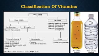
DEPARTMENT OF BIOCHEMISTRY
Prepared by:-
Supriya singh
Definition
• Vitamins are organic nutrients that are required in small quantities
for a variety of biochemical functions, normal maintenance of
optimum growth and health of the organism and its deficiency can
lead to many complications and must be supplied by the diet.
Classification Of Vitamins
Comparison Of Two Types Of Vitamins
Fat Soluble Vitamins Water Soluble Vitamins
Major Vitamins A,D,E, and K Vitamins B and Vitamin C
Solubility Soluble in fats and
organic solvents
Water Soluble
Digestion and absorption Along with lipids requires
bile salts
Easily absorbed in
intestine
Carrier Protein Present No Carrier Protein
Excretion Not Excreted Excreted
Deficiency Manifests only when
stores are depleted
Manifests rapidly as there
is no storage
Storage Stored in liver No storage
Toxicity Hypervitaminosis may
result
Unlikely, since excess is
excreted
Treatment of deficiency Single large doses may
prevent deficiency
Regular dietary supply is
required
Chemistry Of Vitamin- A
• Vitamin A is fat soluble.
• Required for vision, repair, reproduction, growth and tissue differentiation.
• Important forms of vitamin A are –
1. Retinol (Vitamin A alcohol)
2. Retinal (Vitamin A aldehyde)
3. Retinoic acid (Vitamin A acid)
4. β-Carotene (Pro-vitamin)
• Compounds with Vitamin A activity are referred to as Retinoid.
• Vitamin A is composed of ‘β-IONONE RING’ to which ‘POLYISOPRENOID SIDE CHAIN’ is
attached.
• Polyisoprenoid chain –all trans configuration, contains 4 double bonds, has 2
methyl groups with terminal carbon having ‘R’ group.
• ‘R’ Group –alcohol/aldehyde/acid
• β-Ionone ring –contains 1 double bond with 3 methyl groups.
• The all-trans variety of retinal, also called vitamin A1.
• Biologically important compound is 11-cis-retinal.
β – Ionone ring
All trans-retinal 11-cis-retinal
Unsaturated Isoprenoid side chain (all trans)
Attaching group
Absorption, Storage And Transportation
Wald’s Visual Cycle
• G. Wald was awarded Nobel Prize in 1967
• Studied role of vitamin A in vision.
• The visual cycle is the biological conversion of a photon into an electrical signal in
the retina.
1) Generation of nerve impulse
2) Regeneration of 11-cis-retinal
1) Generation Of Nerve Impulse
• Rhodopsin presents in rods is made up of opsin and 11-cis-retinal, plays pivotal role
in vision and found in the photoreceptor cells of retina.
• The absorption of light by Rhodopsin causes isomerization of the retinaldehyde from
11-cis to all-trans, and a conformational change in opsin.
• The unstable intermediates form of Rhodopsin , batho-rhodopsin, Lumirhodopsin,
Metarhodopsin-I, Metarhodopsin-II and finally Opsin, occurs within picoseconds of
illumination.
• This results in the release of retinaldehyde from the protein, and the initiation of a
nerve impulse.
2) Regeneration of 11-cis-retinal
• Trans-retinal enters the blood circulation.
• Cis-retinal is generated, reaches in retina.
• All-trans-retinal is isomerized to 11-cis-retinal in retina by enzyme retinal isomerase.
• 11-cis-retinal recombine with opsin to regenerate rhodopsin.
• All-trans-retinal transported to liver and reduced to all-trans-retinol by alcohol
dehydrogenase (ADH), an NADPH dependent enzyme.
• All-trans-retinol is isomerized to 11-cis-retinol and then oxidized to 11-cis-retinal in
liver.
Wald’s Visual Cycle
Dark Adaptation Mechanism
• Torsten Wiesel -1981 Nobel prize.
• When a person shifts suddenly from bright light to a dim light, rhodopsin
stores are depleted in rods and vision is impaired.
• After a few minutes, rhodopsin is resynthesized and vision is improved.
• Increased in Vitamin-A deficiency.
Photosensitive Cells In Retina
• Rods
Dim Light
Contains Rhodopsin (opsin+11cis-retinal)
Def. of 11cis-retinal - Increased Dark Adaptation Time
(Night Blindness)
• Cones
• Bright Light & Color Vision
• Contains conopsin (photosensitive protein)
• 3 types of cones – characterised by different conopsin that is maximally sensitive to:
• Blue – Cyanopsin
• Green – Iodopsin
• Red – Porphyropsin
Functions Of Vitamin A
• Vitamin A is essential for vision.
• Cell Growth and Differentiation- Retinoic acid act like steroid hormone.
• Reproduction- Retinol acts like a steroid hormone.
• Maintenance of healthy epithelial cells.
• Synthesis of Glycoproteins- Retinyl phosphate synthesized from Retinol.
• Hematopoiesis- Retinol and Retinoic acid involved in synthesis of
transferrin.
• Maintenance of Immune System- To fight against various infections.
• Anti-oxidant activity- β carotenes reduce the risk of cancer and prevent heart attacks.
Recommended Dietary Allowance (RDA)
• The daily requirement of vitamin A is expressed as retinol equivalents (RE)
rather than International Units ( IU) .
1 IU = 0.3 mg of retinol.
1 RE = 1 μg of retinol or 6 μg of beta carotene.
• The recommended daily allowance (RDA) for
i. Children = 400-600 μg/day
ii. Men = 750-1000 μg /day
iii. Women = 750 μg/day
iv. Pregnancy = 1000 μg/day
Dietary Sources Of Vitamin A
 Animal sources –
Milk, butter, cream, cheese, Liver, fish, and eggs are
excellent food sources for vitamin A.
 Vegetable sources of pro-vitamin A carotenoids-
Dark green and deeply colored fruits and vegetables.
Papaya, mango, pumpkins and green leafy
vegetables (spinach) are other good sources for
vitamin A activity.
Deficiency Manifestations Of Vitamin A
 The deficiency manifestations are related to the eyes, skin and growth.
 Deficiency manifestations of the eyes : -
1. Night Blindness or Nyctalopia
one of the earliest symptoms of vitamin A deficiency. The
individuals have difficulty to see in dim light since the dark
adaptation time is increased.
2. Xerophthalmia
The conjunctiva becomes dry, thick and wrinkled. The
conjunctiva gets keratinized and looses its normal transparency.
Dryness spreads to cornea and became glazy and lustreless due
to keratinization.
Night Blindness
Xerophthalmia
3. Bitot’s Spots
Due to increased thickness of conjunctiva in certain
areas, white triangular plaques are seen.
4. Keratomalacia
When xerophthalmia persists for a long time,
corneal ulceration and degeneration occur. This
results in the destruction of cornea, a condition
referred to as keratomalacia.
Bitot’s Spots
Keratomalacia followed by Blindness
 Effect on growth : -
Vitamin A deficiency results in growth retardation, especially failure of
skeletal formation. This may be due to defective synthesis of chondroitin
sulfate.
 Effect on growth skin and epithelial cells : -
i) Follicular hyperkeratosis or phrynoderma is a condition characterized by
excessive development of keratin in hair follicles. The skin becomes rough
and dry.
ii) Keratinization of epithelial cells of gastrointestinal tract, urinary tract and
respiratory tract have been observed.
Follicular Hyperkeratosis
Causes For Deficiency Of Vitamin A
1. Decreased intake.
2. Obstructive jaundice causing defective absorption.
3. Cirrhosis of liver leading to reduced synthesis of RBP.
4. Severe malnutrition, where amino acids are not available for RBP
synthesis.
5. Chronic nephrosis, where RBP is excreted through urine.
Hypervitaminosis A Or Toxicity
 Excessive intake - Lead to toxicity.
 Symptoms includes –
• Dermatitis – Inflammation of the skin
• Enlargement of liver
• Anorexia- Eating disorder
• Irritability-
• Headache
• Peeling of skin
• Drowsiness and vomiting.
Introduction
• Known as Calciferol, 1,25-dihydroxy vitamin D (calcitriol), vitamin D3 or
Cholecalciferol, vitamin D2 or ergocalciferol.
• It is present in animals, plants & has several important functions in the body.
• Considered as Hormone.
• McCollum,1919 established that Rickets was due to deficiency of a dietary factor & lack
of sunlight.
• Angus and coworkers isolated Vitamin D in 1931 as calciferol, later identified as
Vitamin D3.
• Synthesized by the body(skin) from sunlight. So, called “Sun-Shine
Vitamin”.
Chemistry Of Vitamin D
• Vitamin D is derived from 7-dehydrocholesterol or ergosterol by the action of UV radiations.
• There are two chemical forms of vitamin D : -
1. Vitamin D2 (Ergocalciferol) - found in plants
2. Vitamin D3 (Cholecalciferol) - Found in animals
• Ergocalciferol and cholecalciferol are sources for vitamin D activity and are referred to as
provitamins.
• All the provitamins D possess a certain essential structural characteristics.
• OH group at C3
• Two conjugated double-bonds between C5-C6 and between C7-C8.
• A hydrocarbon chain at C17.
• Transformation from inactive provitamin to the active vitamin is accomplished by the ultraviolet
rays.
Conversion Of Provitamin D2 And D3
To Vitamin D2 And D3
Synthesis Of Cholecalciferol
• Cholesterol biosynthesis involves formation of 7-dehydrocholesterol or ergosterol
as an intermediate.
• Vitamin D synthesized in the body(skin) from sunlight, UV-B light (290-315 nm)
breaks the bond between position 9 and 10 of the steroid ring.
• On exposure to sunlight,7-dehydrocholesterol is converted to vitamin D3 or
cholecalciferol in the skin.
Synthesis Of Calcitriol
Formation of Calcitriol
• The biologically active form of vitamin D is called calcitriol, synthesised in liver and
kidneys.
(a) Synthesis of 25-OH-D3 in Liver (Calcidiol)
• Vitamin D2 and/or D3 binds to specific D binding protein and is transported to liver where
hydroxylation at 25 position occur, to form 25-hydroxyl cholecalciferol by the enzyme 25-
hydroxylase
• Coenzyme/cofactors required are:
• Mg++ • NADPH • Molecular O2
• Two enzymes, an NADPH-dependant cytochrome P450 reductase and a
cytochrome P450 are also involved.
• 25-OH-D3 (calcidiol) is the major storage form of vitamin D in liver.
(b) Synthesis of 1, 25-di –OH-D3 (Calcitriol) in Kidneys
• In kidneys, 25-OH-D3 undergo hydroxylation at 1-position, by the enzyme 1 α-hydroxylase, in mitochondria
of proximal convoluted tubules of kidney.
• coenzymes/ cofactors required are-
• Mg++
• NADPH and
• Molecular O2
• Ferrodoxin reductase • Ferrodoxin, and • Cytochrome P450
• 1,25-dihydroxy cholecalciferol (DHCC) or calcitriol is generated which is the active form of vitamin D.
Regulation: Regulation of calcitriol synthesis is done by:
• Its own concentration—by feedback inhibition of 1 α-hydroxylase.
• Parathyroid hormone (PTH)
• Serum phosphate level.
• PTH is released in low serum Ca and induces the production of calcitriol.
• Calcitriol regulates its own concentration, inhibit “1 α-hydroxylase” and
stimulates 24,25-di-OH-D3, which is inactive.
Mode of action of calcitriol: Calcitriol acts as a steroid hormone receptors. This
binding is specific and reversible. The receptor has a specific binding site on
DNA.
Metabolism and biochemical function
of vitamin D
Points in favour of above statement are:
• Structurally both have “cyclo-pentanoperhydrophenanthrene” nucleus like a
steroid hormone.
• Vitamin D3 (cholecalciferol) is synthesised in human skin by UV irradiation
from its precursor Provitamin D3 (7-dehydrocholesterol).
• Vitamin D3 as such is inactive and is only the storage form.
• It is converted in liver to 25-OH-D3 (calcidiol) and biological active 1,25 {(OH)2-D3
(calcitriol)} in kidney.
• Like hormones, the formation of both the biological active forms 25-OH-D3 and
1,25-di-OH-D3 are subject to “feedback” inhibition.
Vitamin D Is Considered As A “Prohormone” And
Calcitriol (1,25-di-oh-d3) As A Hormone
• Like hormones, calcitriol has definite “target” organs like small intestine, bones and
kidneys to act upon. It is produced in one organ and acts upon distant target organs
for its activity (property of hormone).
• Calcitriol resembles steroid hormone in its mode of action, i.e. nuclear action.
• Calcitriol maintains calcium homeostasis along with—two other protein hormones
parathormone (PTH) and calcitonin.
• Parathormone (PTH) is considered as a “tropic” hormone for calcitriol, it increases
the calcitriol production by stimulating the enzyme “1 α-hydroxylase” in kidney
tubules.
FUNCTION OF VITAMIN D
• Action of calcitriol on the intestine :Calcitriol increases the intestinal absorption of
calcium and phosphate.
• Action of calcitriol on the bone : In the osteoblast of bone, calcitriol stimulate
calcium uptake for deposition and calcium phosphate. Calcitriol is essential for
bone formation.
• Action of calcitriol on the kidney :Calcitriol is also involved in minimizing the
excretion of calcium and phosphate through the kidney, by decreasing their
excretion and enhancing reabsorption.
• Two sources
- 90% synthesised in skin via UVB light exposure
Cholecalciferol (vitamin D3 = inactive)
- 10% from food – Ergocalciferol (vitamin D2= inactive)
• Dietary Sources
• Fatty fish, like tuna, mackerel, and salmon.
• Cod liver oil
• Foods fortified with vitamin D, like dairy products, orange juice, soy milk, and
cereals.
• Cheese.
• Egg yolks
Sources of Vitamin D
Recommended Dietary allowance
Of Vitamin D
1microgram of vitamin D = 40 International Units
Children –10µg/day (400 IU)/day
Adults – 5-10 μg/ day (200IU)/day
Pregnancy and lactation – 10µg/day
Over 70years- 600IU /day
Deficiency Of Vitamin D
• Concentration of Vitamin D LEVEL (ng / ml)
• Normal level of vitamin D - > 30 ng/ml
• Vitamin D insufficiency -- 20-29 ng/ml
• Vitamin D deficiency -- 10-19 ng/ml
• Severe deficiency -- <10 ng/ml
• Concentration >150 ng/ml is toxic.
• Causes for Vitamin D Deficiency –
• People who are not exposed to sunlight properly.
• Nutritional deficiency of calcium or phosphate
• Malabsorption of vitamin (obstructive jaundice and steatorrhea) and high phytate content
in diet reduce the absorption of vitamin.
• Deficient renal absorption of phosphates.
a) Clinical Features of Rickets
• Seen in children.
• Bone become soft and pliable.
• Bone deformities. Weight bearing bones are bent.
• Clinical manifestations include- bow legs, Pot belly, knock knee,
bossing of frontal bones, and pigeon chest.
• Plasma level of calcitriol is decreased and alkaline phosphatase
activity is elevated.
• An enlargement of epiphysis at the lower end of ribs and
costochondral junction leads to beading ribs or rickety rosary.
• Harrison’s sulcus – Transverse depression passing outwards from
the coastal cartilage to axilla.
Different types of Rickets
• Hypophosphatemic rickets result from defective renal tubular reabsorption of
phosphate.
• Vitamin D resistant rickets, associated with Fanconi syndrome.
• Renal Rickets- In patients with Chronic renal failure.
• Due to decreased synthesis of calcitriol in kidney.
• Treated by administration of calcitriol.
• End organ refractories to 1,25-DHCC also lead to rickets.
b) Clinical Features of Osteomalacia
• Seen in adults.
• Bone become soft due to insufficient mineralization, osteoporosis, hypocalcemia
, hypophosphatemia & vitamin D deficiency.
• Low serum calcium and low serum phosphate.
• Serum alkaline phosphatase is increased.
• Demineralization occurs mainly in spine, pelvis & lower extremities.
Hypervitaminosis D
Toxic effects of hypervitaminosis D include-
• Doses above 10,000IU/day for long period causes toxicity.
• Demineralization of bone (resorption) and increased calcium absorption from the
intestine, leading to elevated calcium in plasma (hypercalcemia).
• Prolonged hypercalcemia is associated with deposition of calcium in many soft
tissues such as kidney and arteries.
• Formation of stones in kidneys (renal calculi).
• Symptoms include weakness, polyuria, intense thirst, difficulty in speaking,
hypertension, loss of appetite, loss of weight etc.
Introduction
• Naturally occurring anti-oxidant.
• Evans & Bishop(1936)-isolated active vitamin.
• Named – Tocopherol.
(Tokos : childbirth, pherein: to bring ,ol : alcohol)
• Anti-sterility vitamin- Essential for normal Reproduction.
Chemistry Of Vitamin E
• Elucidated by Paul Karrer, awarded Nobel Prize in 1937.
• Name given to a group of tocopherols & tocotrienols.
• About eight tocopherols have been identified- α, β, γ, δ etc.
• α-tocopherol is the most active.
• Derivatives of 6-hydroxy chromane (tocol) ring with isoprenoid
side chain.
• antioxidant property is due to the chromane ring.
α-tocopherol
Absorption, Transportation And Storage
• Normal plasma level of tocopherol - 0.5-1mg/dl.
• Absorbed along with fat in Small intestine.
• Bile salts is necessary.
• Absorbed and transported as chylomicrons.
• In liver, incorporated in Lipoproteins &
transported.
• Stored in Adipose tissue.
• During catabolism, chromane ring & side chain
may be oxidized.
• Excreted in bile- conjugation with glucuronic
acid.
Functions Of Vitamin E
• Powerful Antioxidant- Essential for the membrane structure and integrity of
the cell.
• Free radicals scavenger- Protects RBC from hemolysis.
• Slow down aging process and boost Immune response.
• Protects LDL from oxidation-Reduces the risk of atherosclerosis.
• Associated with reproductive functions and prevents sterility.
• Prevent chronic diseases such as cancer and heart disease.
• Protects liver from being damaged by toxic compounds.
• Required for Proper Hair Growth (Beauty Vitamin along with BIOTIN)
Recommended Dietary Allowance
• Males – 10mg/day.
• Females – 8mg/day.
• Pregnancy - 10mg/day.
• Lactation- 12mg/day.
• 15 mg vitamin E = 33 IU.
• Pharmacological dose = 200-400 IU/day.
Dietary Sources
Richest source –
• Vegetable oils – Wheat Germ Oil, Sunflower Oil, Cotton Seed
Oil, Olive oil.
• Coconut oils – relatively low.
• Fish liver oils – devoid.
Deficiency Manifestations
• Major symptoms-
• Hemolytic anemia – increased red blood cell fragility.
• Acute hepatic necrosis- Diets low in cystine and rich in polyunsaturated
fatty acids can cause hepatic necrosis.
• Muscular dystrophy- Muscular weakness & creatinuria.
Hypervitaminosis E
Doses above 1000 IU/day, cause –
• Tendency of hemorrhage, as it is a mild anti-coagulant.
• Minor Neurological symptoms.
Chemistry Of Vitamin K
• Vitamin K is the only fat soluble vitamin with a specific coenzyme
function.
• Required for production of blood clotting factors, essential for
coagulation.
• Naphthoquinone derivatives, with a long isoprenoid side chain.
• Exists in different forms –
1. Vitamin K1
• Phylloquinone
• Present in Plants
• Has 20C chain
2. Vitamin K2
• Menaquinone
• Present in animal
• produced by intestinal bacteria
• Has 30C chain
3. Vitamin K3
• Mena Dione
• Water soluble Synthetic form
• No Side chain
Absorption Transportation And Storage
• Absorption occurs in the intestine in the presence of bile salts.
• The transportation from intestine is carried out through chylomicrons.
• Storage occur in liver and from liver transportation to peripheral cells
is carried out bound with beta lipoproteins.
FUNCTIONS OF VITAMIN K
a) Necessary for coagulation.
• Factors depended are :-
• Factor II
• Factor VII
• Factor IX
• Factor X
• It brings about the post-translational modification.
• Vitamin K is converted to hydroquinone in liver by dehydrogenase using
NADPH then acts as a coenzyme for the carboxylation of glutamic acid
residue, catalysed by a carboxylase.
Synthesized by Liver as
Zymogens
b) Calcium binding protein
• Vitamin K dependent gamma carboxylation necessary for osteocalcin, structural
protein of Kidney, lung and spleen.
c) γ-carboxyglutamate is inhibited by dicoumarol, an anticoagulant.
d) Warfarin is a synthetic analogue that can inhibit vitamin K action.
Vitamin K cycle
Role of Vitamin K in clotting : -
• The carboxyglutamic acid (Gla) residues are negatively charged (COO-) and
they combine with positively charged calcium ions (Ca2+) to form a complex.
• The prothrombin-Ca complex binds to the phospholipids on the membrane
surface of the platelets.
• Leads to the increased conversion of prothrombin to thrombin.
Dietary Sources Of Vitamin K
• Green leafy vegetables are good dietary sources.
• Vitamin K is present in vegetable oils and is particularly rich in olive,
canola, and soybean oils.
• Some amount is contributed by intestinal bacteria
Recommended Dietary Allowance
(RDA)
• The recommended daily allowance (RDA) is 50-100 mg/day.
• Available in Normal Diet.
• Requirement increases in –
• Liver disorders
• Patients on prolonged antibiotic therapy, bile acid sequestrants.
• Prolonged use of antibiotics and sulfa drugs: suppresses the growth of vitamin K2 producing
bacteria thus making vitamin K2 not available.
• Malabsorption and biliary tract obstruction: Sprue, Steatorrhoea, obstructive jaundice and Coeliac
disease can lead to vitamin K deficiency.
• In immediate post-natal infants: Hypoprothrombinemia and bleeding occurs in vitamin K
deficiency. If prothrombin is significantly low this may result in hemorrhagic disease of the
newborn, especially premature infants.
• Fetal Warfarin syndrome- Fetal exposure to maternal ingestion of warfarin during pregnancy.
• Warfarin and dicoumarol inhibit gamma carboxylation, hence widely used as anticoagulants.
Deficiency Of Vitamin K
Causes For Deficiency Of Vitamin K
1. Decreased intake.
2. Malabsorption of lipids-
• Results from obstructive Jaundice, chronic pancreatitis, sprue, etc.
3. Prolonged antibiotic therapy and gastrointestinal infections with diarrhea.
4. Treatment of pregnant women with warfarin can lead to fetal bone
abnormalities.
5. Prolongation of prothrombin time and delayed clotting time, is vitamin k
deficiency.
• Hemolysis
• Hyperbilirubinemia
• Kernicterus and brain damage
• Large quantities of menadione result in toxicity.
Hypervitaminosis K
Questions
Q1. What is Visual Cycle ? What is the role of Vitamin A in Visual Cycle ?
Q2. Name Fat- Soluble Vitamins. Describe the chemistry and function of Vitamin A.
Q3. Describe the source, daily requirements, functions and deficiency symptoms of
Vitamin A.
Q4. Define Vitamins? Differentiate between fat soluble & water soluble vitamins.
Describe chemistry and functions of vitamin D ?
Q5. Describe the sources, RDA and metabolic role of Vitamin D. Comment on why
vitamin D considered to be a prohormone . Write a note on vitamin D
deficiency.
Q6. Write short notes on : -
a) Wald’s Visual Cycle
b) Dark adaptation Time
c) Vitamin acting as a Anti-oxidant
d) Keratomalacia
e) Xerophthalmia
f) Biological Importance of Vitamin A
g) Rickets
h) Hypervitaminosis
i) 1,25-dihydroxy cholecalciferol Vitamin K
j) Vitamin acting as a Anti-oxidant
k) Osteoporosis
Fat Soluble Vitamins ppt

Fat Soluble Vitamins ppt

  • 1.
  • 2.
    Definition • Vitamins areorganic nutrients that are required in small quantities for a variety of biochemical functions, normal maintenance of optimum growth and health of the organism and its deficiency can lead to many complications and must be supplied by the diet.
  • 3.
  • 4.
    Comparison Of TwoTypes Of Vitamins Fat Soluble Vitamins Water Soluble Vitamins Major Vitamins A,D,E, and K Vitamins B and Vitamin C Solubility Soluble in fats and organic solvents Water Soluble Digestion and absorption Along with lipids requires bile salts Easily absorbed in intestine Carrier Protein Present No Carrier Protein Excretion Not Excreted Excreted Deficiency Manifests only when stores are depleted Manifests rapidly as there is no storage Storage Stored in liver No storage Toxicity Hypervitaminosis may result Unlikely, since excess is excreted Treatment of deficiency Single large doses may prevent deficiency Regular dietary supply is required
  • 6.
    Chemistry Of Vitamin-A • Vitamin A is fat soluble. • Required for vision, repair, reproduction, growth and tissue differentiation. • Important forms of vitamin A are – 1. Retinol (Vitamin A alcohol) 2. Retinal (Vitamin A aldehyde) 3. Retinoic acid (Vitamin A acid) 4. β-Carotene (Pro-vitamin) • Compounds with Vitamin A activity are referred to as Retinoid. • Vitamin A is composed of ‘β-IONONE RING’ to which ‘POLYISOPRENOID SIDE CHAIN’ is attached. • Polyisoprenoid chain –all trans configuration, contains 4 double bonds, has 2 methyl groups with terminal carbon having ‘R’ group.
  • 7.
    • ‘R’ Group–alcohol/aldehyde/acid • β-Ionone ring –contains 1 double bond with 3 methyl groups. • The all-trans variety of retinal, also called vitamin A1. • Biologically important compound is 11-cis-retinal. β – Ionone ring All trans-retinal 11-cis-retinal Unsaturated Isoprenoid side chain (all trans) Attaching group
  • 8.
  • 9.
    Wald’s Visual Cycle •G. Wald was awarded Nobel Prize in 1967 • Studied role of vitamin A in vision. • The visual cycle is the biological conversion of a photon into an electrical signal in the retina. 1) Generation of nerve impulse 2) Regeneration of 11-cis-retinal
  • 10.
    1) Generation OfNerve Impulse • Rhodopsin presents in rods is made up of opsin and 11-cis-retinal, plays pivotal role in vision and found in the photoreceptor cells of retina. • The absorption of light by Rhodopsin causes isomerization of the retinaldehyde from 11-cis to all-trans, and a conformational change in opsin. • The unstable intermediates form of Rhodopsin , batho-rhodopsin, Lumirhodopsin, Metarhodopsin-I, Metarhodopsin-II and finally Opsin, occurs within picoseconds of illumination. • This results in the release of retinaldehyde from the protein, and the initiation of a nerve impulse.
  • 11.
    2) Regeneration of11-cis-retinal • Trans-retinal enters the blood circulation. • Cis-retinal is generated, reaches in retina. • All-trans-retinal is isomerized to 11-cis-retinal in retina by enzyme retinal isomerase. • 11-cis-retinal recombine with opsin to regenerate rhodopsin. • All-trans-retinal transported to liver and reduced to all-trans-retinol by alcohol dehydrogenase (ADH), an NADPH dependent enzyme. • All-trans-retinol is isomerized to 11-cis-retinol and then oxidized to 11-cis-retinal in liver.
  • 12.
  • 13.
    Dark Adaptation Mechanism •Torsten Wiesel -1981 Nobel prize. • When a person shifts suddenly from bright light to a dim light, rhodopsin stores are depleted in rods and vision is impaired. • After a few minutes, rhodopsin is resynthesized and vision is improved. • Increased in Vitamin-A deficiency.
  • 14.
    Photosensitive Cells InRetina • Rods Dim Light Contains Rhodopsin (opsin+11cis-retinal) Def. of 11cis-retinal - Increased Dark Adaptation Time (Night Blindness) • Cones • Bright Light & Color Vision • Contains conopsin (photosensitive protein) • 3 types of cones – characterised by different conopsin that is maximally sensitive to: • Blue – Cyanopsin • Green – Iodopsin • Red – Porphyropsin
  • 15.
    Functions Of VitaminA • Vitamin A is essential for vision. • Cell Growth and Differentiation- Retinoic acid act like steroid hormone. • Reproduction- Retinol acts like a steroid hormone. • Maintenance of healthy epithelial cells. • Synthesis of Glycoproteins- Retinyl phosphate synthesized from Retinol. • Hematopoiesis- Retinol and Retinoic acid involved in synthesis of transferrin. • Maintenance of Immune System- To fight against various infections. • Anti-oxidant activity- β carotenes reduce the risk of cancer and prevent heart attacks.
  • 16.
    Recommended Dietary Allowance(RDA) • The daily requirement of vitamin A is expressed as retinol equivalents (RE) rather than International Units ( IU) . 1 IU = 0.3 mg of retinol. 1 RE = 1 μg of retinol or 6 μg of beta carotene. • The recommended daily allowance (RDA) for i. Children = 400-600 μg/day ii. Men = 750-1000 μg /day iii. Women = 750 μg/day iv. Pregnancy = 1000 μg/day
  • 17.
    Dietary Sources OfVitamin A  Animal sources – Milk, butter, cream, cheese, Liver, fish, and eggs are excellent food sources for vitamin A.  Vegetable sources of pro-vitamin A carotenoids- Dark green and deeply colored fruits and vegetables. Papaya, mango, pumpkins and green leafy vegetables (spinach) are other good sources for vitamin A activity.
  • 18.
    Deficiency Manifestations OfVitamin A  The deficiency manifestations are related to the eyes, skin and growth.  Deficiency manifestations of the eyes : - 1. Night Blindness or Nyctalopia one of the earliest symptoms of vitamin A deficiency. The individuals have difficulty to see in dim light since the dark adaptation time is increased. 2. Xerophthalmia The conjunctiva becomes dry, thick and wrinkled. The conjunctiva gets keratinized and looses its normal transparency. Dryness spreads to cornea and became glazy and lustreless due to keratinization. Night Blindness Xerophthalmia
  • 19.
    3. Bitot’s Spots Dueto increased thickness of conjunctiva in certain areas, white triangular plaques are seen. 4. Keratomalacia When xerophthalmia persists for a long time, corneal ulceration and degeneration occur. This results in the destruction of cornea, a condition referred to as keratomalacia. Bitot’s Spots Keratomalacia followed by Blindness
  • 20.
     Effect ongrowth : - Vitamin A deficiency results in growth retardation, especially failure of skeletal formation. This may be due to defective synthesis of chondroitin sulfate.  Effect on growth skin and epithelial cells : - i) Follicular hyperkeratosis or phrynoderma is a condition characterized by excessive development of keratin in hair follicles. The skin becomes rough and dry. ii) Keratinization of epithelial cells of gastrointestinal tract, urinary tract and respiratory tract have been observed. Follicular Hyperkeratosis
  • 21.
    Causes For DeficiencyOf Vitamin A 1. Decreased intake. 2. Obstructive jaundice causing defective absorption. 3. Cirrhosis of liver leading to reduced synthesis of RBP. 4. Severe malnutrition, where amino acids are not available for RBP synthesis. 5. Chronic nephrosis, where RBP is excreted through urine.
  • 22.
    Hypervitaminosis A OrToxicity  Excessive intake - Lead to toxicity.  Symptoms includes – • Dermatitis – Inflammation of the skin • Enlargement of liver • Anorexia- Eating disorder • Irritability- • Headache • Peeling of skin • Drowsiness and vomiting.
  • 24.
    Introduction • Known asCalciferol, 1,25-dihydroxy vitamin D (calcitriol), vitamin D3 or Cholecalciferol, vitamin D2 or ergocalciferol. • It is present in animals, plants & has several important functions in the body. • Considered as Hormone. • McCollum,1919 established that Rickets was due to deficiency of a dietary factor & lack of sunlight. • Angus and coworkers isolated Vitamin D in 1931 as calciferol, later identified as Vitamin D3. • Synthesized by the body(skin) from sunlight. So, called “Sun-Shine Vitamin”.
  • 25.
    Chemistry Of VitaminD • Vitamin D is derived from 7-dehydrocholesterol or ergosterol by the action of UV radiations. • There are two chemical forms of vitamin D : - 1. Vitamin D2 (Ergocalciferol) - found in plants 2. Vitamin D3 (Cholecalciferol) - Found in animals • Ergocalciferol and cholecalciferol are sources for vitamin D activity and are referred to as provitamins. • All the provitamins D possess a certain essential structural characteristics. • OH group at C3 • Two conjugated double-bonds between C5-C6 and between C7-C8. • A hydrocarbon chain at C17. • Transformation from inactive provitamin to the active vitamin is accomplished by the ultraviolet rays.
  • 26.
    Conversion Of ProvitaminD2 And D3 To Vitamin D2 And D3
  • 27.
    Synthesis Of Cholecalciferol •Cholesterol biosynthesis involves formation of 7-dehydrocholesterol or ergosterol as an intermediate. • Vitamin D synthesized in the body(skin) from sunlight, UV-B light (290-315 nm) breaks the bond between position 9 and 10 of the steroid ring. • On exposure to sunlight,7-dehydrocholesterol is converted to vitamin D3 or cholecalciferol in the skin.
  • 28.
    Synthesis Of Calcitriol Formationof Calcitriol • The biologically active form of vitamin D is called calcitriol, synthesised in liver and kidneys. (a) Synthesis of 25-OH-D3 in Liver (Calcidiol) • Vitamin D2 and/or D3 binds to specific D binding protein and is transported to liver where hydroxylation at 25 position occur, to form 25-hydroxyl cholecalciferol by the enzyme 25- hydroxylase • Coenzyme/cofactors required are: • Mg++ • NADPH • Molecular O2 • Two enzymes, an NADPH-dependant cytochrome P450 reductase and a cytochrome P450 are also involved. • 25-OH-D3 (calcidiol) is the major storage form of vitamin D in liver.
  • 29.
    (b) Synthesis of1, 25-di –OH-D3 (Calcitriol) in Kidneys • In kidneys, 25-OH-D3 undergo hydroxylation at 1-position, by the enzyme 1 α-hydroxylase, in mitochondria of proximal convoluted tubules of kidney. • coenzymes/ cofactors required are- • Mg++ • NADPH and • Molecular O2 • Ferrodoxin reductase • Ferrodoxin, and • Cytochrome P450 • 1,25-dihydroxy cholecalciferol (DHCC) or calcitriol is generated which is the active form of vitamin D.
  • 30.
    Regulation: Regulation ofcalcitriol synthesis is done by: • Its own concentration—by feedback inhibition of 1 α-hydroxylase. • Parathyroid hormone (PTH) • Serum phosphate level. • PTH is released in low serum Ca and induces the production of calcitriol. • Calcitriol regulates its own concentration, inhibit “1 α-hydroxylase” and stimulates 24,25-di-OH-D3, which is inactive. Mode of action of calcitriol: Calcitriol acts as a steroid hormone receptors. This binding is specific and reversible. The receptor has a specific binding site on DNA.
  • 31.
    Metabolism and biochemicalfunction of vitamin D
  • 32.
    Points in favourof above statement are: • Structurally both have “cyclo-pentanoperhydrophenanthrene” nucleus like a steroid hormone. • Vitamin D3 (cholecalciferol) is synthesised in human skin by UV irradiation from its precursor Provitamin D3 (7-dehydrocholesterol). • Vitamin D3 as such is inactive and is only the storage form. • It is converted in liver to 25-OH-D3 (calcidiol) and biological active 1,25 {(OH)2-D3 (calcitriol)} in kidney. • Like hormones, the formation of both the biological active forms 25-OH-D3 and 1,25-di-OH-D3 are subject to “feedback” inhibition. Vitamin D Is Considered As A “Prohormone” And Calcitriol (1,25-di-oh-d3) As A Hormone
  • 33.
    • Like hormones,calcitriol has definite “target” organs like small intestine, bones and kidneys to act upon. It is produced in one organ and acts upon distant target organs for its activity (property of hormone). • Calcitriol resembles steroid hormone in its mode of action, i.e. nuclear action. • Calcitriol maintains calcium homeostasis along with—two other protein hormones parathormone (PTH) and calcitonin. • Parathormone (PTH) is considered as a “tropic” hormone for calcitriol, it increases the calcitriol production by stimulating the enzyme “1 α-hydroxylase” in kidney tubules.
  • 34.
    FUNCTION OF VITAMIND • Action of calcitriol on the intestine :Calcitriol increases the intestinal absorption of calcium and phosphate. • Action of calcitriol on the bone : In the osteoblast of bone, calcitriol stimulate calcium uptake for deposition and calcium phosphate. Calcitriol is essential for bone formation. • Action of calcitriol on the kidney :Calcitriol is also involved in minimizing the excretion of calcium and phosphate through the kidney, by decreasing their excretion and enhancing reabsorption.
  • 35.
    • Two sources -90% synthesised in skin via UVB light exposure Cholecalciferol (vitamin D3 = inactive) - 10% from food – Ergocalciferol (vitamin D2= inactive) • Dietary Sources • Fatty fish, like tuna, mackerel, and salmon. • Cod liver oil • Foods fortified with vitamin D, like dairy products, orange juice, soy milk, and cereals. • Cheese. • Egg yolks Sources of Vitamin D
  • 36.
    Recommended Dietary allowance OfVitamin D 1microgram of vitamin D = 40 International Units Children –10µg/day (400 IU)/day Adults – 5-10 μg/ day (200IU)/day Pregnancy and lactation – 10µg/day Over 70years- 600IU /day
  • 37.
    Deficiency Of VitaminD • Concentration of Vitamin D LEVEL (ng / ml) • Normal level of vitamin D - > 30 ng/ml • Vitamin D insufficiency -- 20-29 ng/ml • Vitamin D deficiency -- 10-19 ng/ml • Severe deficiency -- <10 ng/ml • Concentration >150 ng/ml is toxic. • Causes for Vitamin D Deficiency – • People who are not exposed to sunlight properly. • Nutritional deficiency of calcium or phosphate • Malabsorption of vitamin (obstructive jaundice and steatorrhea) and high phytate content in diet reduce the absorption of vitamin. • Deficient renal absorption of phosphates.
  • 38.
    a) Clinical Featuresof Rickets • Seen in children. • Bone become soft and pliable. • Bone deformities. Weight bearing bones are bent. • Clinical manifestations include- bow legs, Pot belly, knock knee, bossing of frontal bones, and pigeon chest. • Plasma level of calcitriol is decreased and alkaline phosphatase activity is elevated. • An enlargement of epiphysis at the lower end of ribs and costochondral junction leads to beading ribs or rickety rosary. • Harrison’s sulcus – Transverse depression passing outwards from the coastal cartilage to axilla.
  • 39.
    Different types ofRickets • Hypophosphatemic rickets result from defective renal tubular reabsorption of phosphate. • Vitamin D resistant rickets, associated with Fanconi syndrome. • Renal Rickets- In patients with Chronic renal failure. • Due to decreased synthesis of calcitriol in kidney. • Treated by administration of calcitriol. • End organ refractories to 1,25-DHCC also lead to rickets.
  • 40.
    b) Clinical Featuresof Osteomalacia • Seen in adults. • Bone become soft due to insufficient mineralization, osteoporosis, hypocalcemia , hypophosphatemia & vitamin D deficiency. • Low serum calcium and low serum phosphate. • Serum alkaline phosphatase is increased. • Demineralization occurs mainly in spine, pelvis & lower extremities.
  • 41.
    Hypervitaminosis D Toxic effectsof hypervitaminosis D include- • Doses above 10,000IU/day for long period causes toxicity. • Demineralization of bone (resorption) and increased calcium absorption from the intestine, leading to elevated calcium in plasma (hypercalcemia). • Prolonged hypercalcemia is associated with deposition of calcium in many soft tissues such as kidney and arteries. • Formation of stones in kidneys (renal calculi). • Symptoms include weakness, polyuria, intense thirst, difficulty in speaking, hypertension, loss of appetite, loss of weight etc.
  • 43.
    Introduction • Naturally occurringanti-oxidant. • Evans & Bishop(1936)-isolated active vitamin. • Named – Tocopherol. (Tokos : childbirth, pherein: to bring ,ol : alcohol) • Anti-sterility vitamin- Essential for normal Reproduction.
  • 44.
    Chemistry Of VitaminE • Elucidated by Paul Karrer, awarded Nobel Prize in 1937. • Name given to a group of tocopherols & tocotrienols. • About eight tocopherols have been identified- α, β, γ, δ etc. • α-tocopherol is the most active. • Derivatives of 6-hydroxy chromane (tocol) ring with isoprenoid side chain. • antioxidant property is due to the chromane ring.
  • 45.
  • 46.
    Absorption, Transportation AndStorage • Normal plasma level of tocopherol - 0.5-1mg/dl. • Absorbed along with fat in Small intestine. • Bile salts is necessary. • Absorbed and transported as chylomicrons. • In liver, incorporated in Lipoproteins & transported. • Stored in Adipose tissue. • During catabolism, chromane ring & side chain may be oxidized. • Excreted in bile- conjugation with glucuronic acid.
  • 47.
    Functions Of VitaminE • Powerful Antioxidant- Essential for the membrane structure and integrity of the cell. • Free radicals scavenger- Protects RBC from hemolysis. • Slow down aging process and boost Immune response. • Protects LDL from oxidation-Reduces the risk of atherosclerosis. • Associated with reproductive functions and prevents sterility. • Prevent chronic diseases such as cancer and heart disease. • Protects liver from being damaged by toxic compounds. • Required for Proper Hair Growth (Beauty Vitamin along with BIOTIN)
  • 48.
    Recommended Dietary Allowance •Males – 10mg/day. • Females – 8mg/day. • Pregnancy - 10mg/day. • Lactation- 12mg/day. • 15 mg vitamin E = 33 IU. • Pharmacological dose = 200-400 IU/day.
  • 49.
    Dietary Sources Richest source– • Vegetable oils – Wheat Germ Oil, Sunflower Oil, Cotton Seed Oil, Olive oil. • Coconut oils – relatively low. • Fish liver oils – devoid.
  • 50.
    Deficiency Manifestations • Majorsymptoms- • Hemolytic anemia – increased red blood cell fragility. • Acute hepatic necrosis- Diets low in cystine and rich in polyunsaturated fatty acids can cause hepatic necrosis. • Muscular dystrophy- Muscular weakness & creatinuria.
  • 51.
    Hypervitaminosis E Doses above1000 IU/day, cause – • Tendency of hemorrhage, as it is a mild anti-coagulant. • Minor Neurological symptoms.
  • 53.
    Chemistry Of VitaminK • Vitamin K is the only fat soluble vitamin with a specific coenzyme function. • Required for production of blood clotting factors, essential for coagulation. • Naphthoquinone derivatives, with a long isoprenoid side chain.
  • 54.
    • Exists indifferent forms – 1. Vitamin K1 • Phylloquinone • Present in Plants • Has 20C chain 2. Vitamin K2 • Menaquinone • Present in animal • produced by intestinal bacteria • Has 30C chain 3. Vitamin K3 • Mena Dione • Water soluble Synthetic form • No Side chain
  • 55.
    Absorption Transportation AndStorage • Absorption occurs in the intestine in the presence of bile salts. • The transportation from intestine is carried out through chylomicrons. • Storage occur in liver and from liver transportation to peripheral cells is carried out bound with beta lipoproteins.
  • 56.
    FUNCTIONS OF VITAMINK a) Necessary for coagulation. • Factors depended are :- • Factor II • Factor VII • Factor IX • Factor X • It brings about the post-translational modification. • Vitamin K is converted to hydroquinone in liver by dehydrogenase using NADPH then acts as a coenzyme for the carboxylation of glutamic acid residue, catalysed by a carboxylase. Synthesized by Liver as Zymogens
  • 57.
    b) Calcium bindingprotein • Vitamin K dependent gamma carboxylation necessary for osteocalcin, structural protein of Kidney, lung and spleen. c) γ-carboxyglutamate is inhibited by dicoumarol, an anticoagulant. d) Warfarin is a synthetic analogue that can inhibit vitamin K action. Vitamin K cycle
  • 58.
    Role of VitaminK in clotting : - • The carboxyglutamic acid (Gla) residues are negatively charged (COO-) and they combine with positively charged calcium ions (Ca2+) to form a complex. • The prothrombin-Ca complex binds to the phospholipids on the membrane surface of the platelets. • Leads to the increased conversion of prothrombin to thrombin.
  • 59.
    Dietary Sources OfVitamin K • Green leafy vegetables are good dietary sources. • Vitamin K is present in vegetable oils and is particularly rich in olive, canola, and soybean oils. • Some amount is contributed by intestinal bacteria
  • 60.
    Recommended Dietary Allowance (RDA) •The recommended daily allowance (RDA) is 50-100 mg/day. • Available in Normal Diet. • Requirement increases in – • Liver disorders • Patients on prolonged antibiotic therapy, bile acid sequestrants.
  • 61.
    • Prolonged useof antibiotics and sulfa drugs: suppresses the growth of vitamin K2 producing bacteria thus making vitamin K2 not available. • Malabsorption and biliary tract obstruction: Sprue, Steatorrhoea, obstructive jaundice and Coeliac disease can lead to vitamin K deficiency. • In immediate post-natal infants: Hypoprothrombinemia and bleeding occurs in vitamin K deficiency. If prothrombin is significantly low this may result in hemorrhagic disease of the newborn, especially premature infants. • Fetal Warfarin syndrome- Fetal exposure to maternal ingestion of warfarin during pregnancy. • Warfarin and dicoumarol inhibit gamma carboxylation, hence widely used as anticoagulants. Deficiency Of Vitamin K
  • 62.
    Causes For DeficiencyOf Vitamin K 1. Decreased intake. 2. Malabsorption of lipids- • Results from obstructive Jaundice, chronic pancreatitis, sprue, etc. 3. Prolonged antibiotic therapy and gastrointestinal infections with diarrhea. 4. Treatment of pregnant women with warfarin can lead to fetal bone abnormalities. 5. Prolongation of prothrombin time and delayed clotting time, is vitamin k deficiency.
  • 63.
    • Hemolysis • Hyperbilirubinemia •Kernicterus and brain damage • Large quantities of menadione result in toxicity. Hypervitaminosis K
  • 64.
    Questions Q1. What isVisual Cycle ? What is the role of Vitamin A in Visual Cycle ? Q2. Name Fat- Soluble Vitamins. Describe the chemistry and function of Vitamin A. Q3. Describe the source, daily requirements, functions and deficiency symptoms of Vitamin A. Q4. Define Vitamins? Differentiate between fat soluble & water soluble vitamins. Describe chemistry and functions of vitamin D ? Q5. Describe the sources, RDA and metabolic role of Vitamin D. Comment on why vitamin D considered to be a prohormone . Write a note on vitamin D deficiency.
  • 65.
    Q6. Write shortnotes on : - a) Wald’s Visual Cycle b) Dark adaptation Time c) Vitamin acting as a Anti-oxidant d) Keratomalacia e) Xerophthalmia f) Biological Importance of Vitamin A g) Rickets h) Hypervitaminosis i) 1,25-dihydroxy cholecalciferol Vitamin K j) Vitamin acting as a Anti-oxidant k) Osteoporosis