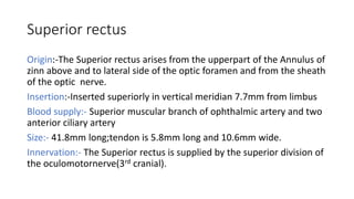
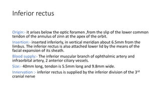
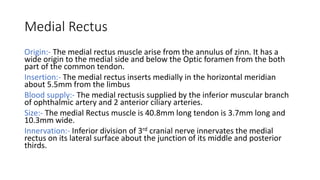
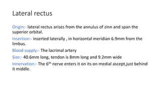
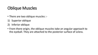
ExtraOcular Muscle
Shahana
Nethradhama school of optometry
Orbital muscle
Intrinsic muscle:
• Controls the shape of lens and size of pupil
Extrinsic muscle:
• Involved in movement of eyeball
Intrinsic muscle
1)Iris sphincter
2)Radial Pupilodilator muscle
3)Ciliary muscle
• Controlled by autonomic nervous system,work in response to amount
of light,closness of an object etc.
• Serves to focus the eye and Control the amount of light entering it.
Extrinsic muscle
Voluntary Involuntary
Superiortarsal muscle Levator palpebral superioris
Inferiortarsal muscle Superior rectus
Orbitalis Inferior rectus
Lateral rectus
Medial rectus
Superior oblique
Inferior oblique
Introduction
• The extraocular muscles are located within the orbit,but are extrinsic and
separate from the eyeball itself. They act to control the movement of
eyeball and superior eyelid.
• There are seven extraocular muscle.
• Functionally they can be divided into two groups:
1)Responsible for eye movement:
- Recti muscles
- Oblique muscles
2)Responsible for Superior eyelid elevation:
- Levator palpebral superioris.
Annulus Of Zinn
• The four recti muscle arise from a short funnel shaped tendinous ring
called the annulus of zinn.The annulus of zinn encloses the optic
foramen and a part of the medial end of Superior orbital fissure.
• The structures that passing through the annulus:-
-Oculomotornerve
-Abducent nerve
-Optic nerve
-Nasociliary nerve
-Ophthalmic artery
Recti muscles
• The name recti is derived from the latin for ‘straight’ - this represents
the fact that the recti muscles have direct path from origin to
attachment.
• There are 4 recti muscles.
Superior rectus
Origin:-The Superior rectus arises from the upperpart of the Annulus of
zinn above and to lateral side of the optic foramen and from the sheath
of the optic nerve.
Insertion:-Inserted superiorly in vertical meridian 7.7mm from limbus
Blood supply:- Superior muscular branch of ophthalmic artery and two
anterior ciliary artery
Size:- 41.8mm long;tendon is 5.8mm long and 10.6mm wide.
Innervation:- The Superior rectus is supplied by the superior division of
the oculomotornerve(3rd cranial).
Inferior rectus
Origin:- it arises below the optic foramen ,from the slip of the lower common
tendon of the annulus of zinn at the apex of the orbit.
Insertion:- inserted inferiorly, in vertical meridian about 6.5mm from the
limbus. The inferior rectus is also attached lower lid by the means of the
facial expansion of its sheath.
Blood supply:- The inferior muscular branch of ophthalmic artery and
infraorbital artery, 2 anterior ciliary vessels.
Size:- 40mm long, tendon is 5.5mm long and 9.8mm wide.
Innervation :- inferior rectus is supplied by the inferior division of the 3rd
cranial nerve
Medial Rectus
Origin:- The medial rectus muscle arise from the annulus of zinn. It has a
wide origin to the medial side and below the Optic foramen from the both
part of the common tendon.
Insertion:- The medial rectus inserts medially in the horizontal meridian
about 5.5mm from the limbus
Blood supply:- The medial rectusis supplied by the inferior muscular branch
of ophthalmic artery and 2 anterior ciliary arteries.
Size:- The medial Rectus muscle is 40.8mm long tendon is 3.7mm long and
10.3mm wide.
Innervation:- Inferior division of 3rd cranial nerve innervates the medial
rectus on its lateral surface about the junction of its middle and posterior
thirds.
Lateral rectus
Origin:- lateral rectus arises from the annulus of zinn and span the
superior orbital.
Insertion:- inserted laterally , in horizontal meridian 6.9mm from the
limbus.
Blood supply:- The lacrimal artery
Size:- 40.6mm long, tendon is 8mm long and 9.2mm wide
Innervation:- The 6th nerve enters it on its on medial ascept,just behind
it middle.
Oblique Muscles
• There are two oblique muscles :-
1) Superior oblique
2) Inferior oblique
• From there origin, the oblique muscles take an angular approach to
the eyeball. They are attached to the posterior surface of sclera.
Superior oblique
Origin:- above and medial to the opticforamen by a narrow tendon
which partially overlaps the origin of the levator.
Insertion:- inserted to the trochlea at orbital rim on the medial wall of
antero-superior-medial orbit on the frontal bone
Blood supply:-The superior muscular branch of ophthalmic artery
supply blood
Size:- 40mm long ,tendon is 20mm long and 10 mm wide.
Innervation:- The superior oblique is supplied by the 4th cranial nerve
which is divided into 3 or 4 branches,enters the muscle on the upper
surface near its lateral broader
Inferior oblique
Origin:- The inferior oblique is the only muscle that take origin from the front
of the orbit,arises from the rounded tendon in a depression on orbital floor
near orbital rim.
Insertion:- Inserted posterior Inferior temporal quadrant at level of macula.
Blood supply:- The inferior branch of ophthalmic artery and infraorbital
artery
Size:- 37mm long,the shortest tendon of insertion and it is 9.6mm wide at
insertion.
Innervation :- The inferior division of oculomotornerve, crosses above the
posterior border to enter the muscle on it’s upper surface at about the
middle of the muscle.
Levator palpebral superioris
• It is a striated muscle to elevate the Eyelid.
• The levator palpebral superioris arises from the under surface of the
lesser wing of the sphenoid above and infront of the Optic foramen
by a short tendon which is blend with the underlying Superior rectus.
Innervation:- it is supplied by 3rd nerve.
The mnemonics to remember the nerve
supply of EOM:-
LR6 SO4 O3.
THANK YOU

ExtraOcularMuscles

  • 1.
  • 2.
    Orbital muscle Intrinsic muscle: •Controls the shape of lens and size of pupil Extrinsic muscle: • Involved in movement of eyeball
  • 3.
    Intrinsic muscle 1)Iris sphincter 2)RadialPupilodilator muscle 3)Ciliary muscle • Controlled by autonomic nervous system,work in response to amount of light,closness of an object etc. • Serves to focus the eye and Control the amount of light entering it.
  • 4.
    Extrinsic muscle Voluntary Involuntary Superiortarsalmuscle Levator palpebral superioris Inferiortarsal muscle Superior rectus Orbitalis Inferior rectus Lateral rectus Medial rectus Superior oblique Inferior oblique
  • 5.
    Introduction • The extraocularmuscles are located within the orbit,but are extrinsic and separate from the eyeball itself. They act to control the movement of eyeball and superior eyelid. • There are seven extraocular muscle. • Functionally they can be divided into two groups: 1)Responsible for eye movement: - Recti muscles - Oblique muscles 2)Responsible for Superior eyelid elevation: - Levator palpebral superioris.
  • 7.
    Annulus Of Zinn •The four recti muscle arise from a short funnel shaped tendinous ring called the annulus of zinn.The annulus of zinn encloses the optic foramen and a part of the medial end of Superior orbital fissure. • The structures that passing through the annulus:- -Oculomotornerve -Abducent nerve -Optic nerve -Nasociliary nerve -Ophthalmic artery
  • 9.
    Recti muscles • Thename recti is derived from the latin for ‘straight’ - this represents the fact that the recti muscles have direct path from origin to attachment. • There are 4 recti muscles.
  • 10.
    Superior rectus Origin:-The Superiorrectus arises from the upperpart of the Annulus of zinn above and to lateral side of the optic foramen and from the sheath of the optic nerve. Insertion:-Inserted superiorly in vertical meridian 7.7mm from limbus Blood supply:- Superior muscular branch of ophthalmic artery and two anterior ciliary artery Size:- 41.8mm long;tendon is 5.8mm long and 10.6mm wide. Innervation:- The Superior rectus is supplied by the superior division of the oculomotornerve(3rd cranial).
  • 11.
    Inferior rectus Origin:- itarises below the optic foramen ,from the slip of the lower common tendon of the annulus of zinn at the apex of the orbit. Insertion:- inserted inferiorly, in vertical meridian about 6.5mm from the limbus. The inferior rectus is also attached lower lid by the means of the facial expansion of its sheath. Blood supply:- The inferior muscular branch of ophthalmic artery and infraorbital artery, 2 anterior ciliary vessels. Size:- 40mm long, tendon is 5.5mm long and 9.8mm wide. Innervation :- inferior rectus is supplied by the inferior division of the 3rd cranial nerve
  • 12.
    Medial Rectus Origin:- Themedial rectus muscle arise from the annulus of zinn. It has a wide origin to the medial side and below the Optic foramen from the both part of the common tendon. Insertion:- The medial rectus inserts medially in the horizontal meridian about 5.5mm from the limbus Blood supply:- The medial rectusis supplied by the inferior muscular branch of ophthalmic artery and 2 anterior ciliary arteries. Size:- The medial Rectus muscle is 40.8mm long tendon is 3.7mm long and 10.3mm wide. Innervation:- Inferior division of 3rd cranial nerve innervates the medial rectus on its lateral surface about the junction of its middle and posterior thirds.
  • 13.
    Lateral rectus Origin:- lateralrectus arises from the annulus of zinn and span the superior orbital. Insertion:- inserted laterally , in horizontal meridian 6.9mm from the limbus. Blood supply:- The lacrimal artery Size:- 40.6mm long, tendon is 8mm long and 9.2mm wide Innervation:- The 6th nerve enters it on its on medial ascept,just behind it middle.
  • 14.
    Oblique Muscles • Thereare two oblique muscles :- 1) Superior oblique 2) Inferior oblique • From there origin, the oblique muscles take an angular approach to the eyeball. They are attached to the posterior surface of sclera.
  • 16.
    Superior oblique Origin:- aboveand medial to the opticforamen by a narrow tendon which partially overlaps the origin of the levator. Insertion:- inserted to the trochlea at orbital rim on the medial wall of antero-superior-medial orbit on the frontal bone Blood supply:-The superior muscular branch of ophthalmic artery supply blood Size:- 40mm long ,tendon is 20mm long and 10 mm wide. Innervation:- The superior oblique is supplied by the 4th cranial nerve which is divided into 3 or 4 branches,enters the muscle on the upper surface near its lateral broader
  • 17.
    Inferior oblique Origin:- Theinferior oblique is the only muscle that take origin from the front of the orbit,arises from the rounded tendon in a depression on orbital floor near orbital rim. Insertion:- Inserted posterior Inferior temporal quadrant at level of macula. Blood supply:- The inferior branch of ophthalmic artery and infraorbital artery Size:- 37mm long,the shortest tendon of insertion and it is 9.6mm wide at insertion. Innervation :- The inferior division of oculomotornerve, crosses above the posterior border to enter the muscle on it’s upper surface at about the middle of the muscle.
  • 18.
    Levator palpebral superioris •It is a striated muscle to elevate the Eyelid. • The levator palpebral superioris arises from the under surface of the lesser wing of the sphenoid above and infront of the Optic foramen by a short tendon which is blend with the underlying Superior rectus. Innervation:- it is supplied by 3rd nerve.
  • 20.
    The mnemonics toremember the nerve supply of EOM:- LR6 SO4 O3.
  • 23.