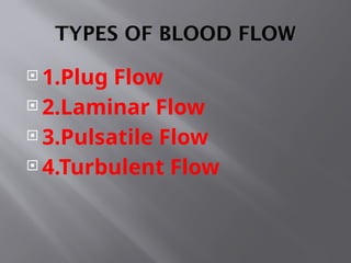
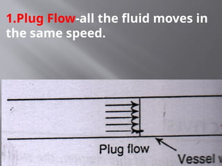
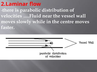
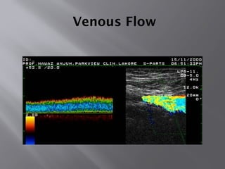
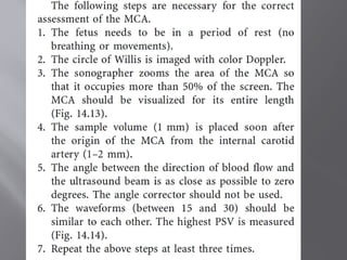
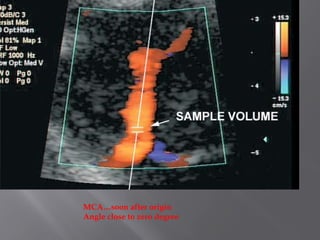
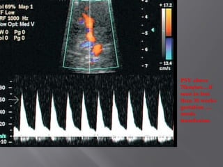
2.Laminar flow
-there isparabolic distribution of
velocities ….Fluid near the vessel wall
moves slowly while in the centre moves
faster.
31.
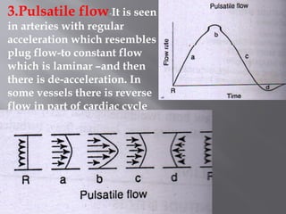
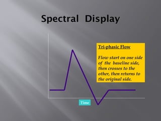
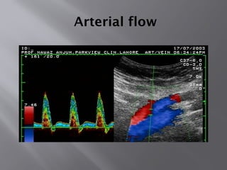
3.Pulsatile flow-It isseen
in arteries with regular
acceleration which resembles
plug flow-to constant flow
which is laminar –and then
there is de-acceleration. In
some vessels there is reverse
flow in part of cardiac cycle
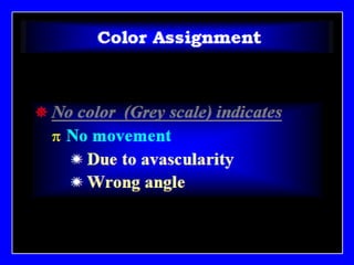
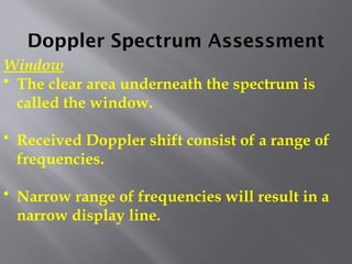
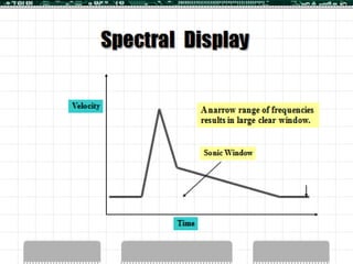
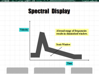
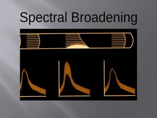
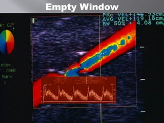
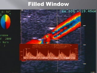
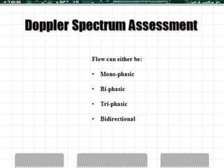
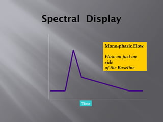
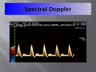
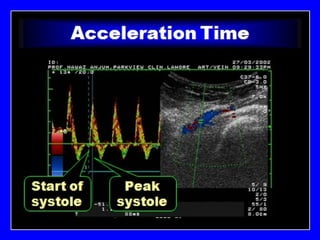
Doppler Spectrum Assessment
Window
•The clear area underneath the spectrum is
called the window.
• Received Doppler shift consist of a range of
frequencies.
• Narrow range of frequencies will result in a
narrow display line.
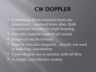
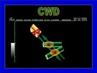
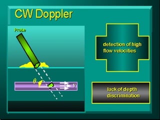
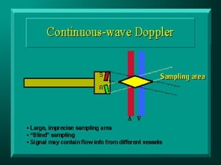
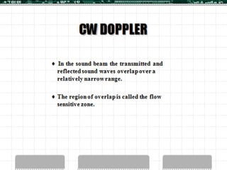
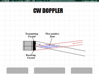
CW DOPPLER
Continuousbeam emission from one
transducer …received from other. Both
transducers encased in single housing.
Can only examine superficial vessels
Image can not be formed.
Used by vascular surgeons…usually not used
in radiology department.
Gynecologists use to monitor umb art flow
A simple, cost effective system.
75.
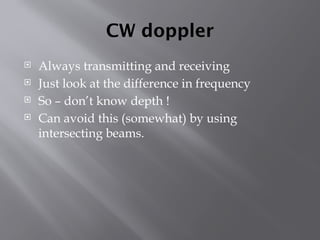
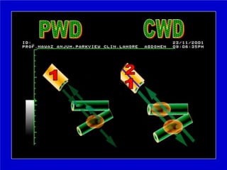
CW doppler
Alwaystransmitting and receiving
Just look at the difference in frequency
So – don’t know depth !
Can avoid this (somewhat) by using
intersecting beams.
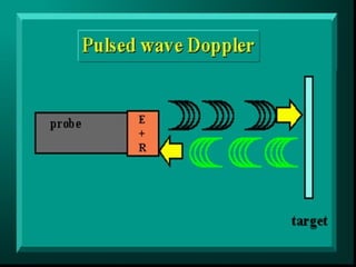
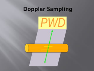
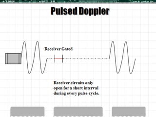
Pulsed wave Doppler
Pulses – just like real time scanning
Can find depth
Need to “gate” analysis of received pulse, so
we know where the moving objects are…
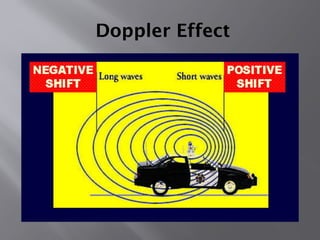
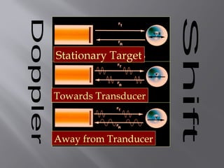
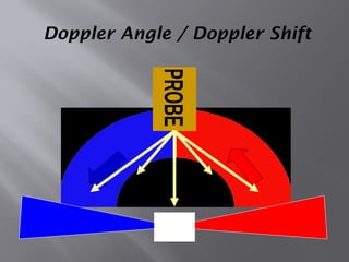
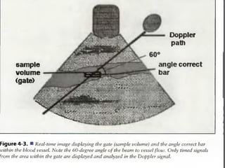
Angle effects
MaximumDoppler shift at 0 degrees
minimum at 90 degrees – proportional to the
Cosine of the angle between the beam and
direction of travel
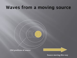
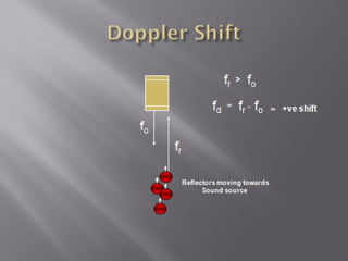
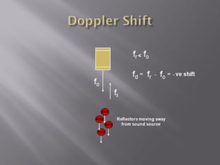
Direction of movement
Alignm
ent of beam
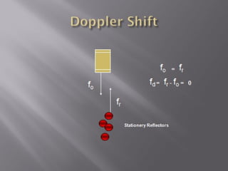
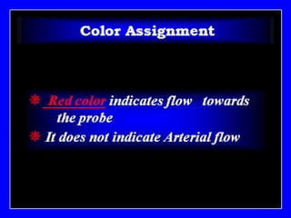
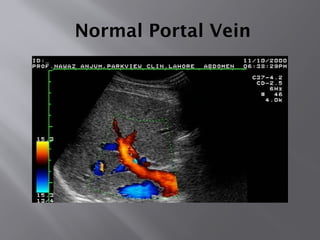
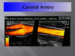
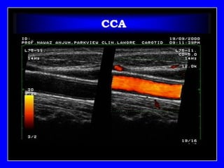
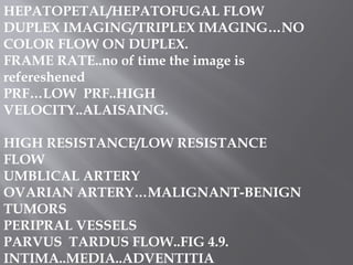
HEPATOPETAL/HEPATOFUGAL FLOW
DUPLEX IMAGING/TRIPLEXIMAGING…NO
COLOR FLOW ON DUPLEX.
FRAME RATE..no of time the image is
refereshened
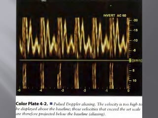
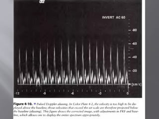
PRF…LOW PRF..HIGH
VELOCITY..ALAISAING.
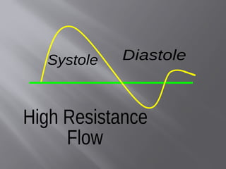
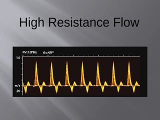
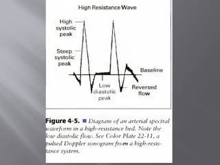
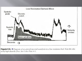
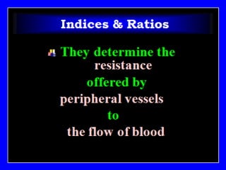
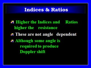
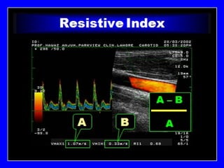
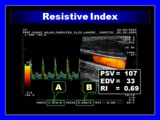
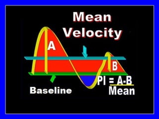
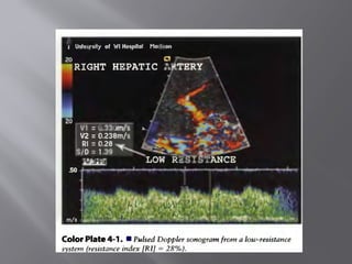
HIGH RESISTANCE/LOW RESISTANCE
FLOW
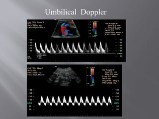
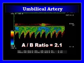
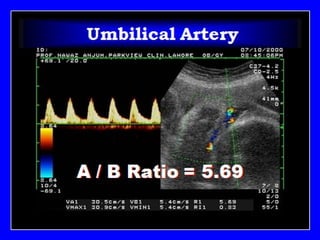
UMBLICAL ARTERY
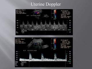
OVARIAN ARTERY…MALIGNANT-BENIGN
TUMORS
PERIPRAL VESSELS
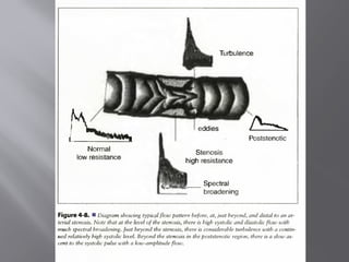
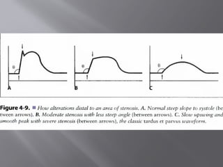
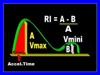
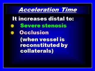
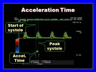
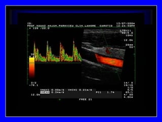
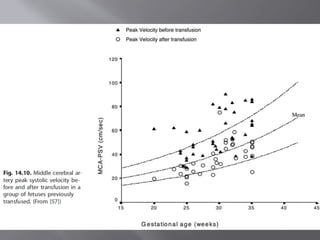
PARVUS TARDUS FLOW..FIG 4.9.
INTIMA..MEDIA..ADVENTITIA
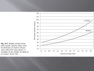
RULE OF THUMB..DECLAREMILD TO MODERATE
ANAEMIA IF
AT 20WEEKS….MORE THAN 40CM/SEC
AT 25WEEKS…. MORE THAN 50CM/SEC
AT 30WEEKS … MORE THAN 60CM/SEC
AT 35WEEKS … MORE THAN 70CM/SEC
AT 40WEEKS… MORE THAN 80-100CM/SEC
NORMAL VALUE IS 30% LESS
211.
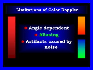
1.How can youreduce aliasing in color Doppler
study.
2.How CW Doppler differs from PW Doppler.
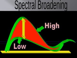
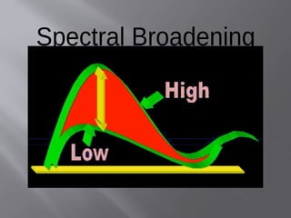
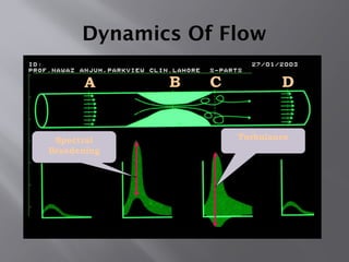
3.What is spectral broadening…Why it is seen
in Doppler study.
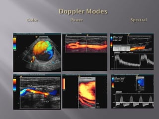
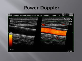
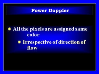
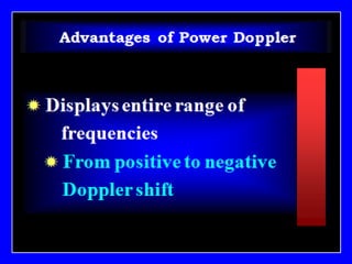
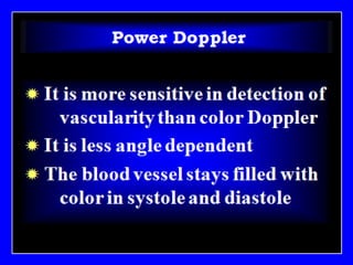
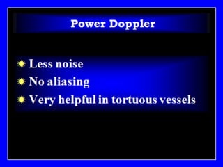
4.How Power Doppler differs from Color
Doppler.
5.What is the difference between low resistance
and high resistance blood flow…How Doppler
study could help in measuring blood flow
resistance.
212.
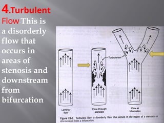
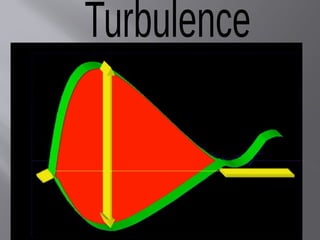
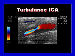
1.What is turbulentflow. How it will
appear on spectral Doppler waveform.
2.What is a reverberation artifact. How can
it be reduced/minimized.
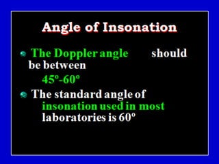
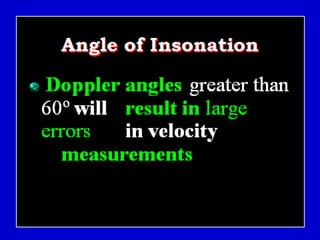
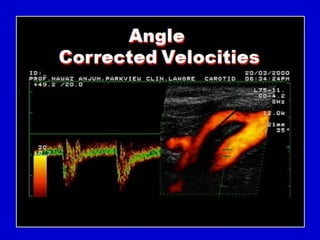
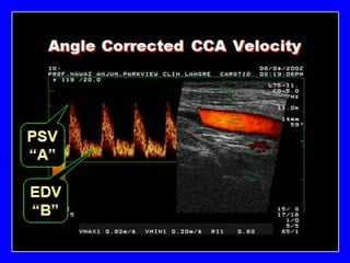
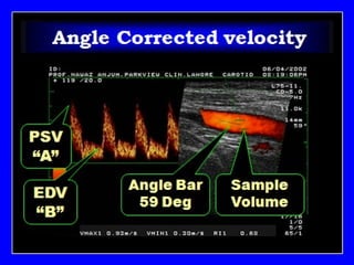
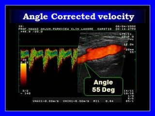
3.What is meant by angle correction in
Doppler study. What is its role in RI and
velocity measurements.
4.How Power Doppler differs from Color
Doppler.
5.How can you reduce aliasing in color
Doppler study.
#37 Spectral Doppler Modes
Pulsed Wave Doppler
Continuous Wave Doppler (with cardiac option)
In Pulsed Wave and Continuous Wave Doppler, you select the area of interest by positioning a Doppler cursor within the 2-D image. Pulsed Wave Doppler displays velocities within the range bounded by the Doppler gate on the cursor. Both let you simultaneously display the 2-D image and Doppler spectral information or independently display spectral Doppler information.
Auxiliary Continuous Wave Doppler (with cardiac option)
Auxiliary Continuous Wave Doppler uses a special auxiliary transducer. Because of its small size, the auxiliary transducer can access any area requiring documentation of blood flow. You position the auxiliary transducer independently of the transducer you are using for the 2-D image. Auxiliary Continuous Wave Doppler displays only a full-screen spectral display.
HPRF (High Pulse Repetition Frequency)
High Pulse Repetition Frequency (HPRF) allows increased velocity detection at depth and localization of flow. Use HPRF Pulsed Doppler mode when you want the Doppler velocity scale higher than the system can provide in standard Pulsed Doppler mode at a specific gate depth. HPRF generates multiple sample volumes displayed with smaller secondary gates in addition to the standard gate size of the primary sample volume. Excellent Sensitivity.
#40 Spectral Doppler Modes
Pulsed Wave Doppler
Continuous Wave Doppler (with cardiac option)
In Pulsed Wave and Continuous Wave Doppler, you select the area of interest by positioning a Doppler cursor within the 2-D image. Pulsed Wave Doppler displays velocities within the range bounded by the Doppler gate on the cursor. Both let you simultaneously display the 2-D image and Doppler spectral information or independently display spectral Doppler information.
Auxiliary Continuous Wave Doppler (with cardiac option)
Auxiliary Continuous Wave Doppler uses a special auxiliary transducer. Because of its small size, the auxiliary transducer can access any area requiring documentation of blood flow. You position the auxiliary transducer independently of the transducer you are using for the 2-D image. Auxiliary Continuous Wave Doppler displays only a full-screen spectral display.
HPRF (High Pulse Repetition Frequency)
High Pulse Repetition Frequency (HPRF) allows increased velocity detection at depth and localization of flow. Use HPRF Pulsed Doppler mode when you want the Doppler velocity scale higher than the system can provide in standard Pulsed Doppler mode at a specific gate depth. HPRF generates multiple sample volumes displayed with smaller secondary gates in addition to the standard gate size of the primary sample volume. Excellent Sensitivity.
#99 Spectral Doppler Modes
Pulsed Wave Doppler
Continuous Wave Doppler (with cardiac option)
In Pulsed Wave and Continuous Wave Doppler, you select the area of interest by positioning a Doppler cursor within the 2-D image. Pulsed Wave Doppler displays velocities within the range bounded by the Doppler gate on the cursor. Both let you simultaneously display the 2-D image and Doppler spectral information or independently display spectral Doppler information.
Auxiliary Continuous Wave Doppler (with cardiac option)
Auxiliary Continuous Wave Doppler uses a special auxiliary transducer. Because of its small size, the auxiliary transducer can access any area requiring documentation of blood flow. You position the auxiliary transducer independently of the transducer you are using for the 2-D image. Auxiliary Continuous Wave Doppler displays only a full-screen spectral display.
HPRF (High Pulse Repetition Frequency)
High Pulse Repetition Frequency (HPRF) allows increased velocity detection at depth and localization of flow. Use HPRF Pulsed Doppler mode when you want the Doppler velocity scale higher than the system can provide in standard Pulsed Doppler mode at a specific gate depth. HPRF generates multiple sample volumes displayed with smaller secondary gates in addition to the standard gate size of the primary sample volume. Excellent Sensitivity.
#100 Spectral Doppler Modes
Pulsed Wave Doppler
Continuous Wave Doppler (with cardiac option)
In Pulsed Wave and Continuous Wave Doppler, you select the area of interest by positioning a Doppler cursor within the 2-D image. Pulsed Wave Doppler displays velocities within the range bounded by the Doppler gate on the cursor. Both let you simultaneously display the 2-D image and Doppler spectral information or independently display spectral Doppler information.
Auxiliary Continuous Wave Doppler (with cardiac option)
Auxiliary Continuous Wave Doppler uses a special auxiliary transducer. Because of its small size, the auxiliary transducer can access any area requiring documentation of blood flow. You position the auxiliary transducer independently of the transducer you are using for the 2-D image. Auxiliary Continuous Wave Doppler displays only a full-screen spectral display.
HPRF (High Pulse Repetition Frequency)
High Pulse Repetition Frequency (HPRF) allows increased velocity detection at depth and localization of flow. Use HPRF Pulsed Doppler mode when you want the Doppler velocity scale higher than the system can provide in standard Pulsed Doppler mode at a specific gate depth. HPRF generates multiple sample volumes displayed with smaller secondary gates in addition to the standard gate size of the primary sample volume. Excellent Sensitivity.
#101 Spectral Doppler Modes
Pulsed Wave Doppler
Continuous Wave Doppler (with cardiac option)
In Pulsed Wave and Continuous Wave Doppler, you select the area of interest by positioning a Doppler cursor within the 2-D image. Pulsed Wave Doppler displays velocities within the range bounded by the Doppler gate on the cursor. Both let you simultaneously display the 2-D image and Doppler spectral information or independently display spectral Doppler information.
Auxiliary Continuous Wave Doppler (with cardiac option)
Auxiliary Continuous Wave Doppler uses a special auxiliary transducer. Because of its small size, the auxiliary transducer can access any area requiring documentation of blood flow. You position the auxiliary transducer independently of the transducer you are using for the 2-D image. Auxiliary Continuous Wave Doppler displays only a full-screen spectral display.
HPRF (High Pulse Repetition Frequency)
High Pulse Repetition Frequency (HPRF) allows increased velocity detection at depth and localization of flow. Use HPRF Pulsed Doppler mode when you want the Doppler velocity scale higher than the system can provide in standard Pulsed Doppler mode at a specific gate depth. HPRF generates multiple sample volumes displayed with smaller secondary gates in addition to the standard gate size of the primary sample volume. Excellent Sensitivity.
#102 Spectral Doppler Modes
Pulsed Wave Doppler
Continuous Wave Doppler (with cardiac option)
In Pulsed Wave and Continuous Wave Doppler, you select the area of interest by positioning a Doppler cursor within the 2-D image. Pulsed Wave Doppler displays velocities within the range bounded by the Doppler gate on the cursor. Both let you simultaneously display the 2-D image and Doppler spectral information or independently display spectral Doppler information.
Auxiliary Continuous Wave Doppler (with cardiac option)
Auxiliary Continuous Wave Doppler uses a special auxiliary transducer. Because of its small size, the auxiliary transducer can access any area requiring documentation of blood flow. You position the auxiliary transducer independently of the transducer you are using for the 2-D image. Auxiliary Continuous Wave Doppler displays only a full-screen spectral display.
HPRF (High Pulse Repetition Frequency)
High Pulse Repetition Frequency (HPRF) allows increased velocity detection at depth and localization of flow. Use HPRF Pulsed Doppler mode when you want the Doppler velocity scale higher than the system can provide in standard Pulsed Doppler mode at a specific gate depth. HPRF generates multiple sample volumes displayed with smaller secondary gates in addition to the standard gate size of the primary sample volume. Excellent Sensitivity.
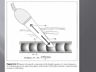
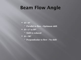
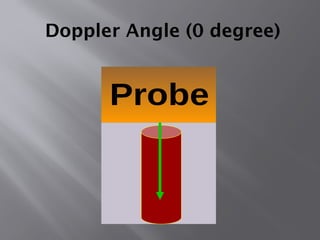
#103 The Cosine Relationship
COS q
-Beam parallel to flow
-COS 0° = 1, optimum sensitivity
-Beam perpendicular to flow
-COS 90° = 0, no sensitivity
[Review: Define cosine and its importance in Doppler.]
[Example]
If sound beam was propagated at the same angle as the blood flow, the COSq would be 1 and there would be maximum velocity sensitivity. If the sound beam-to-blood flow angle is between a 1 and 89 degree angle, the detected Doppler frequency shift is reduced according to the cosq term. If the beam is perpendicular, or 90 degrees to the flow, the cosqis 0 and there will be no detected Doppler shift. This demonstrates how important it is to have a good Doppler angle to the blood flow.
Note: When a sonographer wishes to identify the presence of flow, this interdependence of flow and the cosine is unimportant (as long as the incident angle is something other than 90 degrees).
#104 The Cosine Relationship
COS q
-Beam parallel to flow
-COS 0° = 1, optimum sensitivity
-Beam perpendicular to flow
-COS 90° = 0, no sensitivity
[Review: Define cosine and its importance in Doppler.]
[Example]
If sound beam was propagated at the same angle as the blood flow, the COSq would be 1 and there would be maximum velocity sensitivity. If the sound beam-to-blood flow angle is between a 1 and 89 degree angle, the detected Doppler frequency shift is reduced according to the cosq term. If the beam is perpendicular, or 90 degrees to the flow, the cosqis 0 and there will be no detected Doppler shift. This demonstrates how important it is to have a good Doppler angle to the blood flow.
Note: When a sonographer wishes to identify the presence of flow, this interdependence of flow and the cosine is unimportant (as long as the incident angle is something other than 90 degrees).


![Doppler principles [1]](https://cdn.slidesharecdn.com/ss_thumbnails/dopplerprinciples1-210517111539-thumbnail.jpg?width=640&height=640&fit=bounds)


![Doppler principles [2]](https://cdn.slidesharecdn.com/ss_thumbnails/dopplerprinciples2-210517111747-thumbnail.jpg?width=640&height=640&fit=bounds)


