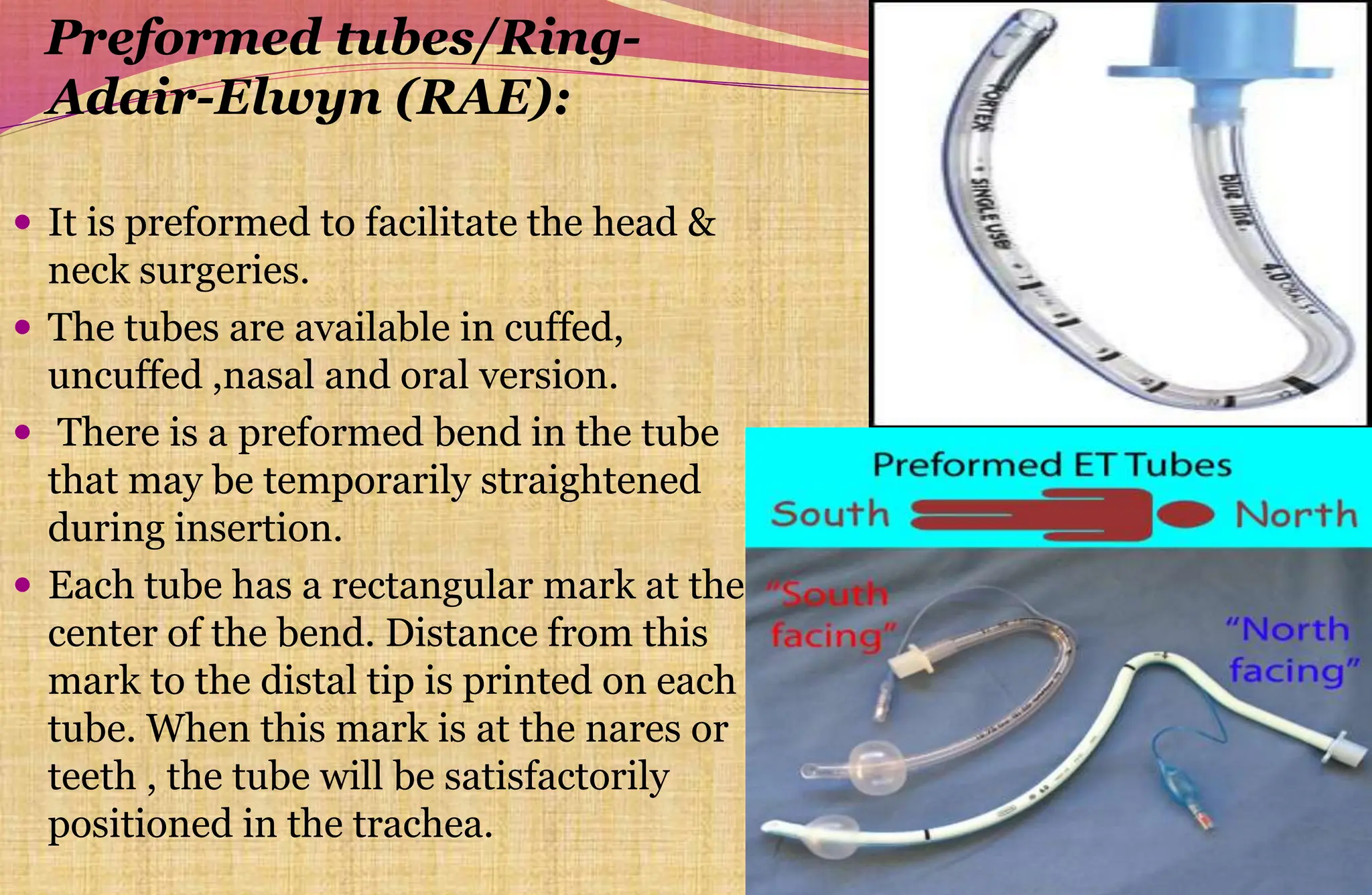
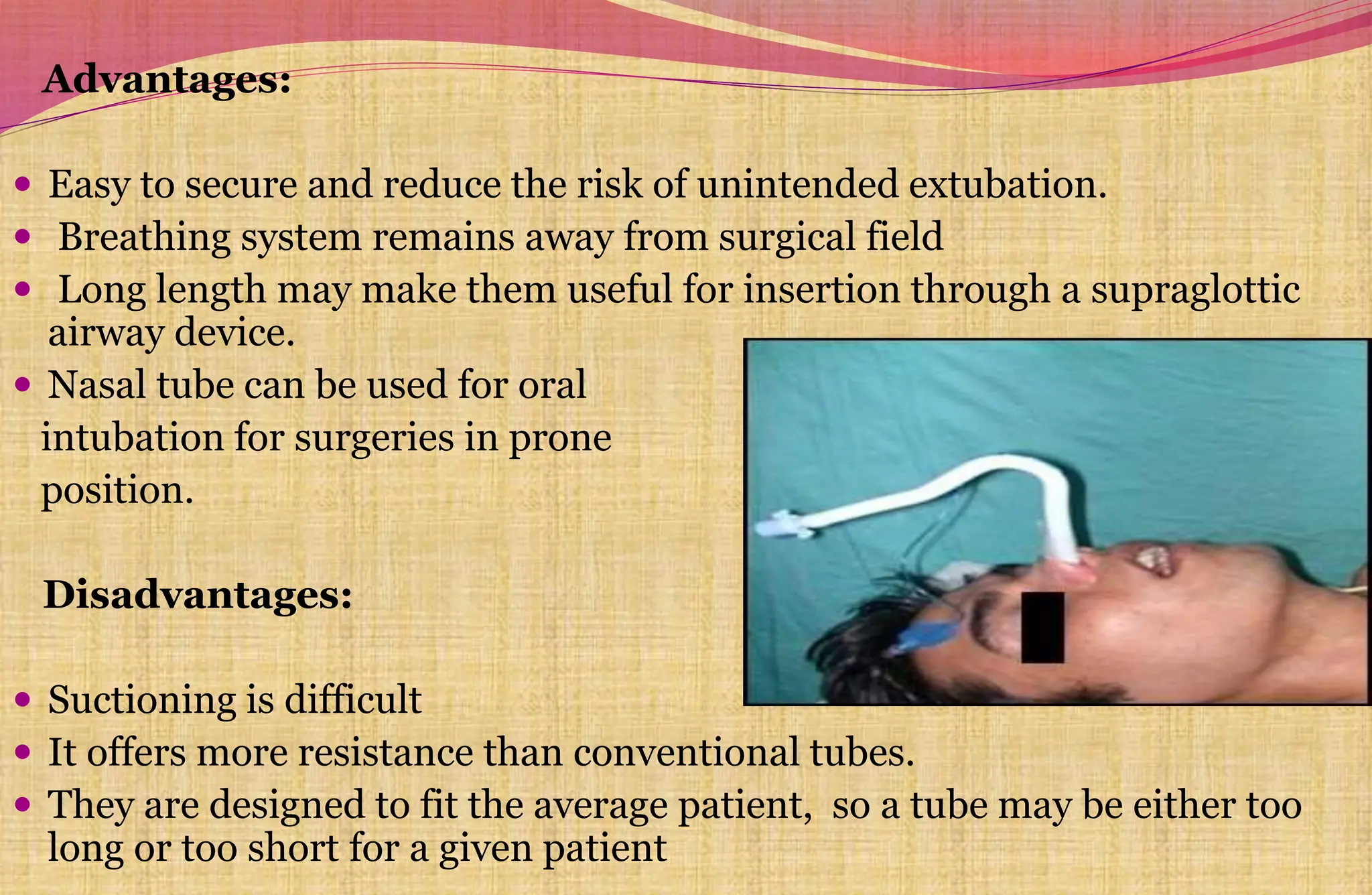
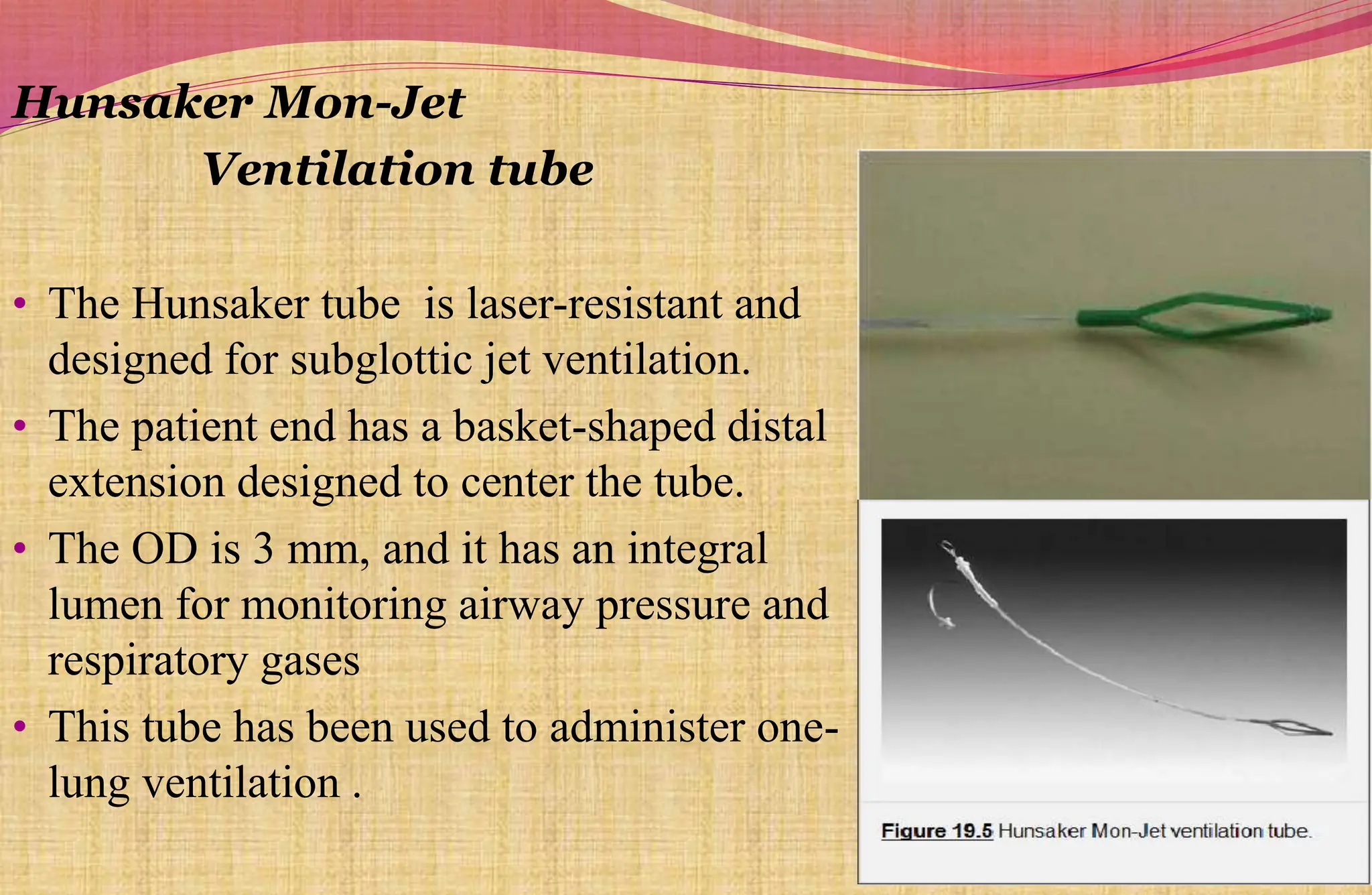
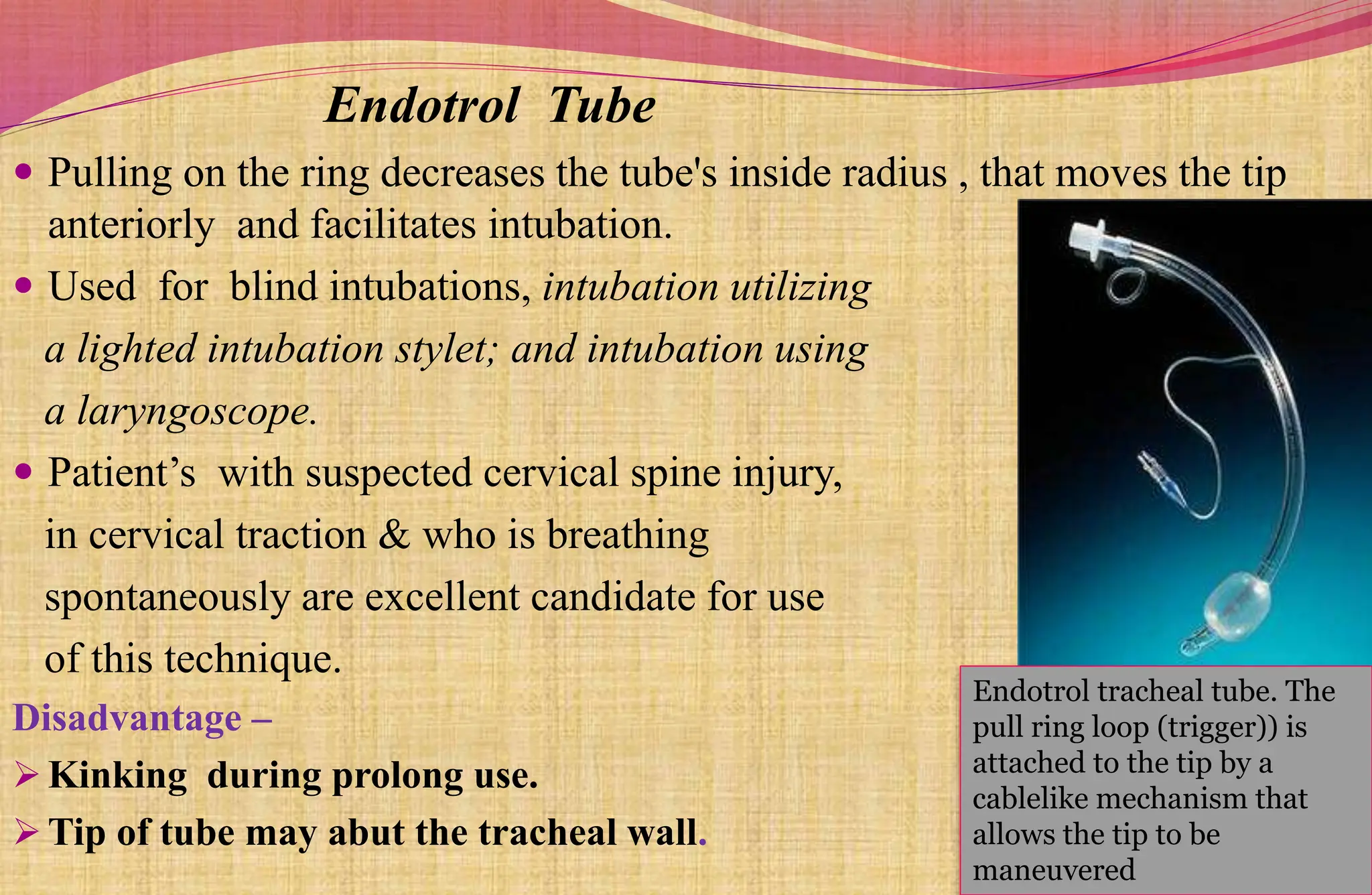
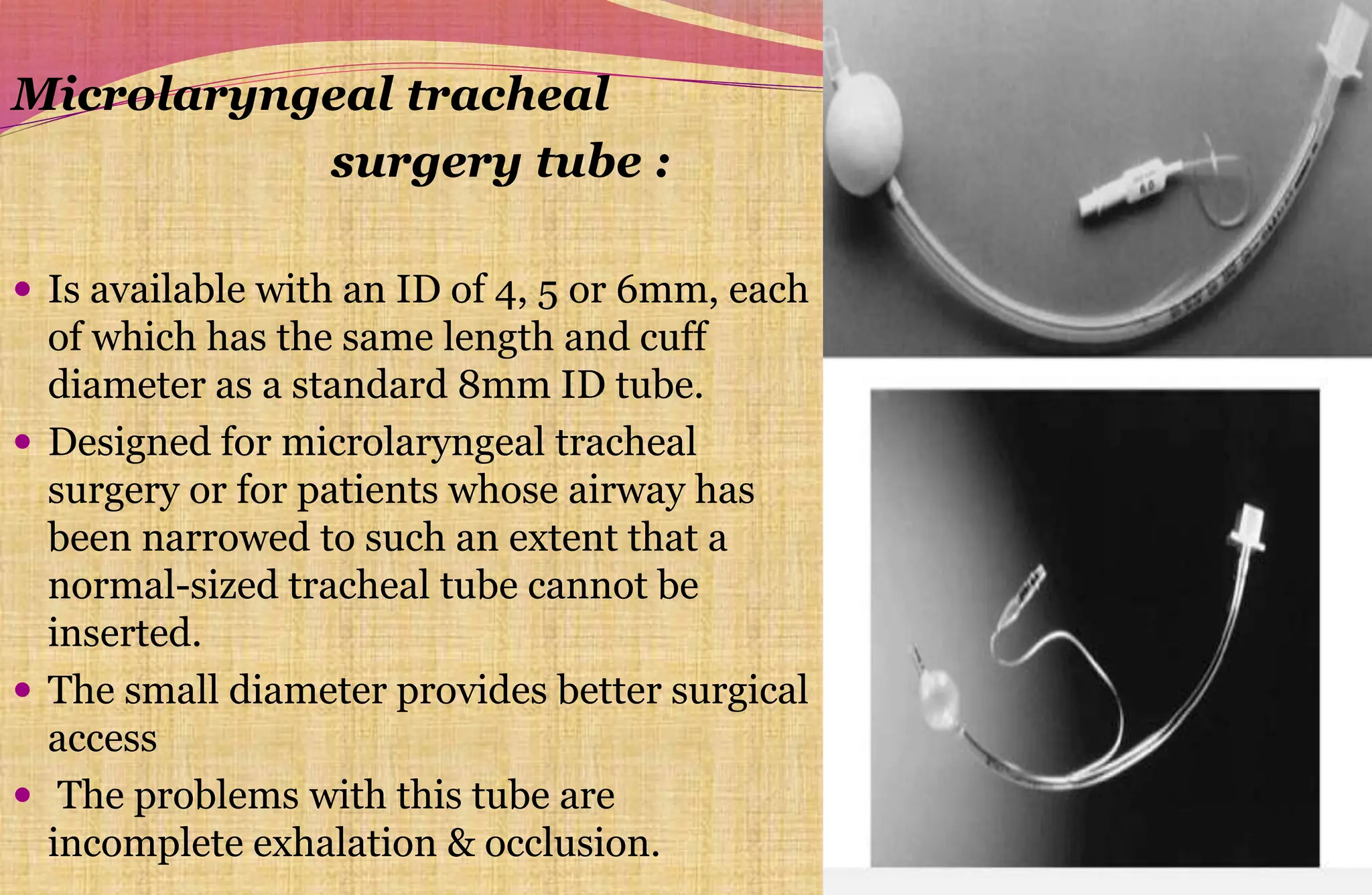
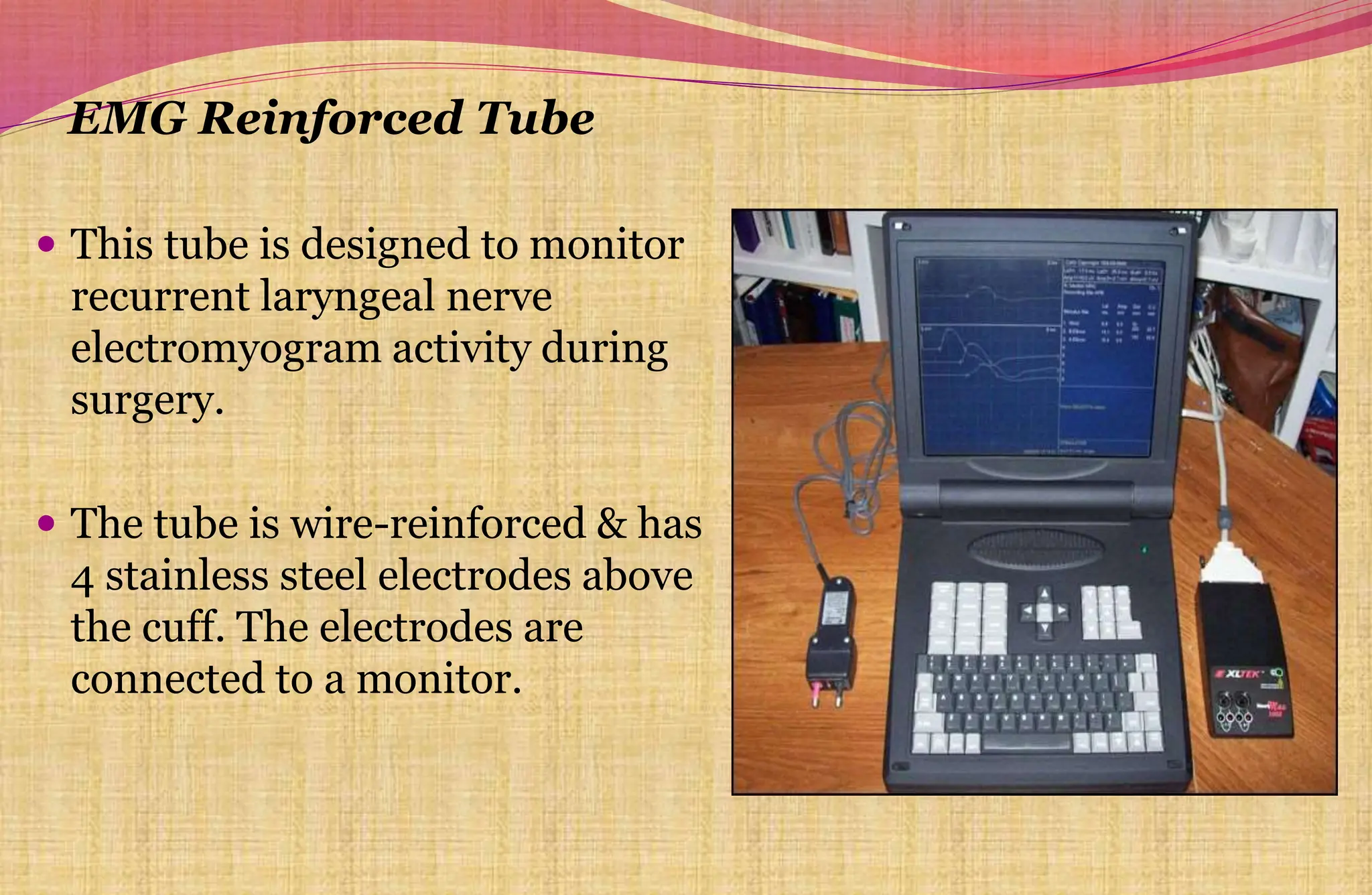
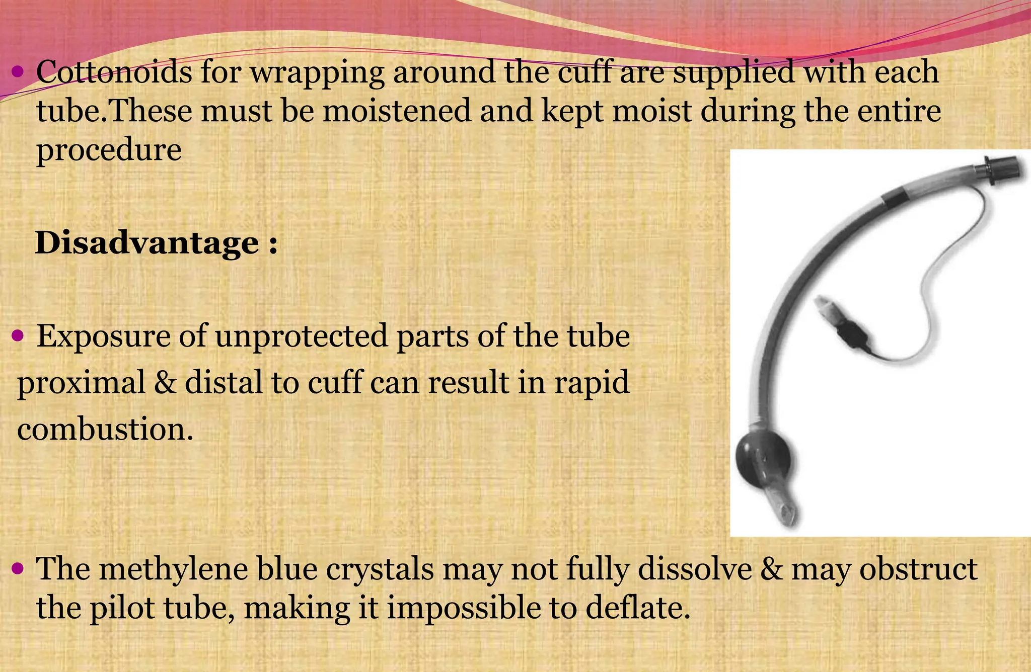
2. DEFINITION:
An endotracheal tube is a device
that is inserted through the larynx
into the trachea to convey gases
and vapors to and from the lungs.
3. PROPERTIES OF AN IDEAL ET TUBE
1) Low cost
2) Inertness, Lack of tissue toxicity
3) Smooth, non-wettable outer surface to avoid damage to mucosa & Inner
surface to prevent building of secretions.
4) Non-inflammable
5) Transparent
6) Easily sterilized
7)Sufficient body to maintain its shape during insertion & to prevent
occlusion by torsion, kinking or compression by the cuff or external
pressure.
8) Sufficient strength to allow thin wall construction
9) Thermoplasticity to conform to anatomic passage and to be self
centering within the trachea.
10) Non reactive with lubricants or anesthetic agents
11) Latex free
4. 1. Red rubber tube
2. PVC tube
3. Silicon tubes-
smooth,
flexible
but expensive
Commonly used tubes
5. RESISTANCE AND WORK OF BREATHING
A tracheal tube places a mechanical burden on spontaneously breathing patient.
The factors determining the resistance include:
1) Internal diameter: The tube with thick wall decreases the ID and
thereby increases the resistance and vice-versa.
2) Length: Decreasing the length of the tube decreases the resistance.
3) Configuration: Abrupt change in the diameter and direction increases
the resistance. Gentle curve connectors offer less resistance than right-
angled ones as there will be increased resistance because of turbulent flow of
gases.
DEAD SPACE
Volume of the tube & connector is usually less than that of the natural
passage so dead space normally reduced by intubation.
In pediatric patients long tubes & connectors may increase the dead space
considerably
7. Bevel
Patient end has a slant
portion called bevel ,at
an acute angle to the
longitudinal axis .
Its opening faces to the left
when viewed from the
concave surface.
Angel of bevel in oral tube is
45 degree and in nasal
tube 30 degree.
During nasal intubation bevel
should be facing up ,to
avoid the tip from
impinging on epiglottis.
Murphy’s eye Area of murphy eye should be less than
80% of the cross- sectional area of the tube.
It reduces trauma during nasal intubation.
Disadvantage: the accumulation of secretions and inadvertent
passing of forceps or fiberoscope
Tracheal tube lacking the murphy eye are k/a Magill-type
tubes
8. STANDARD MARKING OF ETT
The markings are situated on the bevel side above the cuff & are read
from pt end to machine end:
Words- oral or nasal or oral/nasal
Size: ID in mm
External diameter for tubes size 6 and smaller
Manufacturer's name or trade mark
Tube has Graduated markings, showing the distance in cms from the
patient end.
Precautions are usually noted: Disposable/Do Not Reuse
Implantation tests (IT) or Z-79 indicating the tube has been tested
for tissue toxicity & accomplish ANSI standard.
Opaque lines may also be included at the patient end or along full
length.
10. CUFF SYSTEM :
consists of the cuff & inflation system.
It provide a seal between tube & tracheal wall to prevent passage of
pharyngeal contents into the trachea
Ensures no gas leaks past the cuff.
The cuff also serves to center the tube in trachea.
INFLATION SYSTEM
11. 1) Inflation valve: When a syringe tip is inserted, a plunger is displaced
from its seat & gas can be injected into the cuff. Upon removal of the
syringe the valve seals and gas cannot escape.
2) Pilot balloon: Its function is to give an indication of inflation or
deflation of the cuff and a rough idea of the cuff pressure.
3) External inflation tube: The standard specifies ,
a)The external diameter should not exceed 2.5mm
b) The inflation tube should be attached to the ETT at a small angle.
c) The tube should extend at least 3cm beyond the machine end of the
ETT before a pilot balloon or inflation valve is incorporated.
4) Inflation lumen: This connects the inflation tube to the cuff. It is
located within the wall of the tracheal tube.
12. CUFF:
The cuff is a inflatable sleeve
near the patient end of ETT.
The cuff material should be
strong and tear resistant but
thin, soft and pliable.
Cuffs are usually made of the
same material as the ETT.
Type of cuffs
1) Low volume,high pressure cuff
2) High volume,low pressure cuff
3) Foam cuff
4) Lanz cuff
13.
14. a) Low volume high pressure cuff:
They have a small diameter at rest and low residual volume.
Advantages:
Usually reused (nondisposable silicon)
Less expensive
Offers better protection against aspiration
Better visibility during intubation than low pressure cuffs.
There is also low incidence of sore throat.
Disadvantages: The most serious risk associated with these
cuff is ischemic damage to the trachea following prolonged use.
15. b) High volume low pressure cuff:
They have a high residual volume,large diameter & a thin
competent wall.
Advantage :
It is relatively easy to pass devices such as esophageal stethoscope,
temperature probes, nasogastric tubes around low pressure cuffs.
Disadvantage:
These tubes are more difficult to insert,
Obscure the view of the tube tip and larynx
The cuff is more friable and thus more likely to be torn during
intubation.
Incidence of sore throats is greater with these tubes.
It may not effectively prevent fluid leakage into lower airway.
16. FOAM CUFF
Has a large diameter, residual volume, surface area
It is filled with polyurethane foam that is covered with
sheath,applying suction to the inflation tube cause the foam to
shrink.when negative pressure is released the cuff expand
Pressure on the tracheal wall depend on relationship b/w cuff
diameter at residual volume and diameter of trachea
Diffusion of anaesthetic agent
occurs into cuff but pressure does not
increase if the inflating channel is open.
There is no need of pressure
monitoring
17. LANZ CUFF
It consists of a very compliant latex pilot balloon inside a transparent
plastic sheath with an automatic pressure regulating valve b/w balloon &
cuff
Pressure regulating valve permit rapid gas flow from the balloon but only
slow flow from the cuff to balloon, this prevents the gas from being
squeezed back in to the balloon when airway pressure rises rapidly, so
there is no gas leaks during PPV.
Also prevents increase in
cuff pressure & volume due
to nitrous oxide.
18. CUFF PRESSURE
Intracuff pressure and pressure on tracheal wall:
It is desirable that cuff seals the airway without extending so much
pressure on the trachea so that its circulation is not compromised.
So it is recommended that the pressure on the lateral tracheal wall should be kept between 25-
34cm of H20.
19. Intracuff pressure and Nitrous oxide :
The resting intracuff pressure and volume of the cuff inflated with air rise
during nitrous oxide anesthesia, which results in ischaemia of the tracheal
mucosa or compression of the tube, and increase in volume may lead to
cuff herniation. When N2O is stopped,pressure in the cuff decreases
rapidly.
Steps to prevent the increasing pressure includes the following:
a) Filling the cuff with gas mixture to be used
for anesthesia or saline
b) Fitting the cuff system with pressure relief
valve or pressure regulating devices e.g.
Lanz pressure regulating valve, sponge cuff
c) Monitoring cuff pressure and deflating the
cuff as needed.
20. THE GUIDELINES TO DETERMINE THE SIZE OF ETT:
Designated by ID (mm). ISO standard requires Tube size to be marked b/w cuff
and the take off point of the inflation tube for cuffed tubes.
For uncuffed tube size marking should be towards the patient end
Ideal tube in average Adult male – 8.5 & Adult female - 7.5mm ID.
Age is recognized as the most reliable indicator
of appropriate ETT size for children.
3 months & less ------ 3 mm ID
3 - 9 months ------ 3.5 mm ID
Older than 1 year ------ ID in mm = (16 + age in years)/4
21. Younger than 6 years ---3.75 + age in years/3 = ID in mm
Older than 6years --- 4.5 + age in years / 4 = ID in mm
Choosing a tube whose external diameter is same width as the distal
phalanx of the patient's little finger. Not so accurate but can be used
when patient’s age is unknown.
CHECKING OF TUBE
Tube should be examined to defect such as splitting, holes & missing
sections.
Tube should be checked for obstruction, looking into both ends or
by inserting a stylet
Cuff should remain inflated for 1 min to check for any leaks
22. Preparing and Insertion of the ET Tube:
After the sterile wrapping is opened, tube should be handled only at the
connector end.
Using sterile lubricant jelly on low
pressure,high volume cuff may decrease
aspiration by filling the folds.
Insertion of Tube either oral/nasal
Under flexible fiberoptic laryngoscopy
Under direct laryngoscopy
Blind
23. DEPTH OF INSERTION
The tube should be in the middle third of the trachea with the head in
neutral position. The formulas based on subject’s height, age & weight may
be used in children for oral intubation..…
1) Length in cm = age/2 + 12
2) Length in cm = weight in kg/5 + 12
3) Length in cm = height in cms/10 + 5
4) Rule of 7-8-9 for
1 kg infant-depth of 7 cm at the lips
2 kg infant-8cm
3 kg infant-9 cm
Naso tracheal intubation -Multiplying crown heel length
by 0.21
24. In adults, the tube should be passed until the cuff is 2.25 to 2.5cm below
the vocal cords
In uncuffed tubes, tube tip should not be inserted more than
1cm past the cords <6months
2cm past the cords up to 1yr
3-4cm past the cords > 1yr
In average size adult patients, securing the tube at the anterior incisor at
23cm in males and 21cm in females will usually avoid endobronchial
intubation.
For nasal intubation 5cm should be added to these lengths for positioning
at the nares.
25. CONFIRMATION OF ET TUBE POSITION
Visualize the tube passing through vocal cords
Feel the compliance of reservoir bag
Observe the chest wall rise with inspiration
5 point Auscultation
Movement of respiratory mist in ETT
Cuff Palpation over the suprasternal notch
EtCO2 detectors,Capnography(gold standard)
Chest x –ray
Fibreoptic laryngoscope /bronchoscope
Esophageal detector device(rely on aspiration of air rapidly through ETT)
Sonomatic confirmation of tracheal intubation(SCOTI device)
Pulse oximeter value,Good pt. color & Vocal silence after awake intubation
26.
27. INFLATION OF CUFF:
After checking tube position cuff is inflated with minimum amount of gas
that will cause it to seal against the trachea at peak inspiratory pressure.
Cuff pressure adjusted so there is no leak at PIP.
Leak can be detected by
Palpation or auscultation of pretracheal area
Difference b/w inhaled and exhaled volumes
Monitoring for CO2 in upper airway
28. 1) DURING SURGERY-
- To secure & maintain a free airway
- To protect against aspiration of gastric/ oral contents
- To provide positive pressure ventilation
2) NON SURGICAL CONDITIONS- as a life saving measure
- CPR
- Protection of airway in unconscious or semiconscious patient eg. Drug
overdose, poisoning, CNS disease, head injury, polytrauma and impaired
laryngeal reflexes.
- When patient needs mechanical ventilation.
- Respiratory obstruction
- Tracheobronchial toileting in severe sputum retention
INDICATIONS OF ET TUBE
29. PERIOPERATIVE /POSTOPERATIVE
COMPLICATIONS OF ET TUBE
Failure to pass over an Intubating
Device
Trauma
Esophageal Intubation
Inadvertent Bronchial Intubation
Swallowed Tracheal Tube
Foreign Body Aspiration
Leaks
Intubating Device Trapped inside
the Tracheal Tube
Tracheal Tube Fires
Tracheal Tube Obstruction
Aspirating Fluid from above the
Cuff
Misplacement of Equipment into
the Trachea(temp probe,NG tube)
Scan Artifact
Unintended Extubation
Infection
Difficult Extubation
Emergence Phenomena
Postoperative sore throat
Hoarseness
Neurologic Injuries
Upper Airway Edema
Vocal Cord Dysfunction
Vocal Cord Granuloma
Ulcerations
Latex Allergy
Tracheal Stenosis
30. SPECIFIC ET TUBES
Cole tube:
It is uncuffed, stepped wall designed
ETT.
Patient end is smaller in diameter than
the rest of the tube
They are sized according to the internal
diameter of the tracheal portion. It
ranges from 2 to 5mm..
It is recommended for neonatal
resuscitation but not for long term
intubation.
Disadvantage : cannot be used nasally
31. Spiral embedded tubes:
Also known as Armored tube,
flexometallic tube.
These tubes have a metal or nylon spiral
woven reinforcing wire covered both
internally and externally by rubber,
PVC or silicone.
The spiral may not extend into the
distal & proximal ends.
Stylet may needed for intubation
These tubes are esp useful in situations
where the tube is likely to be bent or
compressed as in head & neck surgery
32. Advantages:
Primary advantage of tube is resistance to kinking and compression.
The portion of the tube outside the patient can be easily angled away
from the surgical field without kinking. This makes them useful for
insertion into patient with tracheostomy,for submental intubation &
retromolar positioning.
Pass easily over a fiberscope than a conventional tube.
Disadvantages:
Tube may rotate on the stylet during intubation.
Insertion through nose & intubating LMA is difficult.
Elastic recoil force may increase tendency to unintentional
extubation
If the patient bites the tube it will cause permanent deformity
resulting in obstruction of the tube, so BITE BLOCK b/w molar tooth
should be used to prevent this (not an oral airway)
33. Preformed tubes/Ring-
Adair-Elwyn (RAE):
It is preformed to facilitate the head &
neck surgeries.
The tubes are available in cuffed,
uncuffed ,nasal and oral version.
There is a preformed bend in the tube
that may be temporarily straightened
during insertion.
Each tube has a rectangular mark at the
center of the bend. Distance from this
mark to the distal tip is printed on each
tube. When this mark is at the nares or
teeth , the tube will be satisfactorily
positioned in the trachea.
34. Advantages:
Easy to secure and reduce the risk of unintended extubation.
Breathing system remains away from surgical field
Long length may make them useful for insertion through a supraglottic
airway device.
Nasal tube can be used for oral
intubation for surgeries in prone
position.
Disadvantages:
Suctioning is difficult
It offers more resistance than conventional tubes.
They are designed to fit the average patient, so a tube may be either too
long or too short for a given patient
35. Laryngectomy tube:
Designed for insertion into a tracheostomy site.
The tube is preformed in a ‘J’ configuration at
the pt end. This allows the part of the tube
external to the patient to be directed away from
the surgical field.
The tip may be short and/or without a bevel to
avoid inadvertent advancement into a bronchus.
Disadvantage -The soft cuff and short distance
between the cuff and distal tip of the tube may
cause the bevel to abut the tracheal wall.
36. DOUBLE LUMEN TUBE (DLT)
DLT is essentialy two single lumen tubes bonded together.
Shorter tracheal lumen, designed to terminate above the carina
Longer bronchial lumen, distal portion of this lumen has a
preformed curve that allows preferential entry into
either bronchus.
The internal lumen of each tube is D shaped with the
straight side of the D in the middle of the tube.
Tube has a two inflation system one for bronchial cuff
another for tracheal cuff.
Because of differences in bronchial anatomy
b/w then two sides, tubes are designed
specifically for either the right or left bronchus.
Adult DLT commonly come in sizes 35,37,39,41 Fr
26,28 and 32Fr also available for younger patients.
37. Anatomic variations between individuals in the distance b/w the
right carina & the upper lobe orifice often result in difficulties in
ventilating that lobe with right sided tubes. To overcome this
-on some tubes, the cuff has a slot to allow ventilation of
the right upper lobe.
-some right sided DLTs have two bronchial cuffs with an
opening for the right upper lobe b/w them.
Used to achieve isolation of either the right or the left lung
during thoracotomies,control of contamination or
hemorrhage & in U/L pathology e.g bullae or large cyst.
Problems: Difficulty with insertion and positioning,
obstructed ventilation and hypoxemia d/t
malpositioning, trauma, failure to seal,
difficult extubation.
38. Hunsaker Mon-Jet
Ventilation tube
• The Hunsaker tube is laser-resistant and
designed for subglottic jet ventilation.
• The patient end has a basket-shaped distal
extension designed to center the tube.
• The OD is 3 mm, and it has an integral
lumen for monitoring airway pressure and
respiratory gases
• This tube has been used to administer one-
lung ventilation .
39. Endotrol Tube
Pulling on the ring decreases the tube's inside radius , that moves the tip
anteriorly and facilitates intubation.
Used for blind intubations, intubation utilizing
a lighted intubation stylet; and intubation using
a laryngoscope.
Patient’s with suspected cervical spine injury,
in cervical traction & who is breathing
spontaneously are excellent candidate for use
of this technique.
Disadvantage –
Kinking during prolong use.
Tip of tube may abut the tracheal wall.
Endotrol tracheal tube. The
pull ring loop (trigger)) is
attached to the tip by a
cablelike mechanism that
allows the tip to be
maneuvered
40.
41. Microlaryngeal tracheal
surgery tube :
Is available with an ID of 4, 5 or 6mm, each
of which has the same length and cuff
diameter as a standard 8mm ID tube.
Designed for microlaryngeal tracheal
surgery or for patients whose airway has
been narrowed to such an extent that a
normal-sized tracheal tube cannot be
inserted.
The small diameter provides better surgical
access
The problems with this tube are
incomplete exhalation & occlusion.
42. Designed to aid intubation when the patient has an anterior larynx.
Pulling the white bar toward the connector causes the tube to flex at
the cuff & the tip to move anteriorly
Parker Flex-Tip tube
Easier to advance over an intubating catheter/flexible endoscope
then a conventional tube.
EndoFlex Tube
*The tube has a two murphy eyes & the“hooded” curved,
flexible tapered tip that points toward the center of the
distal lumen on the concave surface of the tube so that the
bevel faces posteriorly during insertion, so less likely to
impinge on the side of the right vocal cord.
43. Tubes with extra lumens:
Tubes are available with one or more separate lumens terminating
near the tip.
Useful for respiratory gas sampling, airway pressure monitoring,
irrigation, suctioning, injection of fluids and drugs & jet ventilation.
*Clear lumen- Jet ventilation, Administration of O2 during Suctioning and Bronchoscopy
*Opaque lumen- Irrigation and Sampling of gases from the trachea
44. Emergency drugs that can be administered to the lung through the ET include:
epinephrine norepinephrine, lidocaine, atropine,naloxone.
Disadvantage: Blood/secretions /moisture can block extra lumen
LITA (Laryngotracheal Instillation of Topical anaesthesia)
Has additional small bore channel
within the concave surface of the tube.
10 small holes at the distal 13cm of the
tube allows the injected medication to
be spread both above & below the cuffs.
This can provide a smooth emergence
from anesthesia without coughing.
45. EMG Reinforced Tube
This tube is designed to monitor
recurrent laryngeal nerve
electromyogram activity during
surgery.
The tube is wire-reinforced & has
4 stainless steel electrodes above
the cuff. The electrodes are
connected to a monitor.
46. Laser-shield II Tracheal Tube:
It is designed for use with CO2 and
KTP lasers.
Made from silicone with an inner aluminium
wrap and a smooth Teflon outer coating.
The cuff is not laser resistant & contains methylene blue crystals as a
marker to identify perforation. It should be inflated with water or saline
solution.
ENDOTRACHEAL TUBES DESIGNED
FOR LASER SURGERIES
47. Cottonoids for wrapping around the cuff are supplied with each
tube.These must be moistened and kept moist during the entire
procedure
Disadvantage :
Exposure of unprotected parts of the tube
proximal & distal to cuff can result in rapid
combustion.
The methylene blue crystals may not fully dissolve & may obstruct
the pilot tube, making it impossible to deflate.
48. REFERENCES
Understanding Anaesthesia Equipment, 5th Edition, Jerry A.
Dorsh and Susan E. Dorsh
Equipment in Anaesthetic Practice, 6th Edition,Arun Kumar Paul
Airway Management , 5th Edition, Rashid M Khan
Miller’s Anesthesia 8th Edition
49.
50. Laser-Flex Tubes:
Laser flex tube is a flexible stainless steel tube
with a smooth plastic surface and matte finish to
reflect a laser beam.
Designed for used with C02& KTP lasers
Adult version has two PVC cuffs and PVC tips with
Murphy eye.
Cuffs are inflated by two separate inflation tubes. Distal cuff should be
filled first until sealing occurs then proximal cuff is filled with saline
coloured with methylene blue.
The distal cuff can be used if proximal one is damage by laser.
Disadvantage: stiffness, roughness, cannot be trimmed.
The double cuff adds to the time of intubation and extubation.
51. NortonTube
- The Norton tube is a reusable, flexible, uncuffed
spiral wound metal tube with stainless
steel connector & has a thick wall.
- Not affected by any laser
- Disadvantage : Air leak
Tissue damage
Special ventillation requires
if cuff is not used.
Sheridan laser tracheal tube
This is a red rubber tube wrapped with copper foil tape. This is
overwrapped with water-absorbent fabric that
should be saturated with water prior to use.
It is designed for use with a CO2 or KTP laser.
Disadvantage It has a thick wall .
High-pressure cuff.
52. Lasertubus:
This is made of white rubber & has a cuff-
within-a cuff design.
If the outer cuff is perforated by a laser beam,
the trachea will still be sealed by inner cuff. The
inner cuff is filled with air & outer with water or
saline.
The shaft above the cuff is covered by a
corrugated silver foil, which is covered by
merocel sponge that should be moistened with
saline before use.
It is recommended for use with argon, NdYAG,
CO2 lasers.
53. This is designed to be inserted through the intubating laryngeal mask (ILM,
ILMA ,LMA-Fastrach) .
It is a straight, wire-reinforced silicone tube with a tapered patient end, blunt
tip, short bevel, and Murphy eye. Can be autoclaved.
Used for submental intubation & tracheal resection and reconstruction after
the tip has been removed.
Easier to advance over a fiberscope .
High-pressure, low-volume cuff .
It is available in sizes 6, 6.5, 7, 7.5, 8
Disadvantage -
eccentric cuff inflation,
internal deformities, and
the tip folding during insertion.
Tube bite
Intubating Laryngeal Mask Tracheal Tube &
Tube Stabilizer
54. Hi-Lo Evac Tube
It incorporates dedicated channel which
can be used to clear secretions below the
vocal cords but above the cuff.
Lumen may be blocked by secretions
Hi-Lo Jet Tube
It is an uncuffed tube with additional
lumen that can be used for Jet
ventilation, monitoring airway
pressure, sampling respiratory gases
or administering local anesthetics.