
Download to read offline

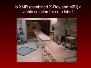
This document discusses the potential for using MRI as an alternative to x-ray guidance in cardiac catheterization labs and interventional procedures. It outlines the history of MRI in cardiovascular imaging and interventions. Recent studies are presented that demonstrate the feasibility of MRI-guided coronary catheterization and balloon angioplasty. However, technological barriers around making equipment MRI-compatible as well as regulatory and reimbursement issues still need to be addressed before MRI could fully replace x-rays in cath labs. The combination of x-ray and MRI, known as XMR, may be a viable solution.


![ONFH[AVN HIP] -TRIPLE REGIME -A NOVAL SURGICAL CONCEPT .pptx](https://cdn.slidesharecdn.com/ss_thumbnails/onfhavnhip2026koaconcalicutdrgokuldevdrmashraf-260210064517-213ec005-thumbnail.jpg?width=640&height=640&fit=bounds)


