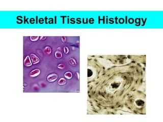
2. To outline the classification of cartilage tissue with
examples.
To state the differences of the different types of cartilage.
To describe the arrangement of cells and matrix in bone.
Objectives – Cartilage & Bones
3. Bone & Cartilage
specialized type of connective
tissue
adapted to bear weight
provide rigidity necessary for
the supporting framework of
the body.
4. • Development - primitive mesenchyme
• structural and functional differences are related to
the nature
the proportion of the ground substance
the fibres in the extracellular matrix.
Bone & Cartilage
5. Bone & Cartilage
Cartilage –
semi rigid nature
predominant ground substance
joints
walls of thorax
larynx, trachea
bronchi, nose and ears.
Bone- mineralized extracellular matrix
6. • chondro refers to cartilage
– chondrocyte
– Perichondrium
• osteo refers to bone
– osteogenesis
– osteocyte
– Periostium
• blast refers to precursor cell or one that produces
something
– Osteoblast
• cyte refers to cell
– osteocyte
Terms
8. Cartilage
Cartilage comprise of
cells - Chondrocytes
ground substances
fibres
Covered with fibrovascular membrane – Perichondrium
outer fibrous layer
Inner chondrogenic & vascular layer
Growth of cartilage
Appositional growth – surface deposition of cells of inner
perichondrial layer
Interstitial growth – multiplication of cells situated within the
matrix of the cartilage.
Intercellular matrix
9. Nutrition of Cartilages
No capillary blood supply of its own
2 ways
Diffusion from capillaries in the adjoining connective
tissue
Synovial fluid from joint cavities
10. Cartilage cells
Young cartilage cells – chondroblasts
smaller in size
irregular in shape
Mature cartilage cells – chondrocytes
in spaces of matrix/lacunae
single cells – spherical shape
cell nest (2-4 cells) – D shape
large, rounded nuclei
basophilic cytoplasm
stained section : shrinkage artifact
11. Cartilage – fibres
Thick collagen fibres
Branching and anastomosing elastic fibres
Cartilage – ground substances
Amorphous gel like substance
Stained in basic dye due to chondroitin sulphates &
keratosulphates
Deep basophilic rim called capsular territorial matrix &
stains darker than the interterritorial matrix.
12. Based on the visibility and nature of fibres in the
ground substances
– hyaline cartilage
– white fibro cartilage
– yellow elastic cartilage.
Classification of Cartilage
13. 2 varieties
Costal cartilage
Present at the ends of growing bones, ribs & respiratory passages
Covered by perichondrium
Articular cartilage
Present at articular ends of the bones (most synovial joints)
No perichondrium covering
H &E : Homogenous basophilic matrix
Collagen type 11 fibres.
Fibres not seen as a distinct entity
cell nest
Hyaline Cartilage
14. Hyaline Cartilage
Red arrow indicates the Perichondrium.
Black arrow indicates the chondroblasts.
Yellow arrow indicates the chondrocytes in lacuna
16. Yellow Elastic Cartilage
Elasticity
network of branching and anastomosing elastic fibres in its
ground substances
Present in Pinna, epiglottis, external auditory meatus,
arytenoid , corniculate & cuneiform cartilage
In fresh state – yellowish colour
Large chondrocytes
single or small groups chondrocytes
Perichondrium present
Collagen type 11 fibres
19. similar to hyaline cartilage
except that - excessive amounts of collagen fibres in
the EC matrix
Parallel bundle of type 1 collagen fibres
Chondrocytes lie singly & smaller number
Chondrocytes arranged in rows.
present in intervertebral discs, pubic symphysis &
intra-articular discs
Perichondrium is absent in adult fibrocartilage
White fibrocartilage
25. Support
Movement
Protection
Mineral storage
Blood cell formation and energy storage
Functions of Bones
26. 2 main forms
woven bone (primary bone )
lamellar bone (secondary bone )
Woven bone :
Newly formed bone , no layers/lamellar
Collagen fibre bundles run randomly in different direction
found in the foetus, tooth sockets ,sutures
in repair of fractures
Lamellar bone: compact or dense bone
cancellous or spongy bone
Bones
27. network of narrow irregular bars or trabecullae
enclosing large intercommunicating spaces
filled with bone marrow.
solid dense mass
spaces are small and cannot be seen with the naked
eye.
Compact bone
Cancellous (spongy bone)
28. Macroscopic appearance
A typical long bone (humerus. radius) cut
longitudinally- shaft- diaphysis
hollow cylinder of compact bone
trabeculae of spongy bone - inner surface
marrow cavity with yellow marrow
Epiphysis-
spongy bone- spaces between the trabeculae
contain red marrow continuous with the
medullary cavity
30. 2 layers.
outer layer- dense connective tissue
collagen and elastic fibres
blood vessels and nerves
inner layer - loose connective tissue.
more cellular
during growth this layer contains
osteogenic cells- osteogenic layer
Periosteum
31. vascular membrane
has the potential to form bone
during bone growth and healing.
easily stripped off in young
bones
In adult bones - firmly adherent
particularly at the site of
insertion of tendons and
ligaments.
Periosteum
32. periosteal fibres penetrate the
surface layers of the bone as
perforating fibres of
Sharpey
Nutrients reach the bone
through vessels that
enter through the nutrient
foramina.
Periosteum
33. arrangement of the bone matrix –
layers / lamellae.
Small ovoid spaces - lacunae
(between and within the lamellae)
contain a single bone cell - osteocyte
radiating from lacunae- canaliculi
join up with canaliculi of adjacent
lacunae.
canaliculi contain the processes of cells
Microscopic appearance - Bone
35. unit structure of the bone.
most of the lamellae are cylindrical units
run parallel to the long axis of the bone.
each osteone consists of 8 to 15 concentric lamellae
around - Haversian canal
it contains a neurovascular bundle.
osteones appear round or oval in transverse section.
Haversian systems or osteones
36. haversian canals communicate with each other by
oblique and transverse channels.
communicate with the periosteal and endosteal
surface by Volkman’s canals
blood vessels communicate with those of marrow
cavity via those canals
canaliculi open into Haversian canals
lacunae communicate with Haversian canal.
Haversian systems or osteones
38. Lamellae may show three different patterns
Haversian systems or osteones
Interstitial lamellae
Circumferential lamellae
Compact bone
39.
40. In between the osteones are irregular areas of
lamellae bone
Osteones and interstitial lamellae- demarcated from
neighbouring systems by a strongly basophilic
cement line or reversal line
it is not traversed by canaliculi.
Interstitial lamellae
41. Outer circumferential lamellae
lies immediately beneath the periosteum
extend almost completely around most of the shaft of
the bone
Inner circumferential lamellae
a less developed system of lamellae lines the
endosteum
Circumferential lamellae
44. bony plates / rods that forms the meshwork = trabeculae
trabeculae and spicules are thin
not traversed by blood vessels & receives nutrition from
blood vessels in the bone marrow
no osteones & contains fragments of lamellar bone
Cancellous Bone