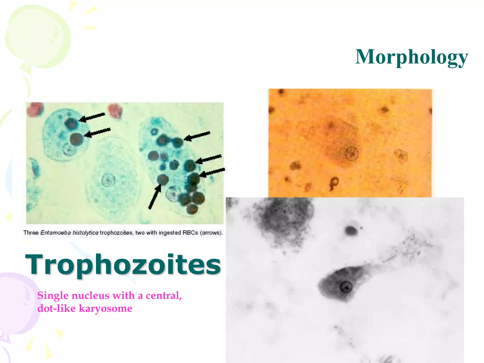
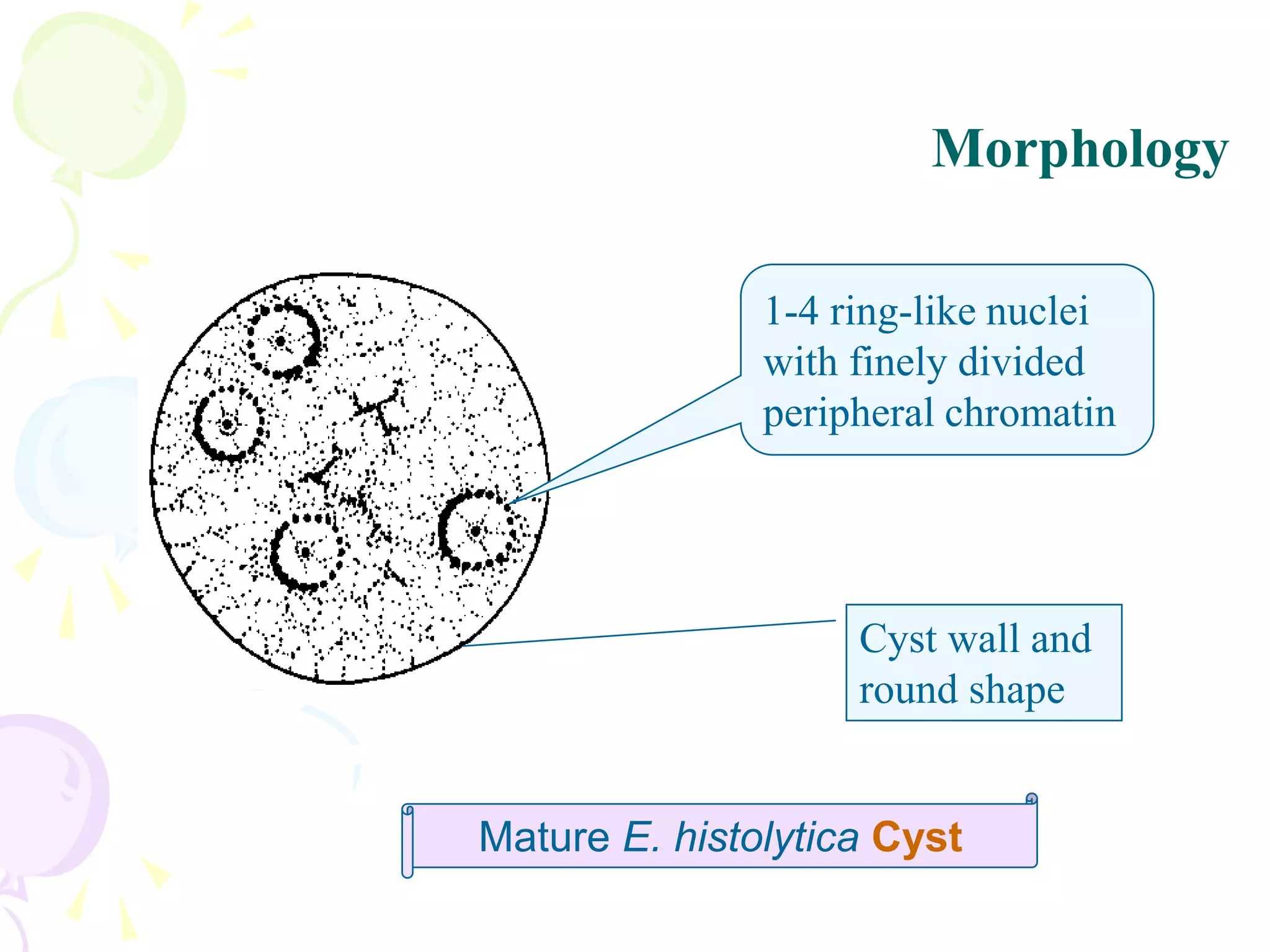
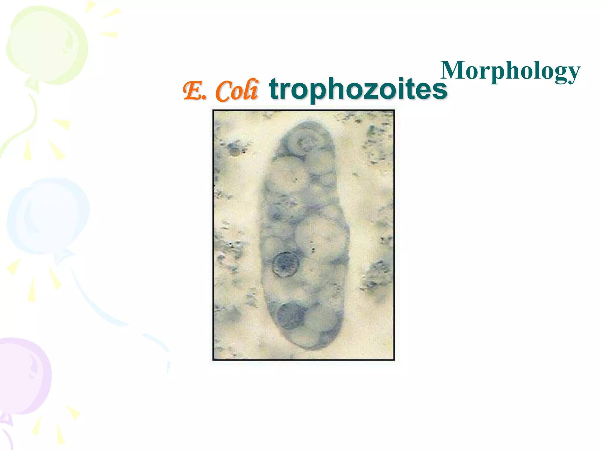
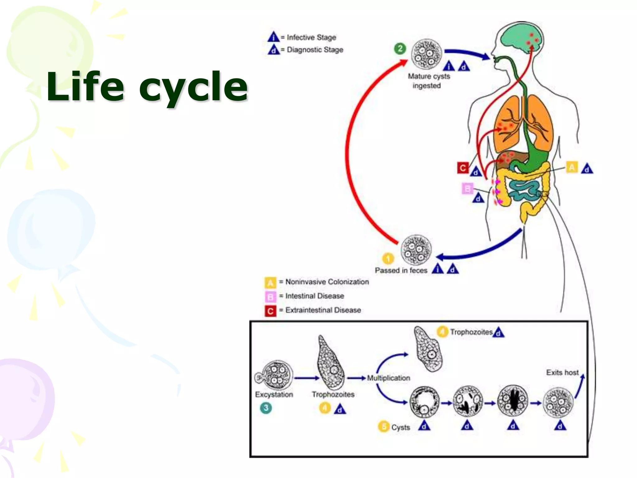
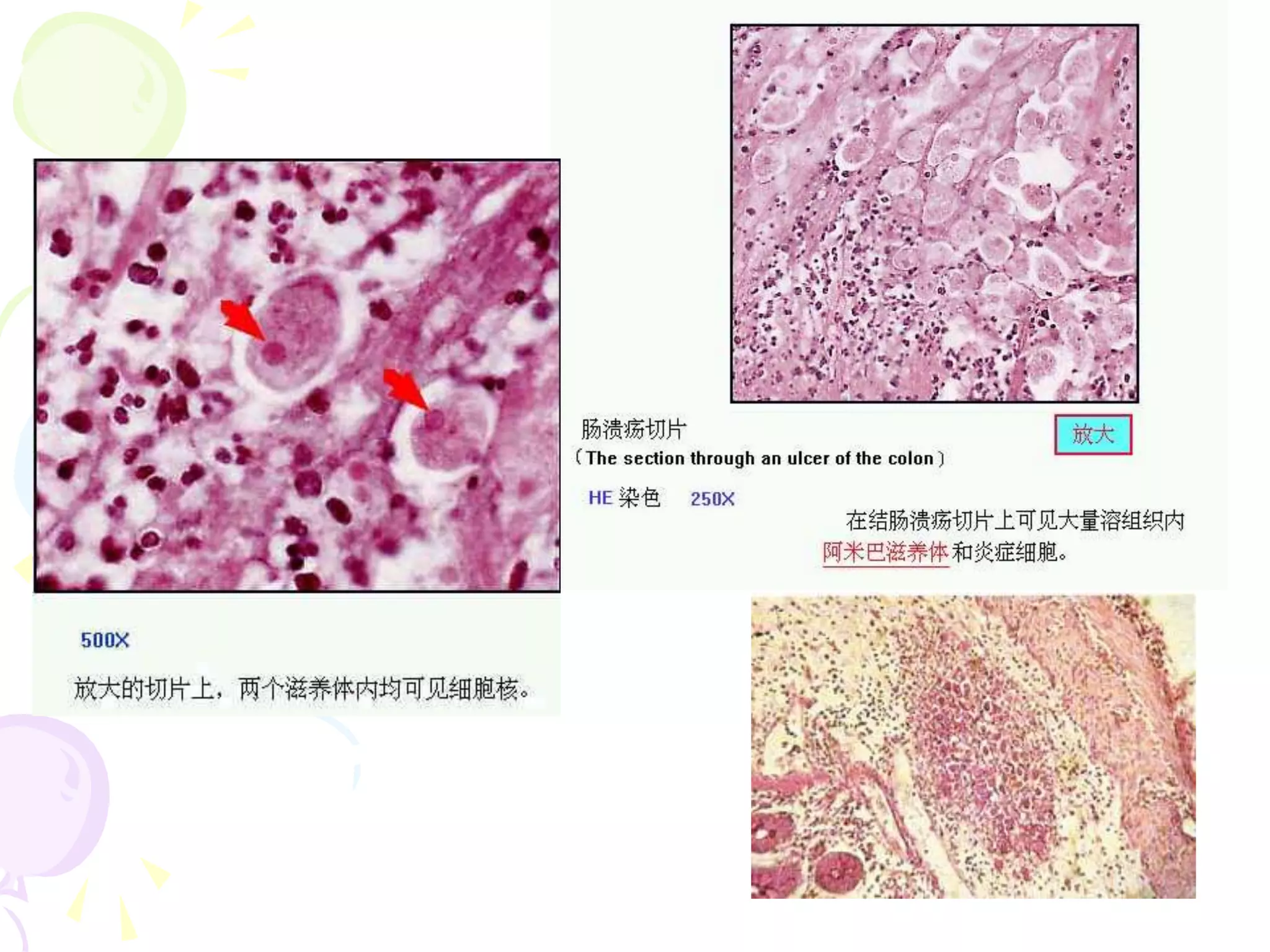
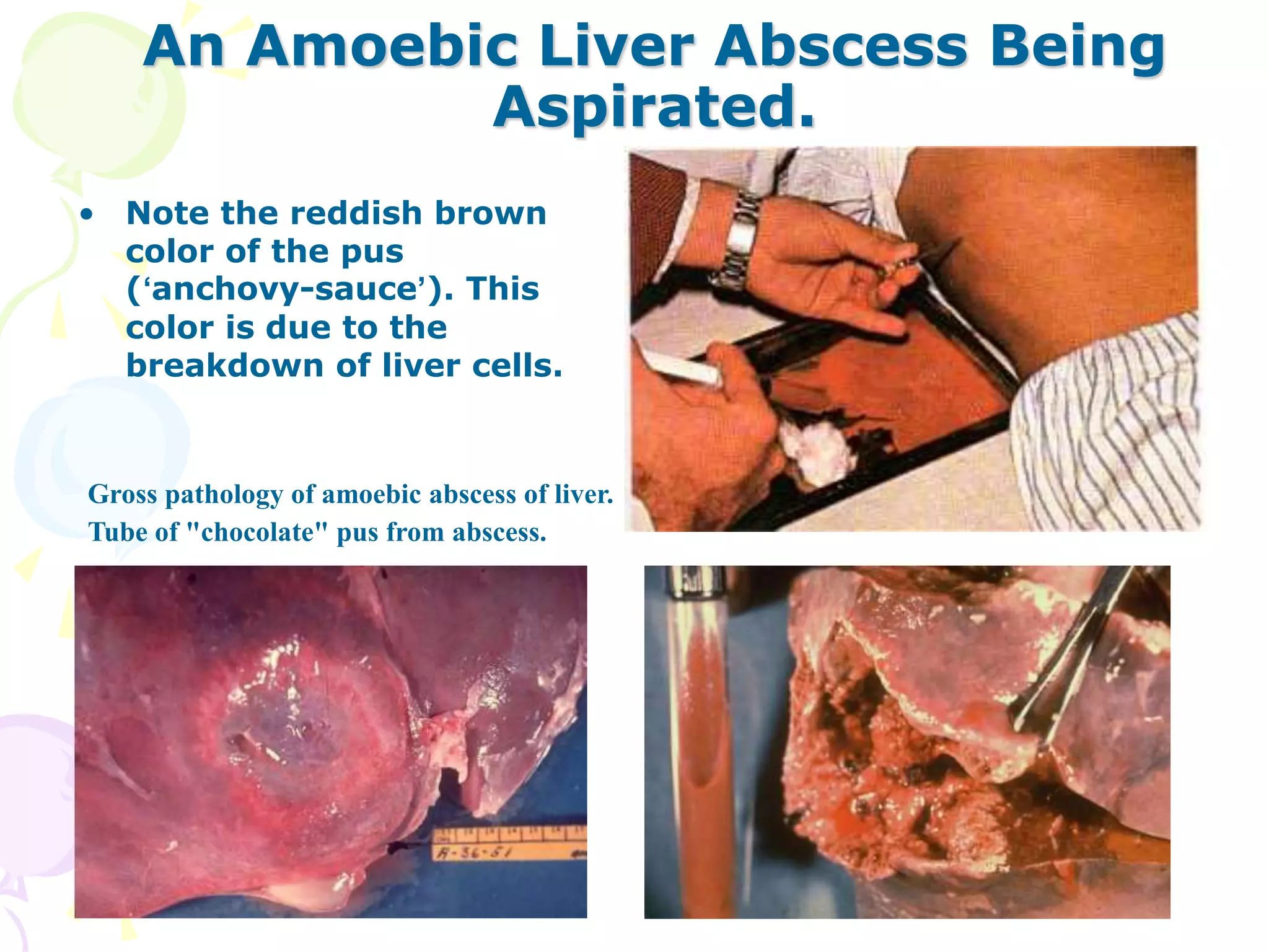
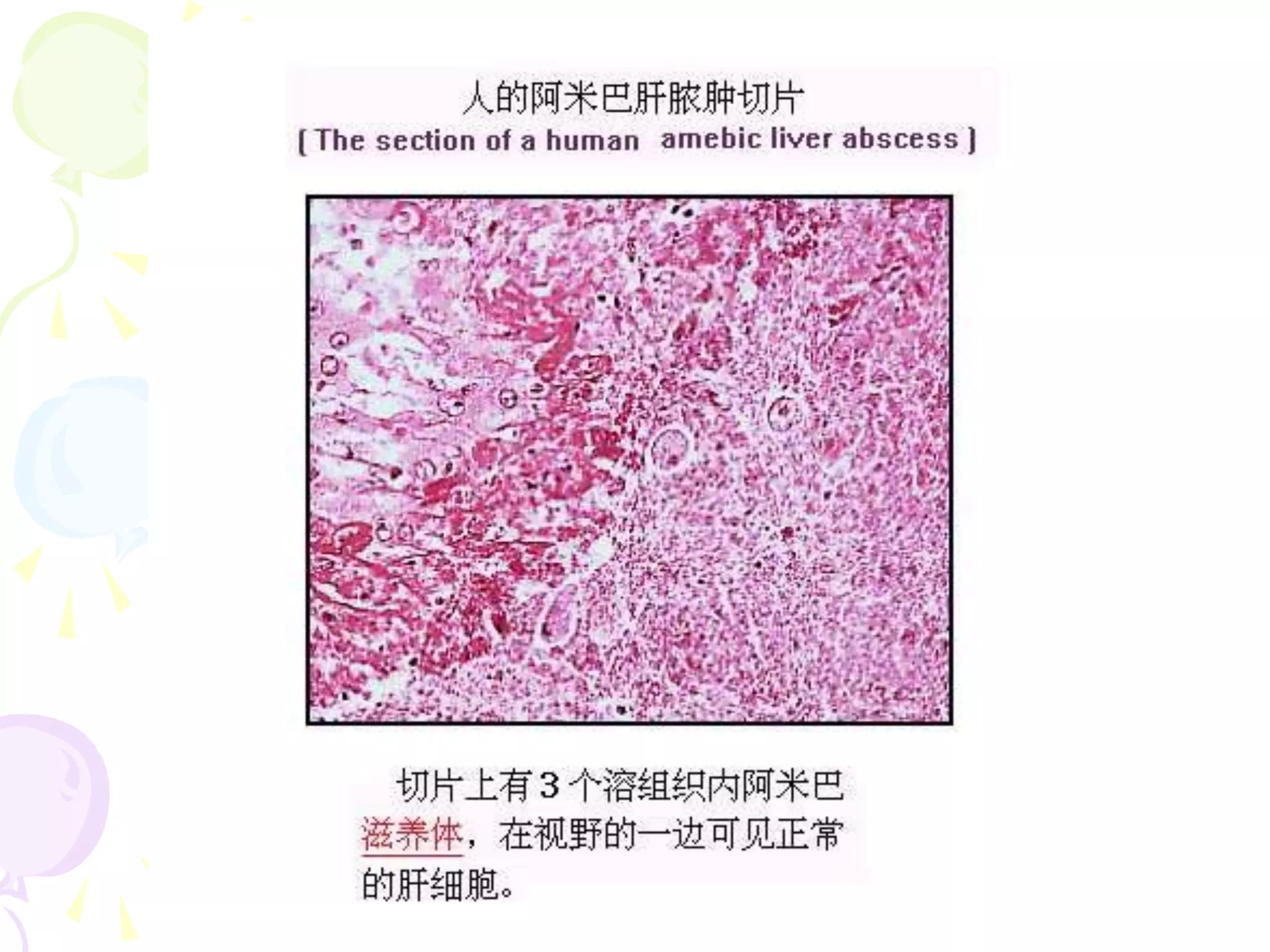
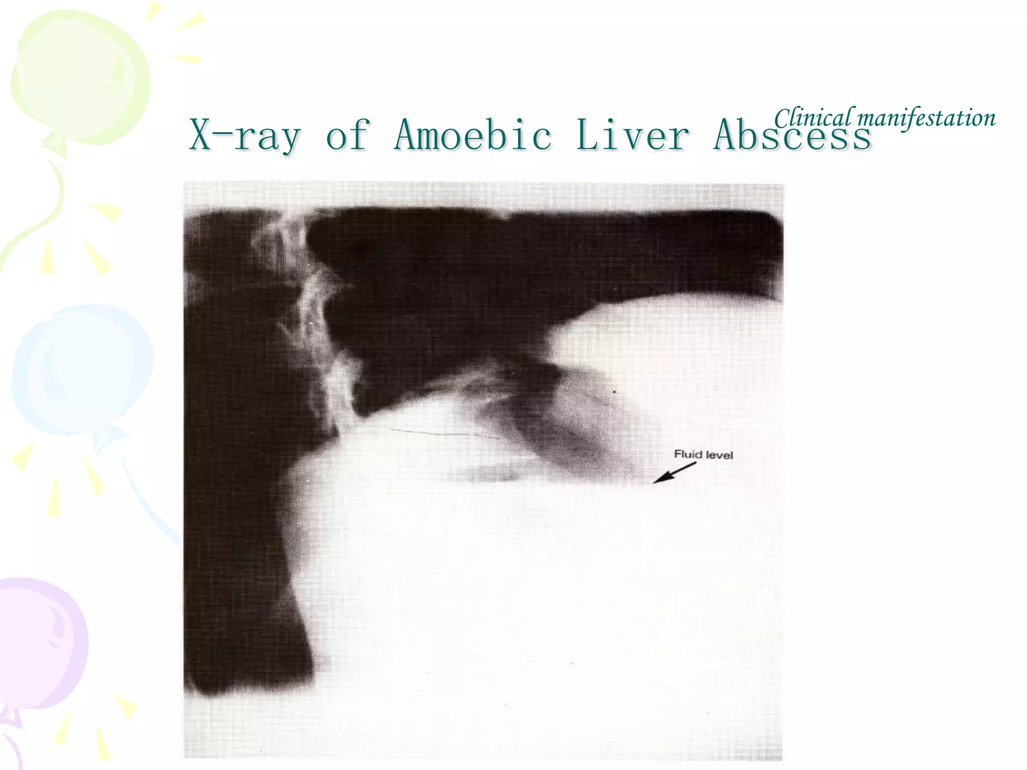
Protozoa
(原虫)
General Account
• One-cell animal – monocellular or
unicellular organisms with full vital
functions
• Species – total named species:65,000;
parasitic: around 10,000
Classification of protozoa
Amoebae
Flagellates
Ciliates
Sporozoa
Life cycle patterns
One-host form
1. One stage form – Trophozoite
2. Two stage form – Trophozoite & Cyst
Two-host form
1. Mammals mammals
2. Mammals insect vectors
Mode of Reproduction
• Asexual Reproduction
 Binary fission – result in 2 daughter cells
 Schizogony – multiple fission result in
multiple cells
 Budding
 Exogenous budding - by external budding result
in multi- cells
 Endodyogony - by internal budding result in 2
cells
• Sexual Reproduction
 Conjugation – exchange of nuclear
material of 2
 Gametogony – sexually differentiated cells
unite -- zygote
Pathogenesis
• Host Resistance
– Innate immunity
– Acquired immunity
• Parasite Invasion
– Toxin
– Mechanically damage
– Immune impair
• Immune inhibition
• hypersentivity
Opportunistic & Accidental
(protozoa) infections
Opportunistic parasites
• Opportunistic infection
– An infection by a microorganism
that normally does not cause
disease but becomes pathogenic
when the body's immune system is
impaired and unable to fight off
infection
Entamoeba histolytica
Acanthamoeba
Naegleria
Amoebic Infections
Epidemiology
• 4th leading cause of death from parasitic diseases
worldwide
Organism # of deaths/yr # infected
Entamoeba ~75,000 ~300 million
Ascaris ~200,000 ~480 million
Schistosoma ~750,000 ~200 million
Plasmodium 2-3 million ~500 million
(Malaria)
• Amoebiasis is not restricted to the tropics and
subtropics, it also occurs in temperate and even in
arctic and antarctic zones
Contaminated water is a source of infection.
Infection is common in developing countries where sanitation is poor.
Amoeba in alimentary tract
• Entamoeba
– E. histolytica (pathogenic)
– E. dispar (non-pathogenic)
– E. coli (big sister)
– E. hartmani (little brother)
– E. gingivalis (oral)
• Endolimax nana (occasionally pathogenic)
• Iodamoeba butschlii
Morphology
Entamoeba histolytica
Cysts Trophozoites
Thick wall Plasmalemma (thin)
1-4 ring-like nuclei 1 ring-like nucleus
Chromatoid body (blunt) Lacking
Round, 10-16 μm Irregular, 10-60 μm
Concentratable Labile
Morphology
Ingested
RBC
Nucleus with central
karyosome and finely
divided chromatin
granules
Pseudopod
E. histolytica trophozoite
Ectoplasma
Endoplasma
Single nucleus with a central,
dot-like karyosome
Morphology
Trophozoites
Micrograph of a trophozoite ingesting a red blood cell deprived from its host.
Mature E. histolytica Cyst
1-4 ring-like nuclei
with finely divided
peripheral chromatin
Cyst wall and
round shape
Morphology
Morphology
E. Coli trophozoites
•
Morphology
E. Coli cysts
•
Morphology
E. histolytica Stages - CYSTS
• Infective Stage for humans
• Resistant walls maintain viability
– If moist can last several weeks
– Killed by desiccation or boiling
• Diagnostic Stage in formed stools
– Can be concentrated and stained easily
– Not seen in liquid (diarrheic) stools or
tissues
E. histolytica Stages - TROPHOZOITES
• Cause amoebiasis (damage tissue)
• Spread throughout the body, but ...
– Rarely transmit the infection to others
• Labile in liquid stools or tissue, and
– must be rapidly found or preserved
(quick fixation & cold storage) for
Diagnosis
Life cycle
Life cycle
• Humans acquire E. histolytica by:
– Ingesting cysts (4 nuclei mature) in
fecally contaminated food or water
– Rarely by directly inoculating
trophozoites into colon or other sites
– (anal sex?)
• Fecal-Oral transmission (hand to
mouth)
Life cycle
• The basic generation-cycle: cyst –
lumen trophozoites – cyst
• Trophozoites may invade intestine
and spread
• Cyst formation – essential factors:
enviroment + time
• Infective cysts and trophozoites
pass in feces
General Types of Virulence Factors:
• Adherence factors
• 260kDa Gal/GalNAc lectin
• Invasion factors
• Amoeba pores
• Cysteine proteinases
• Endotoxins
Pathogenesis
Trophozoites ...
– Attach to mucosal epithelial cells
(MEC)
– Lyse MEC
– Ulcerate and invade mucosa
– Cause dysentery (diarrhea + blood)
– Metastasize via blood &/or lymph to
– Form abscesses in extraintestinal
sites ...
Pathogenesis
Clinical Classification of
Amoebiasis
(World Health Organization)
• Asymptomatic Infection:"Cyst
Passers/carrier”
• Symptomatic Infection:
– Intestinal Amoebiasis: (colon and rectum盲肠、
升结肠、直肠、乙状结肠和阑尾)
• Acute Dysenteric (dysentery)
• Chronic Non-Dysenteric (“self-cured”)
– Extra-Intestinal Amoebiasis:
• Amoebic Liver Abscess (ALA)
• Amoebic Pulmonary Abscess
• Other sites (brain, skin, GU, ?)
Clinical classification
• Asymptomatic infection
(carrier) >90% (E. dispar?)
• Symptomatic cases <10%
– 8% -10% dysentery, colitis, etc
– 2% invasive amoebiasis
– 0.1% deaths
Acute Dysenteric Amoebiasis
Symptoms:
Bloody mucoid diarrhea
RBCs and few WBCs in stools
– Abdominal pain
– weight loss
– bloating, tenesmus(里急后重)
and cramps
Clinical manifestation
Acute Dysenteric Amoebiasis
Signs:
Fever (33%)
Tender (enlarged) liver
Stools positive for trophozoites +/- WBC
NO cyst in loose stools
Clinical manifestation
•
Clinical manifestation
 Pinpoint lesion on
mucous membrane
 Flask-shaped crateriform
ulcers
Pathological changes in large intestine
Chronic Non-Dysenteric Amoebiasis
“self-cured” carrier state
Usually for 1 year, 37%
symptomatic >5 years
Intermittent diarrhea, mucus,
abdominal pain, flatulence and/or
weight loss
E. histolytica trophs in loose stools
Cysts in solid stools
Positive serology and ulcerations on
sigmoidoscopy or pathologic test
Clinical manifestation
Amoebic Liver Abscess (ALA)
• Symptoms
– History of dysentery (1 yr), weight
loss, abdominal pain, chest or
shoulder pain
• Signs
– fever, hepatomegaly
– Diagnostic aspiration:non-odorous, reddish-brown in
color aspirate (chocolate jam) "anchovy paste"
– Might find trophozoites in the aspirate
– Skin inflammation
Extra-Intestinal Amoebiasis
Clinical manifestation
Ulcers caused by invasion of E. histolytica into the liver.
Clinical manifestation
•
Clinical manifestation
An Amoebic Liver Abscess Being
Aspirated.
• Note the reddish brown
color of the pus
(‘anchovy-sauce’). This
color is due to the
breakdown of liver cells.
Gross pathology of amoebic abscess of liver.
Tube of "chocolate" pus from abscess.
X-ray of Amoebic Liver Abscess
•
Clinical manifestation
Diagnosis
• Pathogenic diagnosis
– Stool examination:
• Direct Fecal Smear (trophs and cysts)
• Fecal concentration and iodine dye techniques -
(cysts) ZnSO4 or formalin-ether
– Cultivation
– DNA detection
– Sigmoidoscopy
• Serologic Tests (for chronic disease):
ELISA, IHA (indirect hemagglutination)
• Imaging: X-ray; CT
Stool examination
trophozoite cyst
specimen loose feces solid feces
method direct smear with normal
saline
direct smear with iodine
stain
diseases
amoebic dysentery
chronic intestinal
amoebiasis or carriers
remarks
1.container must clean
2.examined soon after they have
been passed.
3.select bloody and mucous
portion.
Two microscopically
indistinguishable Entamoeba sp.
• E. histolytica
– invades tissues
– should always be treated
• E. dispar
– is non-pathogenic, even in AIDS
– should not be treated
• For invasive forms:
metronidazole
• For luminal forms:
Iodoquinofonum, paromomycin,
diloxanide
• Do not treat asymptomatic
intestinal E. dispar infection
Treatment of Amoebiasis
Location Clinical Class Drug Name Drug Action
Intestinal
Asymptomatic Iodoquinofonnum(喹碘方) lumenal amebicide
Mild to moderate
intestinal disease
Metronidazole(甲硝唑) tissue amebicide
Severe intestinal
disease
Metronidazole plus a lumenal
drug
both
Extraintestinal
Hepatic disease Metronidazole plus a lumenal
drug
both
• Individual measures
• Diagnosis and treatment of E. histolytica patients
• Safe drinking water (boiling or 0.22 µm filtration)
• Cleaning of uncooked fruits and vegetables
• Prevention of contamination of foods
• Chemotherapeutic Trial
Prevention & Control
Community measures
– Public services and utilities
• Adequate disposal of human stools
• Safe and adequate water supply
– Primary health care systems
• Health education (washing hands, cleaning and protecting
food, controlling insects)
• Specific surveillance programs and Control programs
integrated into ongoing sanitation & diarrhea control
– Health Regulations
• Control of food vendors and food handlers
• Control of flies and cockroaches
Prevention & Control
Infections with
Free Living Amoebae
Naegleria 耐格里属
Acanthamoeba 棘阿米巴属
Free Living Amoebae Not seen
in humans
Naegleria
Acanthamoeba
cysts & trophs
are seen in
humans
i
i
10-35 µm (smaller than A. spp.)
with lobate pseudopodia
15-45 µm with filiform pseudopodia
i
Acanthamoeba spp.
Acanthamoeba
trophozoites with
acanthopodia
Primary Amoebic Meningoencephalitis
PAME
An acute suppurative infection of the
brain and meninges that is rapidly fatal
and usually not diagnosed antemortem
– Caused by Naegleria fowleri
– Headache, lethargy and olfactory problems
– Sore throat, runny nose, severe headache,
vomiting, stiff neck, confusion leading to ...
– Coma and death
PAME
• Patient History (child)
– Prior Health Excellent
– Recent History of Swimming (fresh
water/pools)
– Cases peak during HOT months
• Symptoms/Signs
– Sore throat, runny nose, headache,
vomiting, stiff neck, mental confusion,
olfactory problems, lethargy, coma and
death
DIAGNOSIS
– None effective - few patients survive
– Amphoteracin B +/- ?
PAME
Treatment
Granulomatous Amoebic Encephalitis
GAE
A more slowly progressive, chronic form of
the disease not associated with swimming
(except in hot tubs)
• cause: Acanthamoeba castellanii
• history of subcutaneous nodules, eye or
skin infection, progressive nasal congestion,
headache ...
• CNS lesions with negative serology for
toxoplasmosis
• in debilitated/immuno-compromised Pts
with CD4+ TL <200/mm3
• disseminated infection: skin, sinuses, lungs,
CNS/CSF
GAE
• abscesses/lesions (tissues) have
– granulomatous inflammation
– hemorrhagic necrosis and vasculitis
– trophozoites & cysts with wrinkled-walls!
• amoebae rarely seen in CSF
Pathology
• No satisfactory or effective treatment ?
– amphotericin B
GAE
Treatment
Acanthamoeba Keratitis AK
Corneal infection with
Acanthamoeba spp. trophozoites
& cysts
• Ulcerations & “Ring Infiltrate” of
cornea
• Induced by
– trauma to eye, exposure to
contaminated H2O
– contact lens wear with tap water
rinsing
AK
• Diagnosis
– Examine corneal scrapings or smear
– Histopathologic examination of
cornea
• Treatment
– Triple Antiamoebic Therapy
• neomycin-polymyxin-
gramicidin/propamidine/miconazole
– Penetrating keratoplasty (cadaver
cornea)
Summary
• E. histolytica
– Life cycle, pathogenesis, Diagnosis,
treatment
– carrier
– Morphology differences with E. coli
• Free living amoeba
– prevention
QUESTIONS
• How to diagnose hepatic amoebiasis?
• What are the transmission route of E.
histolytica ?
• Who should be treated for amoebic
infection?
• How will one get amoebiasis? What
are the consequences?

amoebiasis ppt.ppt

  • 1.
  • 2.
    General Account • One-cellanimal – monocellular or unicellular organisms with full vital functions • Species – total named species:65,000; parasitic: around 10,000
  • 3.
  • 4.
    Life cycle patterns One-hostform 1. One stage form – Trophozoite 2. Two stage form – Trophozoite & Cyst Two-host form 1. Mammals mammals 2. Mammals insect vectors
  • 5.
    Mode of Reproduction •Asexual Reproduction  Binary fission – result in 2 daughter cells  Schizogony – multiple fission result in multiple cells  Budding  Exogenous budding - by external budding result in multi- cells  Endodyogony - by internal budding result in 2 cells • Sexual Reproduction  Conjugation – exchange of nuclear material of 2  Gametogony – sexually differentiated cells unite -- zygote
  • 6.
    Pathogenesis • Host Resistance –Innate immunity – Acquired immunity • Parasite Invasion – Toxin – Mechanically damage – Immune impair • Immune inhibition • hypersentivity Opportunistic & Accidental (protozoa) infections
  • 7.
    Opportunistic parasites • Opportunisticinfection – An infection by a microorganism that normally does not cause disease but becomes pathogenic when the body's immune system is impaired and unable to fight off infection
  • 8.
  • 9.
    Epidemiology • 4th leadingcause of death from parasitic diseases worldwide Organism # of deaths/yr # infected Entamoeba ~75,000 ~300 million Ascaris ~200,000 ~480 million Schistosoma ~750,000 ~200 million Plasmodium 2-3 million ~500 million (Malaria) • Amoebiasis is not restricted to the tropics and subtropics, it also occurs in temperate and even in arctic and antarctic zones
  • 10.
    Contaminated water isa source of infection.
  • 11.
    Infection is commonin developing countries where sanitation is poor.
  • 12.
    Amoeba in alimentarytract • Entamoeba – E. histolytica (pathogenic) – E. dispar (non-pathogenic) – E. coli (big sister) – E. hartmani (little brother) – E. gingivalis (oral) • Endolimax nana (occasionally pathogenic) • Iodamoeba butschlii
  • 13.
    Morphology Entamoeba histolytica Cysts Trophozoites Thickwall Plasmalemma (thin) 1-4 ring-like nuclei 1 ring-like nucleus Chromatoid body (blunt) Lacking Round, 10-16 μm Irregular, 10-60 μm Concentratable Labile
  • 14.
    Morphology Ingested RBC Nucleus with central karyosomeand finely divided chromatin granules Pseudopod E. histolytica trophozoite Ectoplasma Endoplasma
  • 15.
    Single nucleus witha central, dot-like karyosome Morphology Trophozoites
  • 16.
    Micrograph of atrophozoite ingesting a red blood cell deprived from its host.
  • 17.
    Mature E. histolyticaCyst 1-4 ring-like nuclei with finely divided peripheral chromatin Cyst wall and round shape Morphology
  • 18.
  • 19.
  • 20.
  • 21.
    E. histolytica Stages- CYSTS • Infective Stage for humans • Resistant walls maintain viability – If moist can last several weeks – Killed by desiccation or boiling • Diagnostic Stage in formed stools – Can be concentrated and stained easily – Not seen in liquid (diarrheic) stools or tissues
  • 22.
    E. histolytica Stages- TROPHOZOITES • Cause amoebiasis (damage tissue) • Spread throughout the body, but ... – Rarely transmit the infection to others • Labile in liquid stools or tissue, and – must be rapidly found or preserved (quick fixation & cold storage) for Diagnosis
  • 23.
  • 24.
    Life cycle • Humansacquire E. histolytica by: – Ingesting cysts (4 nuclei mature) in fecally contaminated food or water – Rarely by directly inoculating trophozoites into colon or other sites – (anal sex?) • Fecal-Oral transmission (hand to mouth)
  • 25.
    Life cycle • Thebasic generation-cycle: cyst – lumen trophozoites – cyst • Trophozoites may invade intestine and spread • Cyst formation – essential factors: enviroment + time • Infective cysts and trophozoites pass in feces
  • 26.
    General Types ofVirulence Factors: • Adherence factors • 260kDa Gal/GalNAc lectin • Invasion factors • Amoeba pores • Cysteine proteinases • Endotoxins Pathogenesis
  • 27.
    Trophozoites ... – Attachto mucosal epithelial cells (MEC) – Lyse MEC – Ulcerate and invade mucosa – Cause dysentery (diarrhea + blood) – Metastasize via blood &/or lymph to – Form abscesses in extraintestinal sites ... Pathogenesis
  • 28.
    Clinical Classification of Amoebiasis (WorldHealth Organization) • Asymptomatic Infection:"Cyst Passers/carrier” • Symptomatic Infection: – Intestinal Amoebiasis: (colon and rectum盲肠、 升结肠、直肠、乙状结肠和阑尾) • Acute Dysenteric (dysentery) • Chronic Non-Dysenteric (“self-cured”) – Extra-Intestinal Amoebiasis: • Amoebic Liver Abscess (ALA) • Amoebic Pulmonary Abscess • Other sites (brain, skin, GU, ?)
  • 29.
    Clinical classification • Asymptomaticinfection (carrier) >90% (E. dispar?) • Symptomatic cases <10% – 8% -10% dysentery, colitis, etc – 2% invasive amoebiasis – 0.1% deaths
  • 30.
    Acute Dysenteric Amoebiasis Symptoms: Bloodymucoid diarrhea RBCs and few WBCs in stools – Abdominal pain – weight loss – bloating, tenesmus(里急后重) and cramps Clinical manifestation
  • 31.
    Acute Dysenteric Amoebiasis Signs: Fever(33%) Tender (enlarged) liver Stools positive for trophozoites +/- WBC NO cyst in loose stools Clinical manifestation
  • 32.
    • Clinical manifestation  Pinpointlesion on mucous membrane  Flask-shaped crateriform ulcers Pathological changes in large intestine
  • 34.
    Chronic Non-Dysenteric Amoebiasis “self-cured”carrier state Usually for 1 year, 37% symptomatic >5 years Intermittent diarrhea, mucus, abdominal pain, flatulence and/or weight loss E. histolytica trophs in loose stools Cysts in solid stools Positive serology and ulcerations on sigmoidoscopy or pathologic test Clinical manifestation
  • 35.
    Amoebic Liver Abscess(ALA) • Symptoms – History of dysentery (1 yr), weight loss, abdominal pain, chest or shoulder pain • Signs – fever, hepatomegaly – Diagnostic aspiration:non-odorous, reddish-brown in color aspirate (chocolate jam) "anchovy paste" – Might find trophozoites in the aspirate – Skin inflammation Extra-Intestinal Amoebiasis Clinical manifestation
  • 36.
    Ulcers caused byinvasion of E. histolytica into the liver. Clinical manifestation
  • 37.
  • 38.
  • 39.
    An Amoebic LiverAbscess Being Aspirated. • Note the reddish brown color of the pus (‘anchovy-sauce’). This color is due to the breakdown of liver cells. Gross pathology of amoebic abscess of liver. Tube of "chocolate" pus from abscess.
  • 41.
    X-ray of AmoebicLiver Abscess • Clinical manifestation
  • 42.
    Diagnosis • Pathogenic diagnosis –Stool examination: • Direct Fecal Smear (trophs and cysts) • Fecal concentration and iodine dye techniques - (cysts) ZnSO4 or formalin-ether – Cultivation – DNA detection – Sigmoidoscopy • Serologic Tests (for chronic disease): ELISA, IHA (indirect hemagglutination) • Imaging: X-ray; CT
  • 43.
    Stool examination trophozoite cyst specimenloose feces solid feces method direct smear with normal saline direct smear with iodine stain diseases amoebic dysentery chronic intestinal amoebiasis or carriers remarks 1.container must clean 2.examined soon after they have been passed. 3.select bloody and mucous portion.
  • 44.
    Two microscopically indistinguishable Entamoebasp. • E. histolytica – invades tissues – should always be treated • E. dispar – is non-pathogenic, even in AIDS – should not be treated
  • 45.
    • For invasiveforms: metronidazole • For luminal forms: Iodoquinofonum, paromomycin, diloxanide • Do not treat asymptomatic intestinal E. dispar infection Treatment of Amoebiasis
  • 46.
    Treatment of Amoebiasis LocationClinical Class Drug Name Drug Action Intestinal Asymptomatic Iodoquinofonnum(喹碘方) lumenal amebicide Mild to moderate intestinal disease Metronidazole(甲硝唑) tissue amebicide Severe intestinal disease Metronidazole plus a lumenal drug both Extraintestinal Hepatic disease Metronidazole plus a lumenal drug both
  • 47.
    • Individual measures •Diagnosis and treatment of E. histolytica patients • Safe drinking water (boiling or 0.22 µm filtration) • Cleaning of uncooked fruits and vegetables • Prevention of contamination of foods • Chemotherapeutic Trial Prevention & Control
  • 48.
    Community measures – Publicservices and utilities • Adequate disposal of human stools • Safe and adequate water supply – Primary health care systems • Health education (washing hands, cleaning and protecting food, controlling insects) • Specific surveillance programs and Control programs integrated into ongoing sanitation & diarrhea control – Health Regulations • Control of food vendors and food handlers • Control of flies and cockroaches Prevention & Control
  • 49.
    Infections with Free LivingAmoebae Naegleria 耐格里属 Acanthamoeba 棘阿米巴属
  • 50.
    Free Living AmoebaeNot seen in humans Naegleria Acanthamoeba cysts & trophs are seen in humans i i 10-35 µm (smaller than A. spp.) with lobate pseudopodia 15-45 µm with filiform pseudopodia i
  • 51.
  • 52.
    Primary Amoebic Meningoencephalitis PAME Anacute suppurative infection of the brain and meninges that is rapidly fatal and usually not diagnosed antemortem – Caused by Naegleria fowleri – Headache, lethargy and olfactory problems – Sore throat, runny nose, severe headache, vomiting, stiff neck, confusion leading to ... – Coma and death
  • 53.
    PAME • Patient History(child) – Prior Health Excellent – Recent History of Swimming (fresh water/pools) – Cases peak during HOT months • Symptoms/Signs – Sore throat, runny nose, headache, vomiting, stiff neck, mental confusion, olfactory problems, lethargy, coma and death DIAGNOSIS
  • 54.
    – None effective- few patients survive – Amphoteracin B +/- ? PAME Treatment
  • 55.
    Granulomatous Amoebic Encephalitis GAE Amore slowly progressive, chronic form of the disease not associated with swimming (except in hot tubs) • cause: Acanthamoeba castellanii • history of subcutaneous nodules, eye or skin infection, progressive nasal congestion, headache ... • CNS lesions with negative serology for toxoplasmosis • in debilitated/immuno-compromised Pts with CD4+ TL <200/mm3 • disseminated infection: skin, sinuses, lungs, CNS/CSF
  • 56.
    GAE • abscesses/lesions (tissues)have – granulomatous inflammation – hemorrhagic necrosis and vasculitis – trophozoites & cysts with wrinkled-walls! • amoebae rarely seen in CSF Pathology
  • 57.
    • No satisfactoryor effective treatment ? – amphotericin B GAE Treatment
  • 58.
    Acanthamoeba Keratitis AK Cornealinfection with Acanthamoeba spp. trophozoites & cysts • Ulcerations & “Ring Infiltrate” of cornea • Induced by – trauma to eye, exposure to contaminated H2O – contact lens wear with tap water rinsing
  • 59.
    AK • Diagnosis – Examinecorneal scrapings or smear – Histopathologic examination of cornea • Treatment – Triple Antiamoebic Therapy • neomycin-polymyxin- gramicidin/propamidine/miconazole – Penetrating keratoplasty (cadaver cornea)
  • 60.
    Summary • E. histolytica –Life cycle, pathogenesis, Diagnosis, treatment – carrier – Morphology differences with E. coli • Free living amoeba – prevention
  • 61.
    QUESTIONS • How todiagnose hepatic amoebiasis? • What are the transmission route of E. histolytica ? • Who should be treated for amoebic infection? • How will one get amoebiasis? What are the consequences?