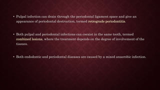
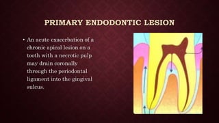
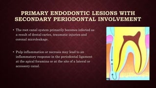
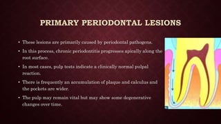
ENDO-PERIO LESIONS
Mohamed Rabie’ Ahmed
INTRODUCTION
• The endodontium and periodontium are closely related and diseases of one tissue
may lead to secondary diseases in the other.
• The differential diagnosis of endodontic and periodontal diseases can sometimes be
difficult but it is important to make a correct diagnosis so that the appropriate
treatment can be provided.
• Pulpal infection can drain through the periodontal ligament space and give an
appearance of periodontal destruction, termed retrograde periodontitis.
• Both pulpal and periodontal infections can coexist in the same tooth, termed
combined lesions, where the treatment depends on the degree of involvement of the
tissues.
• Both endodontic and periodontal diseases are caused by a mixed anaerobic infection.
DEFINITION
• The tooth involved must have pulpal disease
• There must be destruction of the attachment apparatus from gingival sulcus to
either apex of tooth or of an involved lateral canal
• Both root canal treatment & periodontal therapy are required to resolve the entirety
of the lesion
PATHWAYS CONNECTING ENDODONTIC AND
PERIODONTAL TISSUES
• There are two forms of possible pathways for bacteria and their products connecting
the two tissues:
1. Anatomical pathways
2. Non-physiological pathways
ANATOMICAL PATHWAYS
A) Apical foramen
B) Lateral and accessory canals
C) Dentinal tubules
NON-PHYSIOLOGICAL PATHWAYS
“IATROGENIC”
A. Iatrogenic root canal perforations
NON-PHYSIOLOGICAL PATHWAYS
“IATROGENIC”
B. Vertical root fractures
NON-PHYSIOLOGICAL PATHWAYS
“PATHOLOGIC”
A. Externanl Resorption
B. Trauamtic fractures
C. Developmental grooves
A
B
C
CLASSIFICATION
• The most conventional classification used for endodontic-periodontal lesions was
given by Simon et al. (1972):
1. Primary endodontic lesions
2. Primary endodontic lesions with secondary periodontal involvement
3. Primary periodontal lesions
4. Primary periodontal lesions with secondary endodontic involvement
5. True combined lesions
PRIMARY ENDODONTIC LESION
• An acute exacerbation of a
chronic apical lesion on a
tooth with a necrotic pulp
may drain coronally
through the periodontal
ligament into the gingival
sulcus.
PRIMARY ENDODONTIC LESION
• For diagnosis purposes, insert a gutta-percha cone into
the sinus tract and to take one or more radiographs to
determine the origin of the lesion.
• When the pocket is probed, it is narrow and lacks
width.
• Primary endodontic diseases usually heal following
root canal treatment.
PRIMARY ENDODONTIC LESIONS WITH
SECONDARY PERIODONTAL INVOLVEMENT
• The root canal system primarily becomes infected as
a result of dental caries, traumatic injuries and
coronal microleakage.
• Pulp inflammation or necrosis may lead to an
inflammatory response in the periodontal ligament
at the apical foramina or at the site of a lateral or
accessory canal.
PRIMARY ENDODONTIC LESIONS WITH
SECONDARY PERIODONTAL INVOLVEMENT
• Long-term existence of the defect has
resulted in deposits of plaque and calculus
in the pocket with subsequent advancement
of the periodontal disease.
• After adequate root canal treatment, The
integrity of the periodontium will be
reestablished.
PRIMARY PERIODONTAL LESIONS
• These lesions are primarily caused by periodontal pathogens.
• In this process, chronic periodontitis progresses apically along the
root surface.
• In most cases, pulp tests indicate a clinically normal pulpal
reaction.
• There is frequently an accumulation of plaque and calculus and
the pockets are wider.
• The pulp may remain vital but may show some degenerative
changes over time.
PRIMARY PERIODONTAL LESIONS WITH
SECONDARY ENDODONTIC INVOLVEMENT
• The apical progression of a periodontal pocket
may continue until the apical tissues are
involved.
• In this case, the pulp may become necrotic as a
result of infection entering via lateral canals or
the apical foramen.
PRIMARY PERIODONTAL LESIONS WITH
SECONDARY ENDODONTIC INVOLVEMENT
• Although the pulp is exposed to a bacterial challenge
through patent dentinal tubules, it is quite capable of
repair and healing.
• Production of reparative dentin and reduced canal
diameter may result, but pulp tissue remains
relatively unaffected.
TRUE COMBINED LESIONS
• These lesions occur when an endodontically induced
periapical lesion exists at a tooth that is also affected by
marginal periodontitis.
• The tooth has a pulpless, infected root canal system and a co-
existing periodontal defect.
• This is particularly true in single-rooted teeth. In molar
teeth, root resection can be considered as a treatment
alternative if not all roots are severely involved
CLINICAL DIAGNOSTIC PROCEDURES
1. Visual examination
• visual examination of the lips, cheeks, oral mucosa,
tongue, palate and muscles should be carried out .
• The alveolar mucosa and the attached gingiva are
examined for the presence of inflammation,
ulcerations or sinus tracts.
CLINICAL DIAGNOSTIC PROCEDURES
2. Palpation
• With the index finger the mucosa is pressed
against the underlying cortical bone .
• This will detect the presence of periradicular
abnormalities or ''hot'' zones that produce painful
response to digital pressure .
CLINICAL DIAGNOSTIC PROCEDURES
3. Percussion
• An abnormal positive response indicates
inflammation of the periodontal ligament that may
be either from pulpal or periodontal origin .
• The sensitivity of the proprioceptive fibers in an
inflamed periodontal ligament will help identify the
location of the pain .
• This test should be performed gently, especially in
highly sensitive teeth .
CLINICAL DIAGNOSTIC PROCEDURES
4. Mobility
• Tooth mobility is directly proportional to the integrity of the attachment
apparatus or to the extent of inflammation in the periodontal ligament.
• Hypermobility is quite common in cases of primary endodontic involvement
and should not be confused with true mobility caused by periodontal
destruction.
• In cases of primary endodontic pathology, the mobility resolves within a
week of initiating endodontic therapy.
CLINICAL DIAGNOSTIC PROCEDURES
5. Radiographs
• Identification of proximal crestal bone & its
position in relation to CEJ
• To identify the level of bone loss on one side of
the tooth
• Interpretation of discrete periapical/lateral
lesions – suggest cause of lesion
• Radiograph is is of little value when bone loss
extends from crestal bone to/near apex
CLINICAL DIAGNOSTIC PROCEDURES
6. Pulp testing
• Cold test, electric test, blood flow tests and cavity test.
• The presence or absence of vital tissue in a tooth with a single
canal can be determined with confidence with the current
pulp-testing procedures.
• The same degree of confidence cannot be ascribed to positive
pulp test responses in a tooth with multiple canals.
TREATMENT PROTOCOL
• In general, when primary disease of one tissue, i.e. pulp or periodontium, is present
and secondary disease is just starting, treat the primary disease.
• When secondary disease is established and chronic, both primary and secondary
diseases must be treated.
• Periodontal therapy may or may not be required, depending on disease status.
• The complete healing of destroyed periodontal support can be expected following the
treatment of pulpal pathology.
TREATMENT PROTOCOL
• The treatment strategy must be first to focus on the pulpal infection and to perform
debridement and disinfection of the root canal system.
• The second phase includes a period of observation, whereby the extent of
periodontal healing resulting from the endodontic treatment is followed.
• Reduced probing depth can usually be expected within a couple of weeks while bone
regeneration may require several months before it can be radiographically detected.
MANAGEMENT OF PRIMARY ENDO LESION
• Root canal therapy
• Sinus into gingival sulcus / furcation area disappears once root canals cleaned,
shaped & obturated
MANAGEMENT OF PRIMARY PERIO LESION
• Hygiene phase therapy Oral prophylaxis, oral hygiene instructions, Scaling, root
planing, Periodontal surgery
• Removal of cementum will expose dentinal tubules, so minimize use of ultrasonics
and rotary scaling instruments when <2 mm of dentin thickness remaining
MANAGEMENT OF PRIMARY ENDO
SECONDARY PERIO
• Root canal therapy >> Multi visit endo.
• Evaluate 2-3mnths >> Perio therapy if required.
• Intracanal medicament found reduce inflammation & favoring repair.
• Aggressive removal of PDL & cementum during interim endodontic therapy may
adversely affect Healing.
MANAGEMENT - PRIMARY PERIO
SECONDARY ENDO & COMBINED LESIONS
RESECTIVE APPROACHES
A. Root amputation
B. Hemisectioning
C. Bicuspidization
REPARATIVE (REGENERATIVE THERAPY)
A. Guided tissue regeneration (GTR).
B. Bone grafting
REFERENCES
• Jansson L, Ehnevid H, Lindskog S, Blomlφf L. The influence of endodontic infection
on progression of marginal bone loss in periodontitis. J Clin Periodontol
1995;22:729-73.
• Jansson L, Ehnevid H, Blomlφf L, Weintraub A, Lindskog S. Endodontic pathogens
in periodontal disease augmentation. J Clin Periodontol 1995;22:598-602
• Jansson L, Ehnevid H, Lindskog S, Blomlφf L. Relationship between periapical and
periodontal status. A clinical retrospective study. J Clin Periodontol 1993;20:117-
23.
• Simon JH, Glick DH, Frank AL. The relationship of endodontic-periodontic lesions.
J Periodontol 1972;43:202-8.

Endo-Perio Lesions

  • 1.
  • 2.
    INTRODUCTION • The endodontiumand periodontium are closely related and diseases of one tissue may lead to secondary diseases in the other. • The differential diagnosis of endodontic and periodontal diseases can sometimes be difficult but it is important to make a correct diagnosis so that the appropriate treatment can be provided.
  • 3.
    • Pulpal infectioncan drain through the periodontal ligament space and give an appearance of periodontal destruction, termed retrograde periodontitis. • Both pulpal and periodontal infections can coexist in the same tooth, termed combined lesions, where the treatment depends on the degree of involvement of the tissues. • Both endodontic and periodontal diseases are caused by a mixed anaerobic infection.
  • 4.
    DEFINITION • The toothinvolved must have pulpal disease • There must be destruction of the attachment apparatus from gingival sulcus to either apex of tooth or of an involved lateral canal • Both root canal treatment & periodontal therapy are required to resolve the entirety of the lesion
  • 5.
    PATHWAYS CONNECTING ENDODONTICAND PERIODONTAL TISSUES • There are two forms of possible pathways for bacteria and their products connecting the two tissues: 1. Anatomical pathways 2. Non-physiological pathways
  • 6.
    ANATOMICAL PATHWAYS A) Apicalforamen B) Lateral and accessory canals C) Dentinal tubules
  • 7.
  • 8.
  • 9.
    NON-PHYSIOLOGICAL PATHWAYS “PATHOLOGIC” A. ExternanlResorption B. Trauamtic fractures C. Developmental grooves A B C
  • 10.
    CLASSIFICATION • The mostconventional classification used for endodontic-periodontal lesions was given by Simon et al. (1972): 1. Primary endodontic lesions 2. Primary endodontic lesions with secondary periodontal involvement 3. Primary periodontal lesions 4. Primary periodontal lesions with secondary endodontic involvement 5. True combined lesions
  • 11.
    PRIMARY ENDODONTIC LESION •An acute exacerbation of a chronic apical lesion on a tooth with a necrotic pulp may drain coronally through the periodontal ligament into the gingival sulcus.
  • 12.
    PRIMARY ENDODONTIC LESION •For diagnosis purposes, insert a gutta-percha cone into the sinus tract and to take one or more radiographs to determine the origin of the lesion. • When the pocket is probed, it is narrow and lacks width. • Primary endodontic diseases usually heal following root canal treatment.
  • 13.
    PRIMARY ENDODONTIC LESIONSWITH SECONDARY PERIODONTAL INVOLVEMENT • The root canal system primarily becomes infected as a result of dental caries, traumatic injuries and coronal microleakage. • Pulp inflammation or necrosis may lead to an inflammatory response in the periodontal ligament at the apical foramina or at the site of a lateral or accessory canal.
  • 14.
    PRIMARY ENDODONTIC LESIONSWITH SECONDARY PERIODONTAL INVOLVEMENT • Long-term existence of the defect has resulted in deposits of plaque and calculus in the pocket with subsequent advancement of the periodontal disease. • After adequate root canal treatment, The integrity of the periodontium will be reestablished.
  • 15.
    PRIMARY PERIODONTAL LESIONS •These lesions are primarily caused by periodontal pathogens. • In this process, chronic periodontitis progresses apically along the root surface. • In most cases, pulp tests indicate a clinically normal pulpal reaction. • There is frequently an accumulation of plaque and calculus and the pockets are wider. • The pulp may remain vital but may show some degenerative changes over time.
  • 16.
    PRIMARY PERIODONTAL LESIONSWITH SECONDARY ENDODONTIC INVOLVEMENT • The apical progression of a periodontal pocket may continue until the apical tissues are involved. • In this case, the pulp may become necrotic as a result of infection entering via lateral canals or the apical foramen.
  • 17.
    PRIMARY PERIODONTAL LESIONSWITH SECONDARY ENDODONTIC INVOLVEMENT • Although the pulp is exposed to a bacterial challenge through patent dentinal tubules, it is quite capable of repair and healing. • Production of reparative dentin and reduced canal diameter may result, but pulp tissue remains relatively unaffected.
  • 18.
    TRUE COMBINED LESIONS •These lesions occur when an endodontically induced periapical lesion exists at a tooth that is also affected by marginal periodontitis. • The tooth has a pulpless, infected root canal system and a co- existing periodontal defect. • This is particularly true in single-rooted teeth. In molar teeth, root resection can be considered as a treatment alternative if not all roots are severely involved
  • 19.
    CLINICAL DIAGNOSTIC PROCEDURES 1.Visual examination • visual examination of the lips, cheeks, oral mucosa, tongue, palate and muscles should be carried out . • The alveolar mucosa and the attached gingiva are examined for the presence of inflammation, ulcerations or sinus tracts.
  • 20.
    CLINICAL DIAGNOSTIC PROCEDURES 2.Palpation • With the index finger the mucosa is pressed against the underlying cortical bone . • This will detect the presence of periradicular abnormalities or ''hot'' zones that produce painful response to digital pressure .
  • 21.
    CLINICAL DIAGNOSTIC PROCEDURES 3.Percussion • An abnormal positive response indicates inflammation of the periodontal ligament that may be either from pulpal or periodontal origin . • The sensitivity of the proprioceptive fibers in an inflamed periodontal ligament will help identify the location of the pain . • This test should be performed gently, especially in highly sensitive teeth .
  • 22.
    CLINICAL DIAGNOSTIC PROCEDURES 4.Mobility • Tooth mobility is directly proportional to the integrity of the attachment apparatus or to the extent of inflammation in the periodontal ligament. • Hypermobility is quite common in cases of primary endodontic involvement and should not be confused with true mobility caused by periodontal destruction. • In cases of primary endodontic pathology, the mobility resolves within a week of initiating endodontic therapy.
  • 23.
    CLINICAL DIAGNOSTIC PROCEDURES 5.Radiographs • Identification of proximal crestal bone & its position in relation to CEJ • To identify the level of bone loss on one side of the tooth • Interpretation of discrete periapical/lateral lesions – suggest cause of lesion • Radiograph is is of little value when bone loss extends from crestal bone to/near apex
  • 24.
    CLINICAL DIAGNOSTIC PROCEDURES 6.Pulp testing • Cold test, electric test, blood flow tests and cavity test. • The presence or absence of vital tissue in a tooth with a single canal can be determined with confidence with the current pulp-testing procedures. • The same degree of confidence cannot be ascribed to positive pulp test responses in a tooth with multiple canals.
  • 25.
    TREATMENT PROTOCOL • Ingeneral, when primary disease of one tissue, i.e. pulp or periodontium, is present and secondary disease is just starting, treat the primary disease. • When secondary disease is established and chronic, both primary and secondary diseases must be treated. • Periodontal therapy may or may not be required, depending on disease status. • The complete healing of destroyed periodontal support can be expected following the treatment of pulpal pathology.
  • 26.
    TREATMENT PROTOCOL • Thetreatment strategy must be first to focus on the pulpal infection and to perform debridement and disinfection of the root canal system. • The second phase includes a period of observation, whereby the extent of periodontal healing resulting from the endodontic treatment is followed. • Reduced probing depth can usually be expected within a couple of weeks while bone regeneration may require several months before it can be radiographically detected.
  • 27.
    MANAGEMENT OF PRIMARYENDO LESION • Root canal therapy • Sinus into gingival sulcus / furcation area disappears once root canals cleaned, shaped & obturated
  • 28.
    MANAGEMENT OF PRIMARYPERIO LESION • Hygiene phase therapy Oral prophylaxis, oral hygiene instructions, Scaling, root planing, Periodontal surgery • Removal of cementum will expose dentinal tubules, so minimize use of ultrasonics and rotary scaling instruments when <2 mm of dentin thickness remaining
  • 29.
    MANAGEMENT OF PRIMARYENDO SECONDARY PERIO • Root canal therapy >> Multi visit endo. • Evaluate 2-3mnths >> Perio therapy if required. • Intracanal medicament found reduce inflammation & favoring repair. • Aggressive removal of PDL & cementum during interim endodontic therapy may adversely affect Healing.
  • 30.
    MANAGEMENT - PRIMARYPERIO SECONDARY ENDO & COMBINED LESIONS
  • 31.
    RESECTIVE APPROACHES A. Rootamputation B. Hemisectioning C. Bicuspidization
  • 32.
    REPARATIVE (REGENERATIVE THERAPY) A.Guided tissue regeneration (GTR).
  • 33.
  • 34.
    REFERENCES • Jansson L,Ehnevid H, Lindskog S, Blomlφf L. The influence of endodontic infection on progression of marginal bone loss in periodontitis. J Clin Periodontol 1995;22:729-73. • Jansson L, Ehnevid H, Blomlφf L, Weintraub A, Lindskog S. Endodontic pathogens in periodontal disease augmentation. J Clin Periodontol 1995;22:598-602 • Jansson L, Ehnevid H, Lindskog S, Blomlφf L. Relationship between periapical and periodontal status. A clinical retrospective study. J Clin Periodontol 1993;20:117- 23. • Simon JH, Glick DH, Frank AL. The relationship of endodontic-periodontic lesions. J Periodontol 1972;43:202-8.