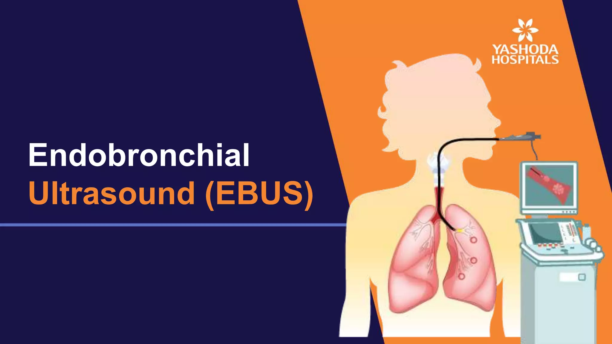
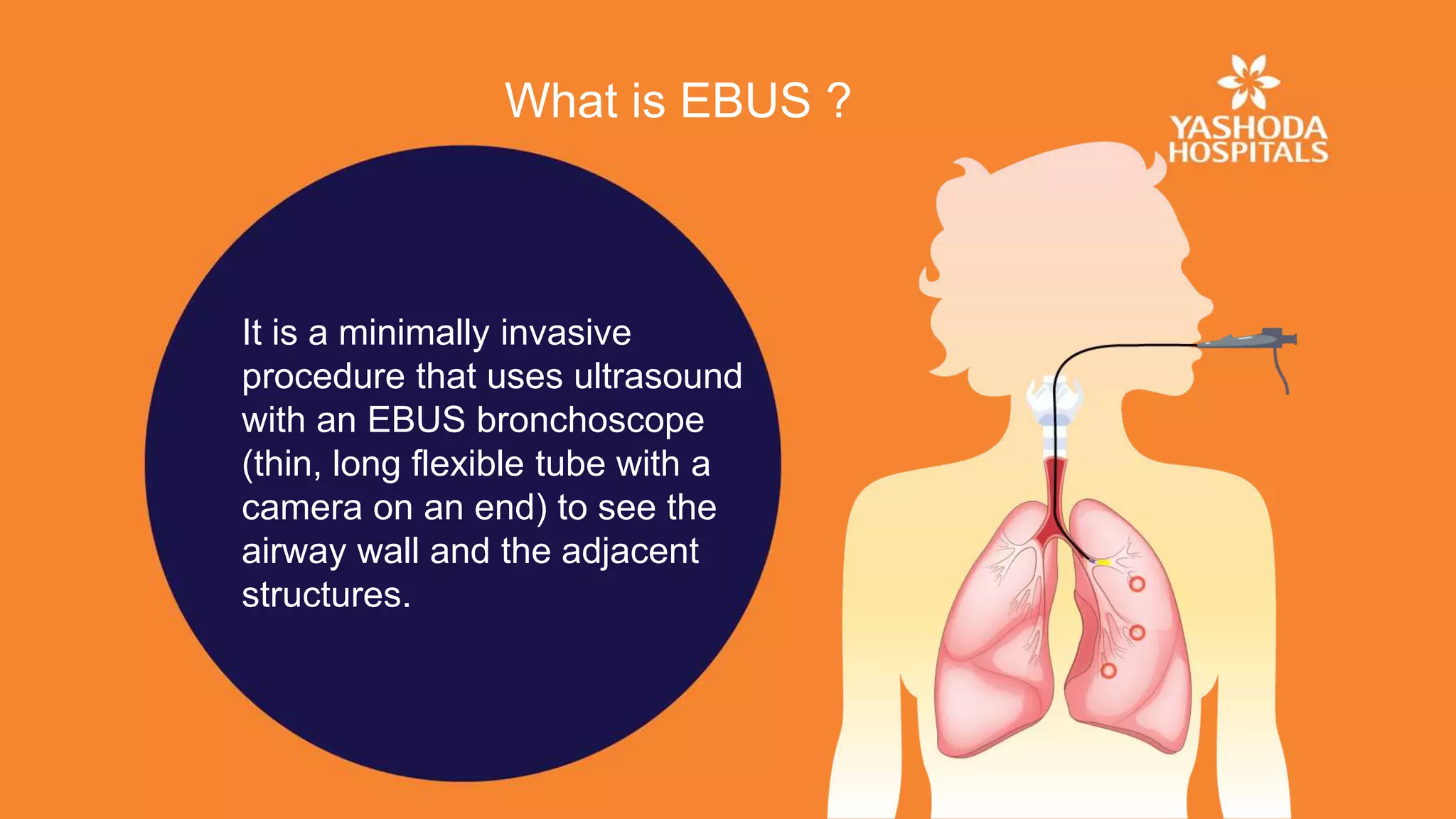
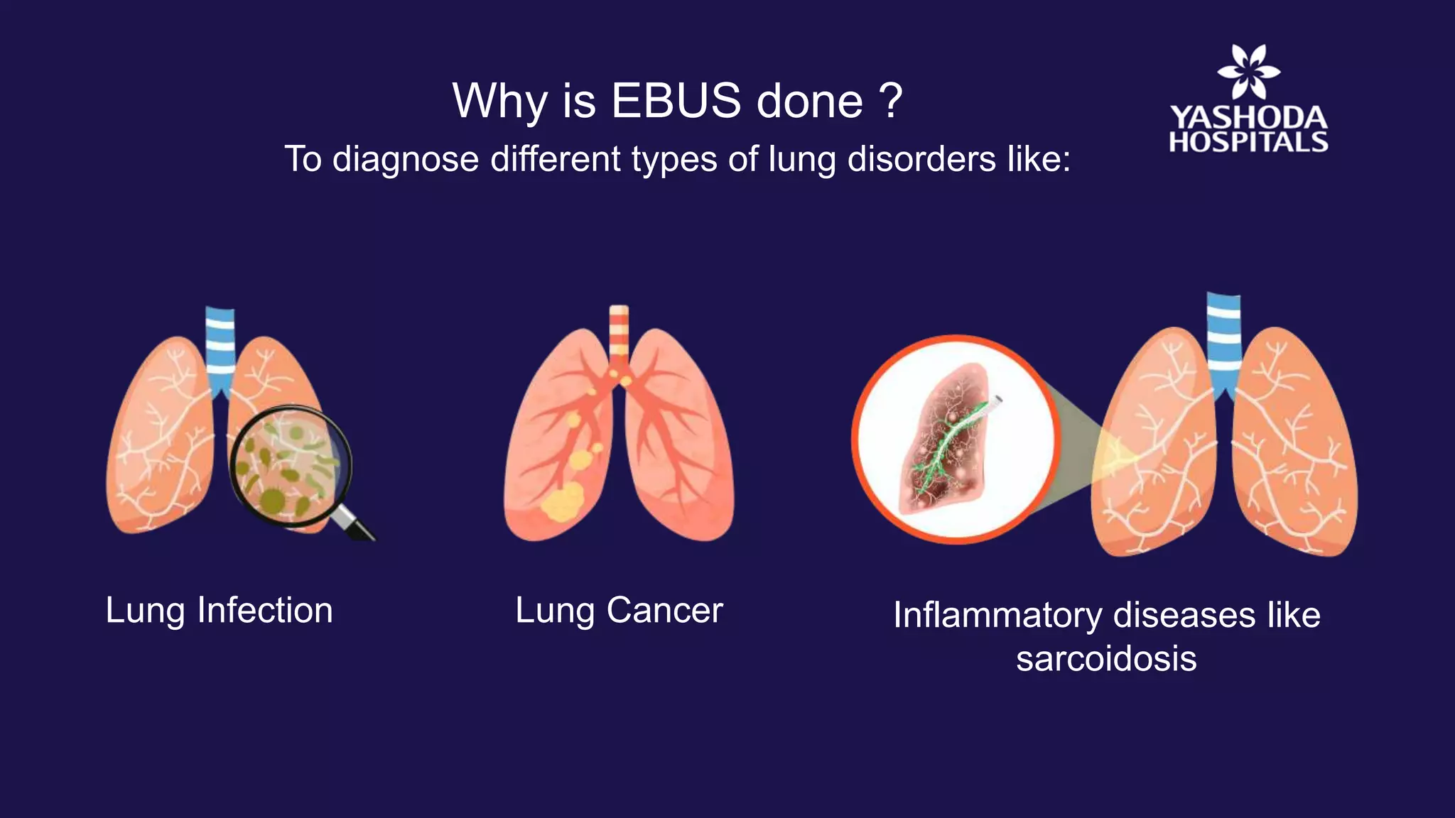
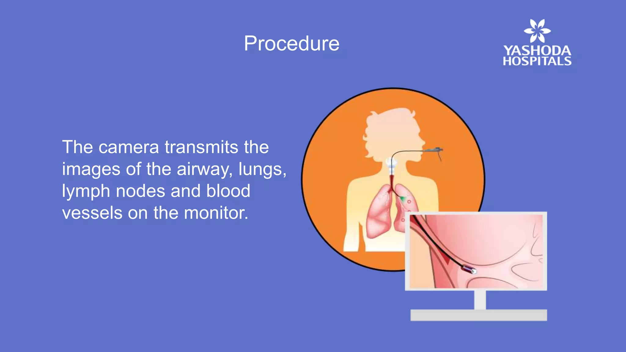
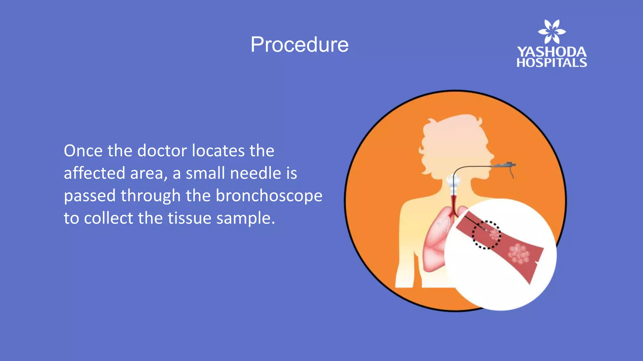
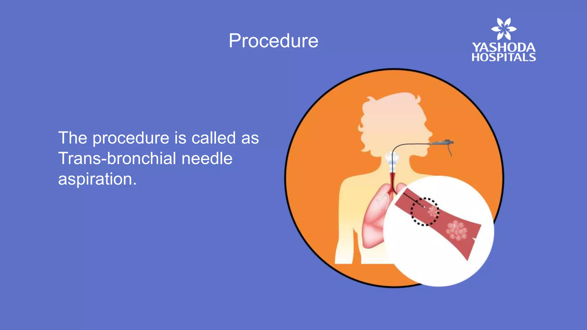
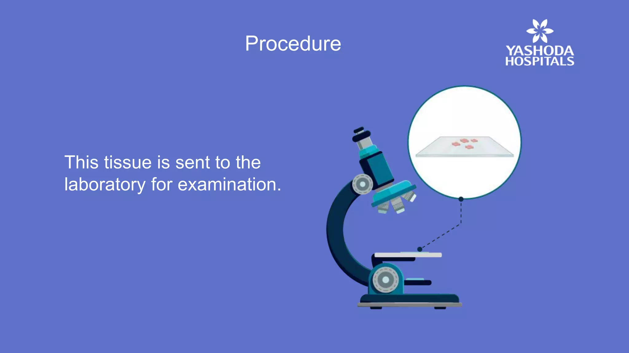
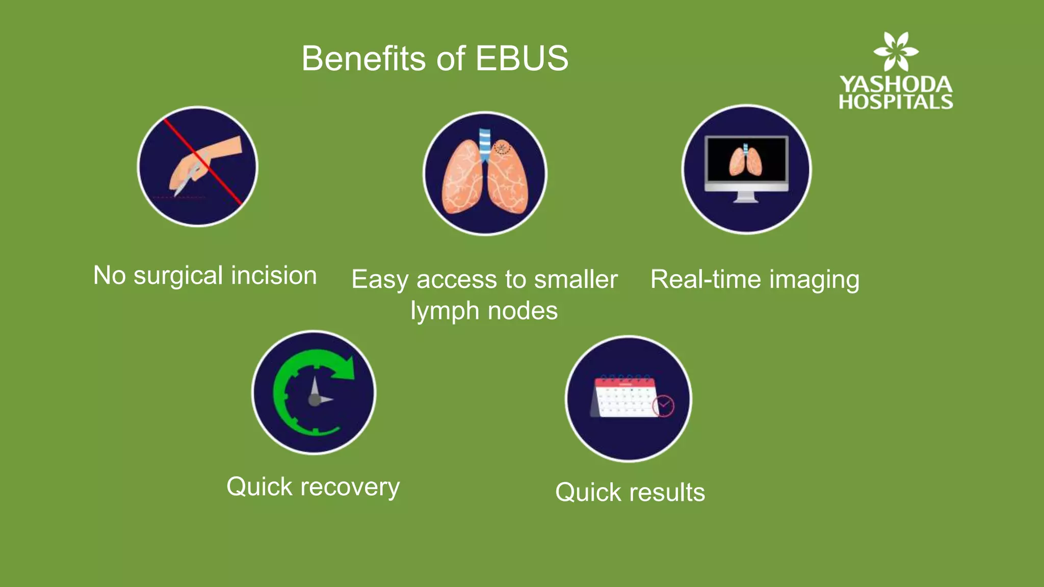
Endobronchial
Ultrasound (EBUS)
What is EBUS ?
It is a minimally invasive
procedure that uses ultrasound
with an EBUS bronchoscope
(thin, long flexible tube with a
camera on an end) to see the
airway wall and the adjacent
structures.
Why is EBUS done ?
Lung Infection Lung Cancer Inflammatory diseases like
sarcoidosis
To diagnose different types of lung disorders like:
Pre-procedure Preparation
Blood tests to evaluate
general health
Nil per oral (fasting) is
advised for few hours before
the procedure
To diagnose different types of lung disorders like:
Procedure
Following anesthesia, an
EBUS bronchoscope is
inserted through the mouth
into the bronchi.
Procedure
The camera transmits the
images of the airway, lungs,
lymph nodes and blood
vessels on the monitor.
Procedure
Once the doctor locates the
affected area, a small needle is
passed through the bronchoscope
to collect the tissue sample.
Procedure
The procedure is called as
Trans-bronchial needle
aspiration.
Procedure
This tissue is sent to the
laboratory for examination.
Pre-procedure Preparation
For one to two days the patient may have:
Mild cough Sore throat
Benefits of EBUS
No surgical incision
Quick results
Easy access to smaller
lymph nodes
Quick recovery
Real-time imaging
Endobronchial Ultrasound Bronchoscopy (EBUS)

Endobronchial Ultrasound Bronchoscopy (EBUS)