MRI scan: Purpose, Procedure and Risks
•Download as PPTX, PDF•
0 likes•1,281 views

What is Magnetic Resonance Imaging (MRI)? is a Strong magnetic field and radio waves are used to produce images of internal organs of the body.
Report
Share
Report
Share

Recommended
MRI Procedure and Preparation
An MRI scan is a painless procedure that uses magnetic fields and radio waves to produce detailed images of organs and tissues. Preparation may involve changing into loose, metal-free clothing and avoiding food, drink, smoking, and medications containing caffeine for several hours prior. During the scan, the patient lies still inside the MRI machine while images are taken, which can take 15-90 minutes. After the scan, the patient can resume normal activities unless sedated, in which case they should avoid driving or drinking for 24 hours.
Myelography
A myelogram is a radiographic study that uses contrast medium and fluoroscopy to evaluate the spinal cord and nerve roots. The contrast medium is injected into the subarachnoid space, usually at the L3-L4 or C1-C2 vertebrae under fluoroscopy. Images are then taken in various positions as the table is tilted to allow the contrast to flow. Myelography can diagnose conditions like tumors or herniated discs but has been replaced by MRI in most cases due to its invasiveness and radiation exposure. Risks include allergic reaction or headache, but benefits include accurate diagnosis when other imaging is not conclusive.
Ct and mri preparation
This document provides preparation instructions for CT, MRI, and contrast administration. It outlines screening requirements like medical history, allergies, medications, and creatinine tests. For CT and MRI, it instructs patients to remove metallic objects and drink water. CT patients may need premedication for contrast allergies. MRI screening identifies absolute contraindications like pacemakers. Gadolinium administration requires creatinine testing in high risk patients due to risks of nephrogenic systemic fibrosis. Pregnant women should only have MRI if absolutely necessary due to risks of intravenous gadolinium.
Myelogram pdf
A myelogram is a radiographic procedure where contrast medium is injected into the spinal subarachnoid space to evaluate the spinal cord, nerve roots, and spinal canal for abnormalities. It is performed by a radiologist under fluoroscopy guidance and involves inserting a needle into the lumbar or cervical spine to inject contrast medium. Images are then taken under fluoroscopy and with conventional radiography in multiple positions to visualize the entire spinal canal and assess for any structural abnormalities. Patients are monitored after the procedure and advised on post-procedure care depending on the type of contrast medium used.
Mri final ppt
Magnetic resonance imaging (MRI) uses strong magnets and radio waves to produce detailed images of the inside of the body without using ionizing radiation. An MRI machine contains a powerful magnet to align hydrogen atoms in the body. Radio waves are then used to excite the atoms, which emit signals as they relax. These signals are detected by antennas and used by a computer to generate 2D or 3D images of tissues and organs. MRI provides excellent soft tissue contrast and is useful for imaging the brain, muscles, joints, and other internal organs. While it has advantages over CT in avoiding radiation, MRI scans can be costly and some patients may find the enclosed scanner space claustrophobic.
Patient preparation for MRI CT and Nuclear Medicine
The document provides information on patient preparation for MRI, CT, and nuclear medicine procedures. For MRI, patients need to fast for 1 hour and remove all metal objects. For CT, fasting is required for 6 hours and metformin needs to be stopped. Nuclear medicine preparation depends on the specific exam, but generally involves fasting, avoiding caffeine and certain medications. The full document provides more detailed guidance for each type of imaging test.
Myelography
This document provides information about myelography, a radiographic examination of the spinal cord. It involves injecting contrast medium to detect spinal cord pathology. The spinal cord extends from the brain down the back and is protected by three meningeal layers. Cerebrospinal fluid surrounds and cushions the spinal cord. A myelogram is performed by puncturing the subarachnoid space and injecting contrast medium before taking radiographic images. Risks include reaction to the contrast medium, increased intracranial pressure, or aggravating existing conditions like arachnoiditis. Patients must stop certain medications beforehand and remain on bed rest afterwards.
CT Scan Vs MRI Scan
CT scans use X-rays to create detailed images of structures inside the body, while MRIs use magnetic fields and radio waves, and both have advantages and disadvantages. CT scans are quicker, cheaper, and better for bone structures, but expose patients to radiation, while MRIs do not use radiation but are more expensive, lengthy, and have stricter safety requirements. Overall, the document compares the uses of CT and MRI scans and their respective benefits and drawbacks.
Contrast media in CT
Contrast media, or contrast, is a liquid used in imaging tests to highlight parts of the body. It contains iodine, which interacts with x-rays and allows differentiation of tissues. Contrast is used in various CT protocols, with timing of administration dependent on the area and structures being imaged, such as 18-22 seconds for CT angiograms of the carotid arteries. Risk factors for receiving contrast include allergies, kidney problems, medications like metformin, and certain medical conditions. Proper screening and potentially pre-medication can help reduce risks.
Myelography
This document provides information about myelography, a radiographic examination of the central nervous system structures in the vertebral canal. It involves injecting contrast material into the subarachnoid space surrounding the spinal cord and brain. The contrast allows visualization of the spinal cord and nerves. A spinal puncture is performed to access the subarachnoid space and inject the contrast. Images are then taken under fluoroscopy to examine the spinal cord, nerves and surrounding structures.
Strength and limitations of mri
The document discusses the components and workings of magnetic resonance imaging (MRI) systems. It explains that MRI systems use incredibly powerful magnets ranging from 0.5 to 3 Tesla to align hydrogen atoms in the body. It describes the dangers posed by metal objects in the strong magnetic field and the screening procedures used. The advantages of MRI are also outlined, including its use of non-ionizing radiation and ability to show soft tissues and blood flow.
Introduction to mri
Dr. Rajesh Venunath Nair teaches radiology at K.S Hegde Medical Academy in Mangalore. His presentation discusses the history, basic principles, hardware, imaging sequences, and clinical applications of magnetic resonance imaging (MRI). It explains how MRI uses radiofrequency pulses and magnetic fields to produce detailed images of internal organs and soft tissues without using ionizing radiation. The presentation covers the main components of MRI scanners, different pulse sequences, tissue contrast mechanisms, use of contrast agents, safety considerations, and recent technical advances that have expanded clinical use of MRI.
Magnetic resonance imaging
MRI uses strong magnetic fields and radio waves to generate images of the inside of the body. It is a medical imaging technique widely used in radiology to visualize anatomy and physiological processes. MRI has many medical uses and applications across different body systems. It is generally a safe technique but there are some risks needing consideration for things like implants, projectile effects, and claustrophobia. Guidelines and certifications aim to standardize roles and ensure safe MRI practices.
MRI Brain
This document outlines an MRI brain protocol. It begins with an introduction to MRI brain imaging and its advantages over CT, such as lack of radiation exposure and greater soft tissue contrast. Common indications for MRI brain are then listed. The document describes patient preparation, contrast usage, coil positioning, imaging sequences including T1, T2, FLAIR, DWI, and advanced techniques like MRS and fMRI. Specific protocols are provided for conditions like MS, trauma, pituitary imaging, and CSF flow studies.
Usg ppt
The perfect diagnostic test to evaluate a patient with severe abdominal pain and vomiting would be ultrasound. Ultrasound uses sound waves to create images of internal organs and tissues and is non-invasive. The history of ultrasound began in 1794 with the study of echolocation in bats. In the 1940s, ultrasound was first used for medical diagnoses to detect brain tumors. The transducer converts electricity into sound waves that penetrate the body and bounce back, creating echoes that are converted back to electricity to form an image on a monitor. Ultrasound provides information about soft tissues without radiation or invasive procedures.
Ct head protocols
Computed tomography (CT) of the head is used to assess head injuries, headaches, dizziness, and symptoms of conditions like aneurysms, bleeding, strokes, and brain tumors. It can also help evaluate the face, sinuses, and skull. CT of the head uses X-rays to generate cross-sectional images of the head and brain which provide more detailed information than regular X-rays, particularly for soft tissues and blood vessels. Common protocols for head CT include non-contrast exams for conditions like trauma or stroke, as well as contrast-enhanced exams to evaluate tumors, aneurysms, or other conditions. Precautions are taken to minimize radiation exposure, especially for children.
Introduction to Magnetic resonance imaging (mri)
An Introduction to Magnetic Resonance Imaging by Dr Ozota Chinenye. A. MVSC University of Ibadan, Nigeria
Mylogram
Myelography is an imaging examination that involves inserting a needle into the spinal canal and injecting contrast dye to visualize the spinal cord, nerve roots, and surrounding structures using fluoroscopy and x-rays. It is used to evaluate herniated discs, tumors, infections, inflammation, spinal lesions, and stenosis. Patients are prepped and positioned for the procedure, a needle is inserted to draw fluid and inject contrast dye, and x-rays are taken. Post-procedure care involves rest, monitoring for side effects, and encouraging fluids to eliminate the dye.
MRI Safety Basics
This document discusses safety considerations for working with MRI machines. It covers several topics:
1. The static magnetic field of an MRI is extremely strong, around 30,000 times stronger than Earth's magnetic field. As a result, there are biological and mechanical risks from the magnetic field.
2. There are different safety zones around an MRI machine, with the innermost zone only allowing screened patients and personnel. Various instructions are provided to patients regarding removing all metal objects before an MRI scan.
3. Both patients and personnel must be screened for any metal implants or other factors that could pose a risk during a scan. Certain metals are considered MRI-safe but patients should always check with a technologist about any objects
Mammography
I have include all the contain about mammography like introduction,principle,anatomy,general views ,mammography physics (x-ray tube, housing,filter ,collimator and generator) and different advance technology about mammography.
Hope it will help your queries.
Thank you....!!
Positron Emission Tomography
PET scans use small amounts of radioactive tracers injected into the body to produce images showing how organs and tissues are functioning. A PET scan works by detecting gamma rays emitted by the tracers, allowing visualization of processes like blood flow, metabolic activity, and biochemical processes. PET scans are used to diagnose and manage conditions like cancer, heart disease, and neurological disorders.
Computer Tomography (CT Scan)
Computed tomography (CT scan) is a medical imaging procedure that uses computer-processed X-rays to produce tomographic images or 'slices' of specific areas of the body. These cross-sectional images are used for diagnostic and therapeutic purposes in various medical disciplines.
A Brief Overview of Mammography
Definition of Mammography
Types of Mammography
Indications of Mammography
Contraindications of Mammography
Mammography Views
Mammogram
Mammography Unit
Additional Views of Mammography
Mammography
Mammography uses low-dose x-rays to examine the breast for early cancer detection. It has advanced from film to digital mammography and tomosynthesis, which creates 3D breast images. Computer-aided detection highlights abnormal areas. Screening mammograms aim to detect cancer in asymptomatic women, while diagnostic mammograms investigate symptoms. Benefits include early detection, but limitations include false positives and negatives due to breast density. Yearly mammograms after age 40 are recommended for breast cancer screening. Ultrasound provides localized breast images without radiation but cannot screen whole breasts. MRI is superior for dense breasts but has no radiation risk.
CT - computed tomography
Computed tomography (CT) uses X-rays and digital geometry processing to generate 3D images of the inside of the body. During a CT scan, an X-ray tube rotates around the patient, emitting beams that are detected and used to construct cross-sectional slices. A radiologist can then analyze these slices to diagnose medical conditions by viewing internal organs, bones, soft tissues, and blood vessels with greater clarity and detail than traditional X-rays. CT scans are commonly used to diagnose cancers, cardiovascular diseases, infections, appendicitis, trauma, and muscular-skeletal disorders.
INTRAVENOUS UROGRAM (IVU)
It is a radio-graphic procedure to access the urinary system.
it also called as intravenous pyelogram (IVP)
CT Scan: Purpose, Procedure and Risk Factors
CT Scan is a Fast, painless, non-invasive test that uses computer and rotating X-rays to determine internal structures of the body
MRI Procedure of Brain
MRI provides detailed images of the brain without exposing patients to radiation. It is useful for evaluating conditions like tumors, strokes, and multiple sclerosis. The document describes the MRI procedure for brain imaging including patient preparation, head coils, sequences, and protocols. Key sequences discussed are T1-weighted, T2-weighted, FLAIR, diffusion weighted, MR angiography, and MR venography.
MRI SCAN ppt
An MRI scan uses radio waves and a strong magnetic field to produce detailed images of the inside of the body. It can be used to detect abnormalities in soft tissues like the brain or cancer. Not everyone can undergo an MRI, as the strong magnetic field is unsafe if the patient has metallic implants like pacemakers. Common reasons doctors recommend an MRI include detecting brain tumors, assessing injuries before surgery, and tracking cancer treatment. The cost of an MRI ranges from Rs. 1500 to Rs. 25,000 depending on the body part and location.
Magnetic resonance imaging (MRI) - medical information
Magnetic resonance imaging (MRI) uses strong magnetic fields and radio waves to produce detailed images of the inside of the body without using ionizing radiation. An MRI scan can be used to diagnose diseases and conditions, monitor treatment, and check for cancer recurrence. During an MRI scan, the patient lies inside the MRI machine, which is a large ring with a central opening. The patient must remain still during the 20-90 minute scan to prevent blurry images. Precautions are taken for any metal objects and some patients may receive an IV contrast agent or medication to improve image quality. A radiologist analyzes the images and sends a report to the referring doctor.
More Related Content
What's hot
Contrast media in CT
Contrast media, or contrast, is a liquid used in imaging tests to highlight parts of the body. It contains iodine, which interacts with x-rays and allows differentiation of tissues. Contrast is used in various CT protocols, with timing of administration dependent on the area and structures being imaged, such as 18-22 seconds for CT angiograms of the carotid arteries. Risk factors for receiving contrast include allergies, kidney problems, medications like metformin, and certain medical conditions. Proper screening and potentially pre-medication can help reduce risks.
Myelography
This document provides information about myelography, a radiographic examination of the central nervous system structures in the vertebral canal. It involves injecting contrast material into the subarachnoid space surrounding the spinal cord and brain. The contrast allows visualization of the spinal cord and nerves. A spinal puncture is performed to access the subarachnoid space and inject the contrast. Images are then taken under fluoroscopy to examine the spinal cord, nerves and surrounding structures.
Strength and limitations of mri
The document discusses the components and workings of magnetic resonance imaging (MRI) systems. It explains that MRI systems use incredibly powerful magnets ranging from 0.5 to 3 Tesla to align hydrogen atoms in the body. It describes the dangers posed by metal objects in the strong magnetic field and the screening procedures used. The advantages of MRI are also outlined, including its use of non-ionizing radiation and ability to show soft tissues and blood flow.
Introduction to mri
Dr. Rajesh Venunath Nair teaches radiology at K.S Hegde Medical Academy in Mangalore. His presentation discusses the history, basic principles, hardware, imaging sequences, and clinical applications of magnetic resonance imaging (MRI). It explains how MRI uses radiofrequency pulses and magnetic fields to produce detailed images of internal organs and soft tissues without using ionizing radiation. The presentation covers the main components of MRI scanners, different pulse sequences, tissue contrast mechanisms, use of contrast agents, safety considerations, and recent technical advances that have expanded clinical use of MRI.
Magnetic resonance imaging
MRI uses strong magnetic fields and radio waves to generate images of the inside of the body. It is a medical imaging technique widely used in radiology to visualize anatomy and physiological processes. MRI has many medical uses and applications across different body systems. It is generally a safe technique but there are some risks needing consideration for things like implants, projectile effects, and claustrophobia. Guidelines and certifications aim to standardize roles and ensure safe MRI practices.
MRI Brain
This document outlines an MRI brain protocol. It begins with an introduction to MRI brain imaging and its advantages over CT, such as lack of radiation exposure and greater soft tissue contrast. Common indications for MRI brain are then listed. The document describes patient preparation, contrast usage, coil positioning, imaging sequences including T1, T2, FLAIR, DWI, and advanced techniques like MRS and fMRI. Specific protocols are provided for conditions like MS, trauma, pituitary imaging, and CSF flow studies.
Usg ppt
The perfect diagnostic test to evaluate a patient with severe abdominal pain and vomiting would be ultrasound. Ultrasound uses sound waves to create images of internal organs and tissues and is non-invasive. The history of ultrasound began in 1794 with the study of echolocation in bats. In the 1940s, ultrasound was first used for medical diagnoses to detect brain tumors. The transducer converts electricity into sound waves that penetrate the body and bounce back, creating echoes that are converted back to electricity to form an image on a monitor. Ultrasound provides information about soft tissues without radiation or invasive procedures.
Ct head protocols
Computed tomography (CT) of the head is used to assess head injuries, headaches, dizziness, and symptoms of conditions like aneurysms, bleeding, strokes, and brain tumors. It can also help evaluate the face, sinuses, and skull. CT of the head uses X-rays to generate cross-sectional images of the head and brain which provide more detailed information than regular X-rays, particularly for soft tissues and blood vessels. Common protocols for head CT include non-contrast exams for conditions like trauma or stroke, as well as contrast-enhanced exams to evaluate tumors, aneurysms, or other conditions. Precautions are taken to minimize radiation exposure, especially for children.
Introduction to Magnetic resonance imaging (mri)
An Introduction to Magnetic Resonance Imaging by Dr Ozota Chinenye. A. MVSC University of Ibadan, Nigeria
Mylogram
Myelography is an imaging examination that involves inserting a needle into the spinal canal and injecting contrast dye to visualize the spinal cord, nerve roots, and surrounding structures using fluoroscopy and x-rays. It is used to evaluate herniated discs, tumors, infections, inflammation, spinal lesions, and stenosis. Patients are prepped and positioned for the procedure, a needle is inserted to draw fluid and inject contrast dye, and x-rays are taken. Post-procedure care involves rest, monitoring for side effects, and encouraging fluids to eliminate the dye.
MRI Safety Basics
This document discusses safety considerations for working with MRI machines. It covers several topics:
1. The static magnetic field of an MRI is extremely strong, around 30,000 times stronger than Earth's magnetic field. As a result, there are biological and mechanical risks from the magnetic field.
2. There are different safety zones around an MRI machine, with the innermost zone only allowing screened patients and personnel. Various instructions are provided to patients regarding removing all metal objects before an MRI scan.
3. Both patients and personnel must be screened for any metal implants or other factors that could pose a risk during a scan. Certain metals are considered MRI-safe but patients should always check with a technologist about any objects
Mammography
I have include all the contain about mammography like introduction,principle,anatomy,general views ,mammography physics (x-ray tube, housing,filter ,collimator and generator) and different advance technology about mammography.
Hope it will help your queries.
Thank you....!!
Positron Emission Tomography
PET scans use small amounts of radioactive tracers injected into the body to produce images showing how organs and tissues are functioning. A PET scan works by detecting gamma rays emitted by the tracers, allowing visualization of processes like blood flow, metabolic activity, and biochemical processes. PET scans are used to diagnose and manage conditions like cancer, heart disease, and neurological disorders.
Computer Tomography (CT Scan)
Computed tomography (CT scan) is a medical imaging procedure that uses computer-processed X-rays to produce tomographic images or 'slices' of specific areas of the body. These cross-sectional images are used for diagnostic and therapeutic purposes in various medical disciplines.
A Brief Overview of Mammography
Definition of Mammography
Types of Mammography
Indications of Mammography
Contraindications of Mammography
Mammography Views
Mammogram
Mammography Unit
Additional Views of Mammography
Mammography
Mammography uses low-dose x-rays to examine the breast for early cancer detection. It has advanced from film to digital mammography and tomosynthesis, which creates 3D breast images. Computer-aided detection highlights abnormal areas. Screening mammograms aim to detect cancer in asymptomatic women, while diagnostic mammograms investigate symptoms. Benefits include early detection, but limitations include false positives and negatives due to breast density. Yearly mammograms after age 40 are recommended for breast cancer screening. Ultrasound provides localized breast images without radiation but cannot screen whole breasts. MRI is superior for dense breasts but has no radiation risk.
CT - computed tomography
Computed tomography (CT) uses X-rays and digital geometry processing to generate 3D images of the inside of the body. During a CT scan, an X-ray tube rotates around the patient, emitting beams that are detected and used to construct cross-sectional slices. A radiologist can then analyze these slices to diagnose medical conditions by viewing internal organs, bones, soft tissues, and blood vessels with greater clarity and detail than traditional X-rays. CT scans are commonly used to diagnose cancers, cardiovascular diseases, infections, appendicitis, trauma, and muscular-skeletal disorders.
INTRAVENOUS UROGRAM (IVU)
It is a radio-graphic procedure to access the urinary system.
it also called as intravenous pyelogram (IVP)
CT Scan: Purpose, Procedure and Risk Factors
CT Scan is a Fast, painless, non-invasive test that uses computer and rotating X-rays to determine internal structures of the body
MRI Procedure of Brain
MRI provides detailed images of the brain without exposing patients to radiation. It is useful for evaluating conditions like tumors, strokes, and multiple sclerosis. The document describes the MRI procedure for brain imaging including patient preparation, head coils, sequences, and protocols. Key sequences discussed are T1-weighted, T2-weighted, FLAIR, diffusion weighted, MR angiography, and MR venography.
What's hot (20)
Similar to MRI scan: Purpose, Procedure and Risks
MRI SCAN ppt
An MRI scan uses radio waves and a strong magnetic field to produce detailed images of the inside of the body. It can be used to detect abnormalities in soft tissues like the brain or cancer. Not everyone can undergo an MRI, as the strong magnetic field is unsafe if the patient has metallic implants like pacemakers. Common reasons doctors recommend an MRI include detecting brain tumors, assessing injuries before surgery, and tracking cancer treatment. The cost of an MRI ranges from Rs. 1500 to Rs. 25,000 depending on the body part and location.
Magnetic resonance imaging (MRI) - medical information
Magnetic resonance imaging (MRI) uses strong magnetic fields and radio waves to produce detailed images of the inside of the body without using ionizing radiation. An MRI scan can be used to diagnose diseases and conditions, monitor treatment, and check for cancer recurrence. During an MRI scan, the patient lies inside the MRI machine, which is a large ring with a central opening. The patient must remain still during the 20-90 minute scan to prevent blurry images. Precautions are taken for any metal objects and some patients may receive an IV contrast agent or medication to improve image quality. A radiologist analyzes the images and sends a report to the referring doctor.
Things to consider before MRI Scan.pptx
Instructions to follow before getting an MRI. Know about different kinds of MRI systems, specific diet before MRI, removing all metallic objects before MRI scan.
Bodymr (2)
MRI uses powerful magnets and radio waves to produce detailed images of the inside of the body without using radiation. It can be used to diagnose or monitor treatment for conditions in the chest, abdomen, and pelvis. Pregnant women may have an MRI to safely monitor the baby. Patients should inform medical staff about any medical devices, implants, or health conditions and follow instructions about eating, drinking and medication prior to the exam. Jewelry should be removed and loose, comfortable clothing worn. The MRI machine uses a large magnet to create images, which are reviewed by radiologists.
ANESTHESIA FOR MRI AND CT SCANs suite room
This document discusses anesthesia considerations for MRI and CT scans. It notes that sedation or anesthesia is often required for infants, uncooperative children, patients with movement or psychological disorders, and critically ill patients. The main challenges include using MRI-compatible monitoring equipment, limited access to patients, and treating medical emergencies safely outside of the scanner. Commonly used sedative agents include oral chloral hydrate, midazolam, and propofol administered with monitoring of ventilation.
Sonography
Sonography is a diagnostic medical test that uses sound waves to assess the internal body organs and capture them in a pictorial form. It is a painless and non-invasive test. Sonography does not use any form of radiation and is hence, relatively a safe test. The image created is known as a sonogram. The test is also referred to as ultrasound imaging.
Presentation: Radiation Protection Week CAH BUKIT RIMAU
The document discusses the history and development of various medical imaging technologies including x-rays, mammography, ultrasound, CT scans, and MRI. It provides information on how each technology works, its applications and limitations, safety considerations, and procedures involved. The key technologies covered include their accidental discovery, early installations in hospitals, advances in equipment and techniques to reduce radiation exposure, and importance in detecting diseases like breast cancer.
MRI Chest
One test can save your life. Know what an MRI Chest is, why you should have it, who should get it, and where can you get tested as well as get your results fast. If you want to read more about MRI Chest, click the link below.
Visit: https://www.labfinder.com/labexams/mri-chest/ and get tested now!
Ultrasound Scan During Pregnancy
Ultrasound scan during pregnancy takes pictures of the unborn baby inside the womb by using high-frequency soundwaves. They are used in pregnancy to examine the baby’s growth and development.
Know more visit our official website :https://windowtothewomb.co.uk/studios/reading-baby-scan-studio/
Patient advice during brain tumor radiation.pptx
1) The document provides advice for patients undergoing brain tumor radiation treatment.
2) Brain tumor radiation typically involves weekly radiation sessions over five days for about fifteen minutes each session.
3) Patients will need a mask made and a planning CT scan to develop the radiation treatment plan before starting radiation.
Transvaginal Ultrasound
Here we talk about the Trans vaginal Ultrasound which is used to get the clear image of the female pelvic organs. We have also discuss about the features, advantages, disadvantages and many more.
Ultrasound Test in Faridabad.docx
Ultrasound testing, also known as sonography, has become an essential tool in modern medicine for diagnosing various medical conditions. In Faridabad, this non-invasive diagnostic marvel is readily available at numerous medical facilities, offering patients a safe and painless way to gain valuable insights into their health.
urography.pdf
Urography uses imaging techniques and contrast material to evaluate the urinary tract for issues like blood in the urine, kidney stones, and cancers. It can be performed using CT, MRI, or conventional x-ray. The procedure involves drinking water to distend the bladder and intravenous injection of contrast material. Images are then taken of the kidneys, ureters, and bladder to detect any abnormalities. Preparation may involve not eating or drinking before the test and removing any metal objects. The test is generally painless and provides detailed images of the urinary tract.
MRI Defecography
One test can save your life. Know what an MRI Defecography is, why you should have it, who should get it, and where can you get tested as well as get your results fast. If you want to read more about MRI Defecography, click the link below.
Visit: https://www.labfinder.com/labexams/mri-defecography-with-contrast/ and get tested now!
AHS-399 Advanced Tech Comparison.FAHR-no crops
Adena uses the most advanced 3T MRI technology in the region, which produces higher resolution images and allows for better diagnoses and more effective treatment plans compared to other MRIs. Adena is the only healthcare facility that has 3T MRI, which can provide better outcomes for patients. Their 3T and 1.5T MRIs allow for more diagnostic confidence and shorter scan times than traditional open MRIs that use weaker magnetic fields.
Understanding The Importance Of A Pre-Abortion Ultrasound.pdf
Conducting an ultrasound before an abortion is a widely accepted practice within the abortion healthcare sector. Pre-abortion ultrasounds are an important step toward guaranteeing safe and successful abortion operations. While they can seem slightly unpleasant, they lead to a better-informed and safer abortion experience. For a safe abortion, you can buy abortion pill pack online.
What Are the Advantages of 3D and 4D Ultrasounds?
www.miamiobgyns.com/blog/advantages-3d-4d-ultrasounds/
The joy parents have when seeing their baby for the first time on the ultrasound screen is one that we can’t help but feel too, no matter how many times we experience it! Watching them hear their child’s rapid heartbeat for the first time is nothing short of electrifying!
"Get a Detailed Full Body MRI Scan at Sanjivini Diagnostics, Chandigarh"
Experience top-notch medical imaging with a comprehensive Full Body MRI Scan at Sanjivini Diagnostics in Chandigarh. Our state-of-the-art facility is equipped with cutting-edge MRI technology and staffed by experienced radiologists. This scan provides detailed insights into your entire body, aiding in early disease detection and precise diagnosis. Trust Sanjivini Diagnostics for accurate results and personalized care. Your health is our priority. Book your appointment today!
Patient advice during breast cancer radiation.pptx
1) Women receiving breast cancer radiation treatment will undergo a mask preparation and planning CT scan. They will then receive weekly radiation treatments over approximately 15-25 sessions.
2) Patients need to cooperate with doctors as radiation treatment involves more than just the radiation. They should follow nutrition and diet charts provided by dieticians.
3) Common side effects include skin blackening and soreness that are usually temporary. Patients should avoid rubbing the skin and use prescribed creams. Regular checkups with doctors are also important.
The Comprehensive Guide for Sonograph.pdf
The Comprehensive Guide for Sonograph.pdfWorld Infertility and IVF Centre : Best IVF Centre in Delhi
Sonography, also known as ultrasound, is a diagnostic test that uses high-frequency sound
waves to create images of various organs, tissues, and blood vessels inside the body. It is a safe, painless, and non-invasive procedure that can help diagnose various medical
conditions, monitor the development of a fetus during pregnancy, and guide certain
procedures such as biopsies or injections.Similar to MRI scan: Purpose, Procedure and Risks (20)
Magnetic resonance imaging (MRI) - medical information
Magnetic resonance imaging (MRI) - medical information
Presentation: Radiation Protection Week CAH BUKIT RIMAU
Presentation: Radiation Protection Week CAH BUKIT RIMAU
Understanding The Importance Of A Pre-Abortion Ultrasound.pdf
Understanding The Importance Of A Pre-Abortion Ultrasound.pdf
"Get a Detailed Full Body MRI Scan at Sanjivini Diagnostics, Chandigarh"
"Get a Detailed Full Body MRI Scan at Sanjivini Diagnostics, Chandigarh"
Patient advice during breast cancer radiation.pptx
Patient advice during breast cancer radiation.pptx
More from YashodaHospitals
LAA Watchman
Left atrial appendage closure device involves a procedure that reduces the risk of stroke in atrial fibrillation patients. It also reduces the risk of bleeding that comes with a long-term use of oral anticoagulants. The WATCHMAN Device is permanently implanted at the site or slightly distal to the ostium of the LAA (the source of more than 90% of stroke-causing bloods clots in atrial fibrillation patients).
Implantable Cardioverter Defibrillators -ICD
It is a small battery-powered
device placed onto the chest
wall to monitor heart rhythm
and detect irregular heartbeats.
Endoscopic spine surgery
Endoscopic spine surgery is a minimally invasive procedure used to treat severe back pain caused by slipped discs, spinal stenosis, or scoliosis when conventional treatments like medication and physical therapy have failed. The surgeon makes a small incision and inserts specialized tools including an endoscope to view and repair the damaged spinal area. Benefits include quicker recovery, shorter hospital stays, less pain and blood loss, and lower risk of complications compared to open spine surgery.
Endobronchial Ultrasound Bronchoscopy (EBUS)
It is a minimally invasive procedure that uses ultrasound with an EBUS bronchoscope (thin, long flexible tube with a camera on an end) to see the airway wall and the adjacent structures.
Spine Disc Surgeries - Discectomy and Decompression
The document discusses various spinal decompression procedures including diskectomy, laminectomy, and foraminotomy. Diskectomy involves removing a portion of a herniated disc to relieve nerve compression. Laminectomy and laminotomy remove part of the lamina bone to widen the spinal canal to relieve pressure on nerves. Foraminotomy enlarges the openings where nerves exit the spine. These procedures provide relief from pain caused by damaged vertebrae or discs pressing on spinal nerves.
Endocytoscopy Procedure
Endocytoscopy is a novel
endoscopic diagnostic procedure
that provides ultra-high magnification
of gastrointestinal tract.
Bronchoscopy For Lung Diseases
Bronchoscopy and pleuroscopy are minimally invasive endoscopic procedures used to diagnose and treat lung diseases. Bronchoscopy involves inserting a thin, lighted tube with a camera through the mouth or nose into the lungs, while pleuroscopy accesses the pleural space between the lungs through a small incision in the chest. Both procedures allow doctors to directly examine the lungs and collect samples, and have benefits of providing quick results without the need for surgical cuts or long hospital stays.
What is Retroperitoneoscopic Surgery
Retroperitoneoscopic surgery (RPS) is a minimally invasive procedure performed through small incisions in the back to access the kidney, adrenals, and ureter for conditions like removing kidney stones or repairing abnormalities. Common RPS procedures include nephrectomies, adrenalectomies, and repairing ureteral obstructions. RPS provides direct access to organs without touching the bowel, allowing for early access to blood vessels and an easy approach to the renal hilum. The benefits of RPS include it being safe, causing less pain, and resulting in quicker recovery times and shorter hospital stays compared to open surgery.
What is Physiotherapy? | Types and Methods of Physiotherapy
Physiotherapy is a non-medical and non-surgical modality to maximize physical strength & function, improve mobility & body balance, improve performance of heart and lungs, heal injuries and reduce pain.
Pacemaker | Implantable Cardiac Devices For Heart Failures
Implantable cardiac devices are electronic, battery-operated medical devices that are implanted to restore the heart's normal rhythm and prevent sudden cardiac death. Implantable cardioverter-defibrillator, Pacemaker and LAVD are such devices that help to maintain rhythm and pumping. A pacemaker is a small implantable cardiac device that is placed under the skin in the chest to help control the heartbeat, improve quality of life and for longevity. It is used to help the heart beat more regularly for irregular heartbeat also known as arrhythmia.
What does it help with?
Pacemaker helps in controlling the rhythm and of the heart by either:
Resynchronizing the rhythm
Correcting the rhythm
Facilitating adequate circulation to support a failing heart
Mechanical Thrombectomy for Acute Ischemic Stroke | Brain Stroke Treatment in...
Mechanical thrombectomy is a minimally invasive procedure to remove blood clots from large brain vessels. It involves inserting a catheter through a small groin incision and using stent-like devices to remove clots causing acute ischemic strokes from large artery occlusion. It can benefit patients who are not candidates for or failed intravenous thrombolysis, especially if performed within 24 hours of symptom onset. Recovery depends on patient factors but may take weeks, with potential for improved outcomes over intravenous thrombolysis alone.
Breast Conserving Surgery in Hyderabad | Breast Cancer Treatment in Hyderabad
Breast-conserving Surgery is also known as lumpectomy or partial lumpectomy, it is a procedure to remove the cancer from the breast and some normal tissue. BCS involves only the part of the breast that has cancer to be removed. BCS is a good option for many women with early-stage cancers. Usually after BCS, radiation therapy is given to destroy cancer cells that may not have been removed during surgery. In some cases, chemotherapy and radiation are both given after BCS.
Who is BCS recommended for?
Not all women with breast cancer are candidates for BCS. However, speak to a doctor to find out whether BCS is an option for you.
BCS might be a good option for the below reason:
1. If the tumor is small and localized.
2. If you are eligible for radiation therapy
3. Do not have inflammatory breast cancer
4. Are not pregnant or, if pregnant, will not need radiation therapy immediately.
5. Do not have a mutation linked to breast cancer.
6. Do not have serious connective tissue diseases such as scleroderma or lupus.
Transcatheter Aortic Valve Replacement (TAVR) | TAVR Procedure at Yashoda Hos...
TAVR is a minimally invasive procedure to replace a severely stenotic aortic valve. The aortic valve regulates blood flow from the left ventricle to the aorta. Aortic valve stenosis occurs when the valve thickens and narrows, reducing blood flow. TAVR can treat aortic stenosis in patients at high risk for open heart surgery or those whose artificial valves have failed. During TAVR, a catheter is used to guide a replacement valve through the body to the heart, where it is deployed to replace the diseased valve. TAVR allows for quicker recovery and shorter hospital stays than open heart surgery.
Peroral Endoscopic Myotomy (POEM) | Treatment For Achalasia of Esophagus and ...
POEM is a minimally invasive procedure that uses a high definition endoscope (a flexible tube with a mounted camera at end) to help treat certain swallowing disorders and stands for Peroral Endoscopic Myotomy.
Who cannot undergo this procedure?
Although POEM is a safe and effective procedure, it may not be a suitable option in certain cases. People with any of the following conditions are generally not recommended to undergo POEM for achalasia:
1. Disorders of coagulation
2. History of any kind of therapy that is likely to compromise the integrity of the mucosa of the esophagus or lead to fibrosis of the submucosa for e.g. radiation for cancer treatment, endoscopic mucosal resection, or radiofrequency ablation
3. Liver cirrhosis with portal hypertension
4. Severe erosive esophagitis
When should it be done?
POEM is generally indicated for treating the following disease:
1. Achalasia: It is a medical condition that affects the muscles of the esophagus. The lower esophageal sphincter (LES), a muscular valve present between the esophagus and the stomach fails to relax during swallowing in this condition.
2. Diffuse esophageal spasm: Esophageal disorders of muscle spasm that are unresponsive to medical therapies
3. Nutcracker esophagus also known as hypertensive peristalsis is a muscle spasm disorder which needs surgical correction.
Piles Types, Causes and Symptoms | Piles Laser Treatment in Hyderabad
Piles or hemorrhoids are the swollen and dilated blood vessels in the lower rectum and around the anus. Minimally invasive laser surgeries allow precisive and safer treatment options with less postoperative pain. Our focus is the safety, convenience and complete care for our patients with the help of an integrated team of experts at Yashoda Hospitals Hyderabad.
Symptoms of Piles:
1. Blood visible after passing stool
1. Itchy, sore and red anus
3. Feeling of bowel being full even after passing stool
4. Slimy mucus in underwear or on toilet paper after wiping your bottom
5. Lumps around the anus
6. Experiencing pain around the anus
7. Fecal Leakage
Risk Factors:
1. Hemorrhoids can be passed on genetically from parent to child
2. Consistent heavy lifting
3. Obesity
4. Constant strain on your body can increase your risk of hemorrhoids.
5. Standing frequently without taking a break
6. Consistent anal sexual intercourse
7. Consistent Diarrhea
8. Consistent constipation
9. During pregnancy when the uterus enlarges.
Capsule Endoscopy & Motorized Spiral Enteroscopy for Small Bowel Imaging
Many conditions of the small intestine affect digestive health and appetite. Intestinal obstruction due to bleeding, malformations or injury or hernia is a major diagnostic related to these problems. See how capsule endoscopy and spiral endoscopy enables the doctors to not only diagnose but treat the condition right away.
First Aid for Falls and Emergency Management
First Aid for Falls and Emergency Management: Learn how to help someone who has fallen, when to keep them still and when to move them
What Is Stereotactic Radiosurgery(SRS) | What Conditions Does It Treat?
A non-invasive radiation therapy where a focused beam of radiation is used to remove tumors or defects in the brain while sparing the healthy tissues.
Robotic surgeries: Procedures, Advantages and Risks
Over 3 million patients have undergone robotic surgery since 2000. Robotic surgery allows surgeons to perform minimally invasive procedures through small incisions using a robotic system that has 3D imaging and wrist-like joints, giving surgeons improved access and control. While the surgeon controls the robotic arms, benefits of robotic surgery include less pain, smaller incisions, shorter hospital stays, and quicker recovery times compared to traditional open or laparoscopic surgeries. The doctor determines if a patient is eligible for robotic surgery based on their individual procedure and medical needs.
PET-CT scan: Uses, Risks and Procedure
PET-CT scan combines positron emission tomography (PET) and computed tomography (CT) to determine the functioning of the cells and tissues.
More from YashodaHospitals (20)
Spine Disc Surgeries - Discectomy and Decompression
Spine Disc Surgeries - Discectomy and Decompression
What is Physiotherapy? | Types and Methods of Physiotherapy
What is Physiotherapy? | Types and Methods of Physiotherapy
Pacemaker | Implantable Cardiac Devices For Heart Failures
Pacemaker | Implantable Cardiac Devices For Heart Failures
Mechanical Thrombectomy for Acute Ischemic Stroke | Brain Stroke Treatment in...
Mechanical Thrombectomy for Acute Ischemic Stroke | Brain Stroke Treatment in...
Breast Conserving Surgery in Hyderabad | Breast Cancer Treatment in Hyderabad
Breast Conserving Surgery in Hyderabad | Breast Cancer Treatment in Hyderabad
Transcatheter Aortic Valve Replacement (TAVR) | TAVR Procedure at Yashoda Hos...
Transcatheter Aortic Valve Replacement (TAVR) | TAVR Procedure at Yashoda Hos...
Peroral Endoscopic Myotomy (POEM) | Treatment For Achalasia of Esophagus and ...
Peroral Endoscopic Myotomy (POEM) | Treatment For Achalasia of Esophagus and ...
Piles Types, Causes and Symptoms | Piles Laser Treatment in Hyderabad
Piles Types, Causes and Symptoms | Piles Laser Treatment in Hyderabad
Capsule Endoscopy & Motorized Spiral Enteroscopy for Small Bowel Imaging
Capsule Endoscopy & Motorized Spiral Enteroscopy for Small Bowel Imaging
What Is Stereotactic Radiosurgery(SRS) | What Conditions Does It Treat?
What Is Stereotactic Radiosurgery(SRS) | What Conditions Does It Treat?
Robotic surgeries: Procedures, Advantages and Risks
Robotic surgeries: Procedures, Advantages and Risks
Recently uploaded
The Importance of Black Women Understanding the Chemicals in Their Personal C...
Certain chemicals, such as phthalates and parabens, can disrupt the body's hormones and have significant effects on health. According to data, hormone-related health issues such as uterine fibroids, infertility, early puberty and more aggressive forms of breast and endometrial cancers disproportionately affect Black women. Our guest speaker, Jasmine A. McDonald, PhD, an Assistant Professor in the Department of Epidemiology at Columbia University in New York City, discusses the scientific reasons why Black women should pay attention to specific chemicals in their personal care products, like hair care, and ways to minimize their exposure.
DAHL APPROACH a novel technique to raise bite
dahl approach is alternative techniue to raise bite with direct restorations
Electrocardiogram_20240614_173859_0000.pdf
Basics of Electrocardiogram
CONTENTS
●Conduction System of the Heart
●What is ECG or EKG?
●ECG Leads
●Normal waves of ECG.
●Dimensions of ECG.
● Abnormalities of ECG
CONDUCTION SYSTEM OF THE HEART
ECG:
●ECG is a graphic record of the electrical activity of the heart.
●Electrical activity precedes the mechanical activity of the heart.
●Electrical activity has two phases:
Depolarization- contraction of muscle
Repolarization- relaxation of muscle
ECG Leads:
●6 Chest leads
●6 Limb leads
1. Bipolar Limb Leads:
Lead 1- Between right arm(-ve) and left arm(+ve)
Lead 2- Between right arm(-ve) and left leg(+ve)
Lead 3- Between left arm(-ve)
and left leg(+ve)
2. Augmented unipolar Limb Leads:
AvR- Right arm
AvL- Left arm
AvF- Left leg
3.Chest Leads:
V1 : Over 4th intercostal
space near right sternal margin
V2: Over 4th intercostal space near left sternal margin
V3:In between V2 and V4
V4:Over left 5th intercostal space on the mid
clavicular line
V5:Over left 5th intercostal space on the anterior
axillary line
V6:Over left 5th intercostal space on the mid
axillary line.
Normal ECG:
Waves of ECG:
P Wave
•P Wave is a positive wave and the first wave in ECG.
•It is also called as atrial complex.
Cause: Atrial depolarisation
Duration: 0.1 sec
QRS Complex:
•QRS’ complex is also called the initial ventricular complex.
•‘Q’ wave is a small negative wave. It is continued as the tall ‘R’ wave, which is a positive wave.
‘R’ wave is followed by a small negative wave, the ‘S’ wave.
Cause:Ventricular depolarization and atrial repolarization
Duration: 0.08- 0.10 sec
T Wave:
•‘T’ wave is the final ventricular complex and is a positive wave.
Cause:Ventricular repolarization Duration: 0.2 sec
Intervals and Segments of ECG:
P-R Interval:
•‘P-R’ interval is the interval
between the onset of ‘P’wave and onset of ‘Q’ wave.
•‘P-R’ interval cause atrial depolarization and conduction of impulses through AV node.
Duration:0.18 (0.12 to 0.2) sec
Q-T Interval:
•‘Q-T’ interval is the interval between the onset of ‘Q’
wave and the end of ‘T’ wave.
•‘Q-T’ interval indicates the ventricular depolarization
and ventricular repolarization,
i.e. it signifies the
electrical activity in ventricles.
Duration:0.4-0.42sec
S-T Segment:
•‘S-T’ segment is the time interval between the end of ‘S’ wave and the onset of ‘T’ wave.
Duration: 0.08 sec
R-R Interval:
•‘R-R’ interval is the time interval between two consecutive ‘R’ waves.
•It signifies the duration of one cardiac cycle.
Duration: 0.8 sec
Dimension of ECG:
How to find heart rhytm of the heart?
Regular rhytm:
Irregular rhytm:
More than or less than 4
How to find heart rate using ECG?
If heart Rhytm is Regular :
Heart rate =
300/No.of large b/w 2 QRS complex
= 300/4
=75 beats/mins
How to find heart rate using ECG?
If heart Rhytm is irregular:
Heart rate = 10×No.of QRS complex in 6 sec 5large box = 1sec
5×6=30
10×7 = 70 Beats/min
Abnormalities of ECG:
Cardiac Arrythmias:
1.Tachycardia
Heart Rate more than 100 beats/min
Research, Monitoring and Evaluation, in Public Health
This is a presentation on the overview of the role of monitoring and evaluation in public health. It describes the various components and how a robust M&E system can possitively impact the results or effectiveness of a public health intervention.
Faridkot ℂ𝕒𝕝𝕝 𝔾𝕚𝕣𝕝𝕤 7742996321 ℂ𝕒𝕝𝕝 𝔾𝕚𝕣𝕝𝕤 Faridkot
Faridkot ℂ𝕒𝕝𝕝 𝔾𝕚𝕣𝕝𝕤 7742996321 ℂ𝕒𝕝𝕝 𝔾𝕚𝕣𝕝𝕤 Faridkot
Simple Steps to Make Her Choose You Every Day
Simple Steps to Make Her Choose You Every Day" and unlock the secrets to building a strong, lasting relationship. This comprehensive guide takes you on a journey to self-improvement, enhancing your communication and emotional skills, ensuring that your partner chooses you without hesitation. Forget about complications and start applying easy, straightforward steps that make her see you as the ideal person she can't live without. Gain the key to her heart and enjoy a relationship filled with love and mutual respect. This isn't just a book; it's an investment in your happiness and the happiness of your partner
Monopoly PCD Pharma Franchise in Tripura
Our company incorporates various drug formulations covering pharma tablets, syrups, capsules, gels, sachets, ointments, creams, injectables.
FACIAL NERVE
The facial nerve, also known as cranial nerve VII, is one of the 12 cranial nerves originating from the brain. It's a mixed nerve, meaning it contains both sensory and motor fibres, and it plays a crucial role in controlling various facial muscles, as well as conveying sensory information from the taste buds on the anterior two-thirds of the tongue.
Sexual Disorders.gender identity disorderspptx
Gender identity disorder, paraphilias , sexual dysfunction
Test bank advanced health assessment and differential diagnosis essentials fo...
Test bank advanced health assessment and differential diagnosis essentials fo...rightmanforbloodline
Test bank advanced health assessment and differential diagnosis essentials for clinical practice 1st edition myrick.
Test bank advanced health assessment and differential diagnosis essentials for clinical practice 1st edition myrick.
Test bank advanced health assessment and differential diagnosis essentials for clinical practice 1st edition myrick.Discover the Perfect Way to Relax - Malayali Kerala Spa Ajman
Malayali Kerala Spa in Ajman, one among the top rated massage centre in ajman, welcomes you to experience high quality massage services from massage staffs from all ove rthe world! Being the best spa massage service providers, we take pride in offering traditional massage services of different countries, like
Indian Massage, Kerala Massage, Thai Massage, Pakistani Massage, Russian Massage etc
If you are seeking relaxation, pain relief, or wellness experience, our ajman spa is here for your unique needs and concerns. The services of our experienced therapists, and personalized attention will ensure that each visit will be memorable for you.
Book your appointment today and let us take you to a world of serenity and self-care. Because you deserves the best.
English Drug and Alcohol Commissioners June 2024.pptx
Presentation made by Mat Southwell to the Harm Reduction Working Group of the English Drug and Alcohol Commissioners. Discuss stimulants, OAMT, NSP coverage and community-led approach to DCRs. Focussing on active drug user perspectives and interests
Data-Driven Dispensing- Rise of AI in Pharmacies.pdf
Imagine AI making your pharmacy experience smoother, safer, and more personalized.
Psychedelic Retreat Portugal - Escape to Lighthouse Retreats for an unforgett...
Our aim is to organise conscious gatherings and retreats for open and inquisitive minds and souls, with and without the assistance of sacred plants.
一比一原版(USF毕业证)旧金山大学毕业证如何办理
USF毕业证offer【微信95270640】《旧金山大学毕业证购买》《如何办理USF毕业证旧金山大学文凭学历》Q微信95270640实体公司,专业可靠,办理毕业证办美国成绩单,做加拿大文凭学历认证,办新西兰学位证,学位证书是什么?《制作旧金山大学毕业证多少钱》《USF学历证书丢了怎么办理》办澳洲文凭认证,办留信网认证(网上可查,实体公司,专业可靠)
专业为留学生办理旧金山大学旧金山大学硕士学位证成绩单【100%存档可查】留学全套申请材料办理。本公司承诺所有毕业证成绩单成品全部按照学校原版工艺对照一比一制作和学校一样的羊皮纸张保证您证书的质量!
如果你回国在学历认证方面有以下难题请联系我们我们将竭诚为你解决认证瓶颈
1所有材料真实但资料不全无法提供完全齐整的原件。【如:成绩单丶毕业证丶回国证明等材料中有遗失的。】
2获得真实的国外最终学历学位但国外本科学历就读经历存在问题或缺陷。【如:国外本科是教育部不承认的或者是联合办学项目教育部没有备案的或者外本科没有正常毕业的。】
3学分转移联合办学等情况复杂不知道怎么整理材料的。时间紧迫自己不清楚递交流程的。
如果你是以上情况之一请联系我们我们将在第一时间内给你免费咨询相关信息。我们将帮助你整理认证所需的各种材料.帮你解决国外学历认证难题。
国外旧金山大学旧金山大学硕士学位证成绩单办理方法:
1客户提供办理信息:姓名生日专业学位毕业时间等(如信息不确定可以咨询顾问:我们有专业老师帮你查询旧金山大学旧金山大学硕士学位证成绩单);
2开始安排制作旧金山大学毕业证成绩单电子图;
3旧金山大学毕业证成绩单电子版做好以后发送给您确认;
4旧金山大学毕业证成绩单电子版您确认信息无误之后安排制作成品;
5旧金山大学成品做好拍照或者视频给您确认;
6快递给客户(国内顺丰国外DHLUPS等快读邮寄)。头贼脑的倒也逗人喜爱日上三竿时山娃总爱窜进自家瓜棚里跟小伙伴们坐着聊天聊着聊着便忍不住往瓜田里逡巡一番抱起一只硕大的西瓜用石刀劈开抑或用拳头砸开每人抱起一大块就啃啃得满嘴满脸猴屁股般的红艳大家一个劲地指着对方吃吃地笑瓜裂得古怪奇形怪状却丝毫不影响瓜味甜丝丝的满嘴生津遍地都是瓜横七竖八的活像掷满了一地的大石块摘走二三只爷爷是断然发现不了的即便发现爷爷也不恼反而教山娃辨认孰熟孰嫩孰甜孰淡名义上是护瓜吃
THE SPECIAL SENCES- Unlocking the Wonders of the Special Senses: Sight, Sound...
Title: Unlocking the Wonders of the Special Senses: Sight, Sound, Smell, Taste, and Balance
Introduction:
Welcome to our captivating SlideShare presentation on the Special Senses, where we delve into the extraordinary capabilities that allow us to perceive and interact with the world around us. Join us on a sensory journey as we explore the intricate structures and functions of sight, sound, smell, taste, and balance.
The special senses are our primary means of experiencing and interpreting the environment, each sense providing unique and vital information that shapes our perceptions and responses. These senses are facilitated by highly specialized organs and complex neural pathways, enabling us to see a vibrant sunset, hear a symphony, savor a delicious meal, detect a fragrant flower, and maintain our equilibrium.
In this presentation, we will:
Visual System (Sight): Dive into the anatomy and physiology of the eye, exploring how light is converted into electrical signals and processed by the brain to create the images we see. Understand common vision disorders and the mechanisms behind corrective measures like glasses and contact lenses.
Auditory System (Hearing): Examine the structures of the ear and the process of sound wave transduction, from the outer ear to the cochlea and auditory nerve. Learn about hearing loss, auditory processing, and the advances in hearing aid technology.
Olfactory System (Smell): Discover the olfactory receptors and pathways that enable the detection of thousands of different odors. Explore the connection between smell and memory and the impact of olfactory disorders on quality of life.
Gustatory System (Taste): Uncover the taste buds and the five basic tastes – sweet, salty, sour, bitter, and umami. Delve into the interplay between taste and smell and the factors influencing our food preferences and eating habits.
Vestibular System (Balance): Investigate the inner ear structures responsible for balance and spatial orientation. Understand how the vestibular system helps maintain posture and coordination, and explore common vestibular disorders and their effects.
Through engaging visuals, interactive diagrams, and insightful explanations, we aim to illuminate the complexities of the special senses and their profound impact on our daily lives. Whether you're a student, educator, or simply curious about how we perceive the world, this presentation will provide valuable insights into the remarkable capabilities of the human sensory system.
Join us as we unlock the wonders of the special senses and gain a deeper appreciation for the intricate mechanisms that allow us to experience the richness of our environment.
National Rural Health Mission(NRHM).pptx
This document describes the NRHM Scheme, its goals, objectives, components, and new initiatives.
Health Tech Market Intelligence Prelim Questions -
The Ultimate Guide to Setting up Market Research in Health Tech part -1
How to effectively start market research in the health tech industry by defining objectives, crafting problem statements, selecting methods, identifying data collection sources, and setting clear timelines. This guide covers all the preliminary steps needed to lay a strong foundation for your research.
This lays foundation of scoping research project what are the
Before embarking on a research project, especially one aimed at scoping and defining parameters like the one described for health tech IT, several crucial considerations should be addressed. Here’s a comprehensive guide covering key aspects to ensure a well-structured and successful research initiative:
1. Define Research Objectives and Scope
Clear Objectives: Define specific goals such as understanding market needs, identifying new opportunities, assessing risks, or refining pricing strategies.
Scope Definition: Clearly outline the boundaries of the research in terms of geographical focus, target demographics (e.g., age, socio-economic status), and industry sectors (e.g., healthcare IT).
3. Review Existing Literature and Resources
Literature Review: Conduct a thorough review of existing research, market reports, and relevant literature to build foundational knowledge.
Gap Analysis: Identify gaps in existing knowledge or areas where further exploration is needed.
4. Select Research Methodology and Tools
Methodological Approach: Choose appropriate research methods such as surveys, interviews, focus groups, or data analytics.
Tools and Resources: Select tools like Google Forms for surveys, analytics platforms (e.g., SimilarWeb, Statista), and expert consultations.
5. Ethical Considerations and Compliance
Ethical Approval: Ensure compliance with ethical guidelines for research involving human subjects.
Data Privacy: Implement measures to protect participant confidentiality and adhere to data protection regulations (e.g., GDPR, HIPAA).
6. Budget and Resource Allocation
Resource Planning: Allocate resources including time, budget, and personnel required for each phase of the research.
Contingency Planning: Anticipate and plan for unforeseen challenges or adjustments to the research plan.
7. Develop Research Instruments
Survey Design: Create well-structured surveys using tools like Google Forms to gather quantitative data.
Interview and Focus Group Guides: Prepare detailed scripts and discussion points for qualitative data collection.
8. Sampling Strategy
Sampling Design: Define the sampling frame, size, and method (e.g., random sampling, stratified sampling) to ensure representation of target demographics.
Participant Recruitment: Plan recruitment strategies to reach and engage the intended participant groups effectively.
9. Data Collection and Analysis Plan
Data Collection: Implement methods for data gathering, ensuring consistency and validity.
Analysis Techniques: Decide on analytical approaches (e.g., statistical
Top 5 Benefits of Cancer Registry Services
Ensure the highest quality care for your patients with Cardiac Registry Support's cancer registry services. We support accreditation efforts and quality improvement initiatives, allowing you to benchmark performance and demonstrate adherence to best practices. Confidence starts with data. Partner with Cardiac Registry Support. For more details visit https://cardiacregistrysupport.com/cancer-registry-services/
Recently uploaded (20)
The Importance of Black Women Understanding the Chemicals in Their Personal C...
The Importance of Black Women Understanding the Chemicals in Their Personal C...
Research, Monitoring and Evaluation, in Public Health
Research, Monitoring and Evaluation, in Public Health
Faridkot ℂ𝕒𝕝𝕝 𝔾𝕚𝕣𝕝𝕤 7742996321 ℂ𝕒𝕝𝕝 𝔾𝕚𝕣𝕝𝕤 Faridkot
Faridkot ℂ𝕒𝕝𝕝 𝔾𝕚𝕣𝕝𝕤 7742996321 ℂ𝕒𝕝𝕝 𝔾𝕚𝕣𝕝𝕤 Faridkot
Test bank advanced health assessment and differential diagnosis essentials fo...
Test bank advanced health assessment and differential diagnosis essentials fo...
Discover the Perfect Way to Relax - Malayali Kerala Spa Ajman
Discover the Perfect Way to Relax - Malayali Kerala Spa Ajman
English Drug and Alcohol Commissioners June 2024.pptx
English Drug and Alcohol Commissioners June 2024.pptx
Data-Driven Dispensing- Rise of AI in Pharmacies.pdf
Data-Driven Dispensing- Rise of AI in Pharmacies.pdf
Psychedelic Retreat Portugal - Escape to Lighthouse Retreats for an unforgett...
Psychedelic Retreat Portugal - Escape to Lighthouse Retreats for an unforgett...
THE SPECIAL SENCES- Unlocking the Wonders of the Special Senses: Sight, Sound...
THE SPECIAL SENCES- Unlocking the Wonders of the Special Senses: Sight, Sound...
Health Tech Market Intelligence Prelim Questions -
Health Tech Market Intelligence Prelim Questions -
MRI scan: Purpose, Procedure and Risks
- 2. Strong magnetic field and radio waves are used to produce images of internal organs of the body. What is Magnetic Resonance Imaging (MRI)?
- 3. Don’t worry! MRI is painless and risk-free procedure.
- 4. MRI scan can be noisy. Children may be sedated for the scan
- 5. Who are not supposed to undergo MRI scan? Pregnant women People with pacemakers
- 6. How long is the MRI procedure? The procedure will take 15 to 90 minutes. 15 - 90 MIN
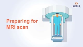
- 7. How to prepare for MRI?
- 8. The doctor may ask the patient to: Undergo screening for allergic reactions.
- 9. The doctor may ask the patient to: Avoid eating or drinking at least 4 hours before the test.
- 10. The doctor may ask the patient to: Remove jewelry or any kind of metals before entering the scan area.
- 11. The doctor may ask the patient to: Inform if you are claustrophobic.