Bronchoscopy.pptx
•Download as PPTX, PDF•
1 like•1,317 views

This document provides tips and instructions for using a PowerPoint presentation on bronchoscopy. It discusses: - Freely editing, modifying, and adding names to the slides - Many slides being blank except for the title to facilitate active learning sessions - Showing blank slides and asking students questions before presenting content - Repeating the process of showing blank slides, asking questions, and then presenting content multiple times - The presentation being useful for self-study as well - Notes providing references and bibliography

Recommended
More Related Content
What's hot
What's hot (20)
Similar to Bronchoscopy.pptx
Similar to Bronchoscopy.pptx (20)
More from Pradeep Pande
More from Pradeep Pande (20)
Recently uploaded
Recently uploaded (20)
Bronchoscopy.pptx
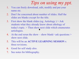
- 1. Tips on using my ppt. 1. You can freely download, edit, modify and put your name etc. 2. Don’t be concerned about number of slides. Half the slides are blanks except for the title. 3. First show the blank slides (eg. Aetiology ) > Ask students what they already know about ethology of today's topic. > Then show next slide which enumerates aetiologies. 4. At the end rerun the show – show blank> ask questions > show next slide. 5. This will be an ACTIVE LEARNING SESSION x three revisions. 6. Good for self study also. 7. See notes for bibliography.
- 3. Introduction & History. • Bronchoscopy is a technique of visualizing the inside of the airways for diagnostic and therapeutic purposes. • Bronchoscope is inserted into the airways, usually through the nose or mouth, or occasionally through a tracheostomy. • Gustav Killian, performed the first bronchoscopy in 1897 • Chevalier Jackson and Victor Negus refined the rigid bronchoscope in the 1920s,
- 4. Introduction & History. • A Japanese, Shigeto Ikeda, invented the flexible bronchoscope in 1966. • Lately fiberoptic scopes have been replaced by bronchoscopes with a charge coupled device (CCD) video chip located at their distal extremity. • More recently EUS Bronchoscopic Sonography.
- 5. Types
- 6. Types • Rigid. • Flexible :Fibreoptic. • Flexible :Video bronchoscope. • EBUS: Endobronchial Ultrasound.
- 8. Indications Diagnostic • Diagnosis of lung cancer • Chest radiographic abnormality • Haemoptysis • Persistent or recurrent cough • paralysed vocal cord • Positive sputum cytology. • Bronchial obstruction • Atelectasis
- 9. Indications Diagnostic • Staging of lung cancer • diagnosis of diffuse lung disease • Identification of infecting agents • Localized wheeze.
- 10. Indications Therapeutic • Insertion of an endotracheal tube for general anesthesia. • Tamponade of endobronchial bleeding . • Removal of foreign bodies. • Aspiration of secretions . • Relief of tracheobronchial narrowing by laser treatment.
- 11. EBUS:. Endobronchial Ultrasound • a technique that uses ultrasound along with bronchoscopy to visualize the airway wall and structures adjacent to it. • It allows real-time guidance of transbronchial needle aspiration (TBNA) of mediastinal and hilar structures and parabronchial lung masses.
- 13. EBUS:Indications. • Staging of non-small cell lung cancer (NSCLC). • Evaluation of mediastinal lesions, intrapulmonary pulmonary nodules, and endobronchial lesions. • Guidance of endobronchial therapy- – resection of endobronchial lesions – stricture dilatation – Stenting – laser therapy – argon plasma coagulation
- 16. Complications
- 17. Complications • Pneumothorax • Haemorrhage • Complications of sedation and topical anaesthesia – Epileptic seizures – Cardiac dysarrythmias, – Hypoventilation – Laryngospasm) • Bronchospasm in asthmatics • Cvs – minor vasovagal episode to serious cardiac dysaaryhtmias, myocardial infarction, pulmonary edema • Hypoxaemia
- 18. Contraindications • Uncooperative patient • Uncorrectable hypoxaemia/ hypercapnia • Unstable myocardium • uncorrectable bleeding tendency • Tracheal stenosis • Poorly controlled asthma
- 19. Get this ppt in mobile 1. Download Microsoft PowerPoint from play store. 2. Open Google assistant 3. Open Google lens. 4. Scan qr code from next slide.
- 20. Get this ppt in mobile
- 21. Get my ppt collection • https://www.slideshare.net/drpradeeppande/ edit_my_uploads • https://www.dropbox.com/sh/x600md3cvj8 5woy/AACVMHuQtvHvl_K8ehc3ltkEa?dl =0 • https://www.facebook.com/doctorpradeeppa nde/?ref=pages_you_manage
Editor's Notes
- drpradeeppande@gmail.com 7697305442