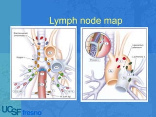
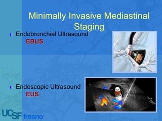
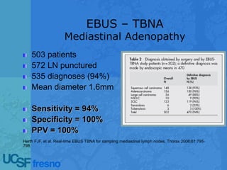
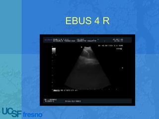
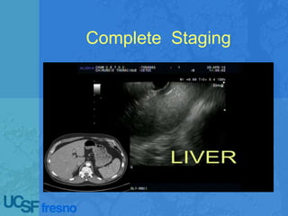
Echoendoscopic lymph node
staging in lung cancer : An
endoscopic alternative
Pravachan Hegde, MD
Interventional Thoracic Endoscopy /
Interventional Pulmonology
Pulmonary & Critical Care Medicine
UCSF - Fresno
Outline
Lymph node map
N1, N2, N3 Nodes
Why do we stage ?
 Staging decides therapy – ?? Surgery
or no surgery
 To identify lymph node metastasis in N2,
N3 nodes and there by prevent “ futile
thoracotomies “ – There are very few
exceptions
 To identify N 1 node metastasis in
candidates with poor lung functions
before deciding on SBRT (Cyberknife)
Who do we stage ?
 Central tumor even if mediastinum normal
radiologically
 Tumor > 3 cm even if peripheral ( Some
experts say 2 cm )
 N1 lymph node on CT / PET
 CT showing lymph nodes > 1 cm
 PET positivity even if lymph nodes < 1 cm
 Low SUV uptake in the primary tumor in the
setting of radiologically normal mediastinum
ACCP 2013 Guidelines
Who doesn't need staging ?
 Peripheral tumor < 3 cm with no lymph
node enlargement on CT and no lymph
node SUV uptake on PET
Minimally Invasive Mediastinal
Staging
Endobronchial Ultrasound
EBUS
Endoscopic Ultrasound
EUS
Invasive Mediastinal
Staging
Mediastinoscopy
Anterior Mediastinotomy
VATS
VAMLA / TEMLA
Background
Testing with minimally invasive tests can prevent surgery
and resection in patients with advanced disease
Ideal Test:
Tissue diagnosis
High accuracy staging
Low procedure-related morbidity
Low cost
Minimally Invasive Lung
Cancer Staging
Staging
Mediastinal LN Staging (N2/N3)
N1 LN Staging
Liver
Adrenal Glands
Celiac axis
Pleura
Chest Wall
T4: Aorta, SVC, Trachea, Atrium
Factors to Consider
Accuracy
Cost
Safety
Completeness
Why EBUS / EUS?
Real-Time Imaging
Portable
No Radiation Exposure
No General Anesthesia
Requirements
Fast
Cheap
Safe
Tissue
Convenience
Outpatient procedure
Fast
No scar
No General Anesthesia
Multiple stations sampled
ASA ok
Real-Time Imaging
Portable
No Radiation Exposure
Cheap
Safe
Tissue
EBUS EUS CM AM VATS
1 – Highest Mediastinal
 
2R – Upper paratracheal
right    
2L – Upper Paratracheal
Left    
3A – Pre-vascular

3P - Retrotracheal
  
4R – Lower Paratracheal
Right   
4L – Lower Paratracheal
Left    
5 – Subaortic (AP window)
  
6 – Para-Aortic NEW
 
7 - Subcarinal
   
8 - Paraesophageal
 
9 – Pulmonary ligament
 
10 - Hilar
  
11 - Interlobar
 
EBUS – TBNA
Mediastinal Adenopathy
503 patients
572 LN punctured
535 diagnoses (94%)
Mean diameter 1.6mm
Sensitivity = 94%
Specificity = 100%
PPV = 100%
Herth FJF, et al. Real-time EBUS TBNA for sampling mediastinal lymph nodes. Thorax 2006;61:795-
798.
EBUS TBNA in CT Negative
Mediastinum
100 NSCLC patients with
no LN > 1cm in the
mediastinum
Identifiable LN in
mediastinum aspirated
2, 4, 7, 10, 11
All patients underwent
surgical staging
119 LN sampled
Malignancy detected in
19
Malignancy missed in 2
Sensitivity = 92.3%
Specificity = 100%
NPV = 96.3%
No complications
Herth FJF, et al. EBUS-guided TBNA of lymph nodes in the radiologically
normal mediastinum. Eur Respir J 2006;28:910-914.
EBUS Published results
EUS Upstaging
44 patients with resectable tumor on CT
scan
EUS changed management in 8 (18%)
Upstaging Tumor to:
Stage IV 7%
Stage IIIA or IIIB 7%
Stage II to IIIA 4%
EUS-FNA led to avoidance of thoracotomy in 13.6% of
patients with CT findings of a resectable tumor
EUS as the First Test in Diagnosis and Staging of Lung Cancer
Singh P, et al. American Journal of Respiratory and Critical Care Medicine.
2007;175:345-354.
EBUS + EUS FNA
160 patients with
enlarged mediastinal LN
EBUS = 85% success
EUS = 78% success
EBUS + EUS = 97%
No Complications
33 patients with med
adenopathy
119 nodes sampled
59 EUS
60 EBUS
11 additional positive nodes by
EBUS over EUS
12 additional positive nodes by
EUS over EBUS
Combination EBUS + EUS =
100% AccuracyHerth FJF, et al. Am J Resp Crit Care Med
2005;171:1164-1167.
Vilman P, et al. Endoscopy 2005;37:833-839.
Combined EBUS + EUS
Suspected Lung
Cancer
TBNA, EBUS-FNA,
EUS-FNA - Combined
138 Patients
Wallace et al. JAMA 2008;299(5):540-546.
Endosonographic Mediastinal
Lymph Node Staging of Lung
Cancer
Prospective comparison of diagnostic test findings
on a consecutive case series
Each subject serving as his or her own control
Sample Size = 166
ClinicalTrials.gov: NCT01011595
Liberman et al Chest. 2014;146(2):389-
397.
Methodology
EBUS
EUS
Cervical Mediastinoscopy
VATS
Anterior Mediastinotomy
Results
30 months
166 patients
Prevalence of N2/N3
disease = 32%
Liberman et al Chest. 2014;146(2):389-
397.
Mean LN
Stations
EBUS 2.2
EUS 1.7
EBUS/EUS 3.9
SMS 3.1
Liberman et al Chest. 2014;146(2):389-397.
Sensitivity NPV Accuracy
EBUS 72% 88% 91%
EUS 62% 85% 88%
EBUS+EUS 91% 96% 97%
EBUS-EUS diagnosed N2/N3/M1 disease in 24
patients in whom SMS was negative
Preventing thoracotomy in an additional 14%
Morbidity
MEDIASTINOSCOPY
2,145 patients
23 complications (1.07%)
Bleeding – 7
Vocal Cord Dysfunction –
12
Tracheal Injury – 2
Pneumothorax – 2
Death – 1
PA Injury
EBUS
100 patients
0 complications
Chest 2004;125:322-325.
1,174 patients
0 major complications
5.5% transient atrial
tachycardia
Eur Resp J 2002;20:118-121.
50 lung biopsies
1 pneumothorax
Eur Resp J 2002;20:972-974.
Lemaire A, et al. Ann Thorac Surg 2006;82:1185-
1190.
Rare but possible tracheal
injury
CETOC - Journal of Bronchology 2010;17:264-265.
COST
Mediastinoscopy =
$2,100* - $12,900**
EUS-FNA = $1,867**
EBUS-FNA =
$2,000**
Pulmonary Resection
= $22,000*
*J Clin Oncol 2001;20:263-273.
**Ann Thorac Surg 2005;80:1231-1239.
RUL tumor with radiologically
normal mediastinum
Left Adrenal Gland
Left Adrenal Gland
SUV = 4.3
EBUS 4 R
Complete Staging
EUS Adrenal biopsy
Take home message
 Standard mediastinoscopy is no longer the gold
standard.
 Combined endosonographic procedures (EBUS/EUS)
are safe,cost-effective, and superior to surgical
mediastinal staging. It allows for the biopsy of lymph
nodes and metastases that are unattainable with
standard mediastinoscopy techniques thereby preventing
futile thoracotomies.
 Combined endosonographic procedures (EBUS/EUS)
are the new gold standard in mediastinal staging of non-
small-cell lung cancer when performed by an
experienced operator.
Thank You !

Echoendoscopic Lymph Node Staging in Lung Cancer: An endoscopic alternative

  • 1.
    Echoendoscopic lymph node stagingin lung cancer : An endoscopic alternative Pravachan Hegde, MD Interventional Thoracic Endoscopy / Interventional Pulmonology Pulmonary & Critical Care Medicine UCSF - Fresno
  • 2.
  • 3.
  • 4.
  • 5.
    Why do westage ?  Staging decides therapy – ?? Surgery or no surgery  To identify lymph node metastasis in N2, N3 nodes and there by prevent “ futile thoracotomies “ – There are very few exceptions  To identify N 1 node metastasis in candidates with poor lung functions before deciding on SBRT (Cyberknife)
  • 6.
    Who do westage ?  Central tumor even if mediastinum normal radiologically  Tumor > 3 cm even if peripheral ( Some experts say 2 cm )  N1 lymph node on CT / PET  CT showing lymph nodes > 1 cm  PET positivity even if lymph nodes < 1 cm  Low SUV uptake in the primary tumor in the setting of radiologically normal mediastinum ACCP 2013 Guidelines
  • 7.
    Who doesn't needstaging ?  Peripheral tumor < 3 cm with no lymph node enlargement on CT and no lymph node SUV uptake on PET
  • 8.
    Minimally Invasive Mediastinal Staging EndobronchialUltrasound EBUS Endoscopic Ultrasound EUS
  • 9.
  • 10.
    Background Testing with minimallyinvasive tests can prevent surgery and resection in patients with advanced disease Ideal Test: Tissue diagnosis High accuracy staging Low procedure-related morbidity Low cost
  • 11.
    Minimally Invasive Lung CancerStaging Staging Mediastinal LN Staging (N2/N3) N1 LN Staging Liver Adrenal Glands Celiac axis Pleura Chest Wall T4: Aorta, SVC, Trachea, Atrium Factors to Consider Accuracy Cost Safety Completeness
  • 12.
    Why EBUS /EUS? Real-Time Imaging Portable No Radiation Exposure No General Anesthesia Requirements Fast Cheap Safe Tissue
  • 13.
    Convenience Outpatient procedure Fast No scar NoGeneral Anesthesia Multiple stations sampled ASA ok Real-Time Imaging Portable No Radiation Exposure Cheap Safe Tissue
  • 14.
    EBUS EUS CMAM VATS 1 – Highest Mediastinal   2R – Upper paratracheal right     2L – Upper Paratracheal Left     3A – Pre-vascular  3P - Retrotracheal    4R – Lower Paratracheal Right    4L – Lower Paratracheal Left     5 – Subaortic (AP window)    6 – Para-Aortic NEW   7 - Subcarinal     8 - Paraesophageal   9 – Pulmonary ligament   10 - Hilar    11 - Interlobar  
  • 15.
    EBUS – TBNA MediastinalAdenopathy 503 patients 572 LN punctured 535 diagnoses (94%) Mean diameter 1.6mm Sensitivity = 94% Specificity = 100% PPV = 100% Herth FJF, et al. Real-time EBUS TBNA for sampling mediastinal lymph nodes. Thorax 2006;61:795- 798.
  • 16.
    EBUS TBNA inCT Negative Mediastinum 100 NSCLC patients with no LN > 1cm in the mediastinum Identifiable LN in mediastinum aspirated 2, 4, 7, 10, 11 All patients underwent surgical staging 119 LN sampled Malignancy detected in 19 Malignancy missed in 2 Sensitivity = 92.3% Specificity = 100% NPV = 96.3% No complications Herth FJF, et al. EBUS-guided TBNA of lymph nodes in the radiologically normal mediastinum. Eur Respir J 2006;28:910-914.
  • 17.
  • 18.
    EUS Upstaging 44 patientswith resectable tumor on CT scan EUS changed management in 8 (18%) Upstaging Tumor to: Stage IV 7% Stage IIIA or IIIB 7% Stage II to IIIA 4% EUS-FNA led to avoidance of thoracotomy in 13.6% of patients with CT findings of a resectable tumor EUS as the First Test in Diagnosis and Staging of Lung Cancer Singh P, et al. American Journal of Respiratory and Critical Care Medicine. 2007;175:345-354.
  • 19.
    EBUS + EUSFNA 160 patients with enlarged mediastinal LN EBUS = 85% success EUS = 78% success EBUS + EUS = 97% No Complications 33 patients with med adenopathy 119 nodes sampled 59 EUS 60 EBUS 11 additional positive nodes by EBUS over EUS 12 additional positive nodes by EUS over EBUS Combination EBUS + EUS = 100% AccuracyHerth FJF, et al. Am J Resp Crit Care Med 2005;171:1164-1167. Vilman P, et al. Endoscopy 2005;37:833-839.
  • 20.
    Combined EBUS +EUS Suspected Lung Cancer TBNA, EBUS-FNA, EUS-FNA - Combined 138 Patients Wallace et al. JAMA 2008;299(5):540-546.
  • 21.
    Endosonographic Mediastinal Lymph NodeStaging of Lung Cancer Prospective comparison of diagnostic test findings on a consecutive case series Each subject serving as his or her own control Sample Size = 166 ClinicalTrials.gov: NCT01011595 Liberman et al Chest. 2014;146(2):389- 397.
  • 22.
  • 23.
    Results 30 months 166 patients Prevalenceof N2/N3 disease = 32% Liberman et al Chest. 2014;146(2):389- 397. Mean LN Stations EBUS 2.2 EUS 1.7 EBUS/EUS 3.9 SMS 3.1
  • 24.
    Liberman et alChest. 2014;146(2):389-397. Sensitivity NPV Accuracy EBUS 72% 88% 91% EUS 62% 85% 88% EBUS+EUS 91% 96% 97%
  • 25.
    EBUS-EUS diagnosed N2/N3/M1disease in 24 patients in whom SMS was negative Preventing thoracotomy in an additional 14%
  • 26.
    Morbidity MEDIASTINOSCOPY 2,145 patients 23 complications(1.07%) Bleeding – 7 Vocal Cord Dysfunction – 12 Tracheal Injury – 2 Pneumothorax – 2 Death – 1 PA Injury EBUS 100 patients 0 complications Chest 2004;125:322-325. 1,174 patients 0 major complications 5.5% transient atrial tachycardia Eur Resp J 2002;20:118-121. 50 lung biopsies 1 pneumothorax Eur Resp J 2002;20:972-974. Lemaire A, et al. Ann Thorac Surg 2006;82:1185- 1190.
  • 27.
    Rare but possibletracheal injury CETOC - Journal of Bronchology 2010;17:264-265.
  • 28.
    COST Mediastinoscopy = $2,100* -$12,900** EUS-FNA = $1,867** EBUS-FNA = $2,000** Pulmonary Resection = $22,000* *J Clin Oncol 2001;20:263-273. **Ann Thorac Surg 2005;80:1231-1239.
  • 29.
    RUL tumor withradiologically normal mediastinum
  • 30.
  • 31.
  • 32.
  • 33.
  • 34.
  • 36.
    Take home message Standard mediastinoscopy is no longer the gold standard.  Combined endosonographic procedures (EBUS/EUS) are safe,cost-effective, and superior to surgical mediastinal staging. It allows for the biopsy of lymph nodes and metastases that are unattainable with standard mediastinoscopy techniques thereby preventing futile thoracotomies.  Combined endosonographic procedures (EBUS/EUS) are the new gold standard in mediastinal staging of non- small-cell lung cancer when performed by an experienced operator.
  • 37.