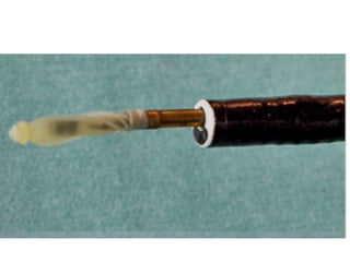
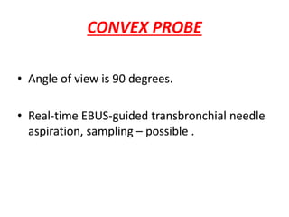
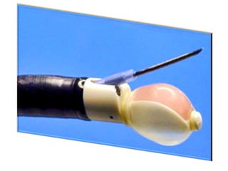
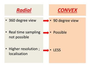
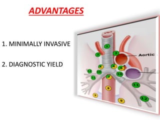
EBUS (ENDOBRONCHIAL ULTRASOUND)
DR.MANOJ MD MEDICINE
• It is a bronchoscopic technique that uses
ultrasound to visualize structures (within the
airway wall, lung, and mediastinum) .
• Used to guide bronchoscopic sampling
EBUS
PROBES USED
• Radial probe (RP-EBUS)
• Convex probe (CP-EBUS)
RADIAL PROBE
• High definition, 360-degree images of the
airway wall and surrounding structures.
• visualize the layers of the airway wall in
greater detail.
• Real time biopsy – not possible.
CONVEX PROBE
• Angle of view is 90 degrees.
• Real-time EBUS-guided transbronchial needle
aspiration, sampling – possible .
CONVEX
• 360 degree view
• Real time sampling
not possible
• Higher resolution ;
localisation
Radial
• 90 degree view
• Possible
• LESS
Technique
• Consious sedation , GA , LA
• TBNA – 40MM
• Discharge if gag reflex comes back.
INDICATION
-diagnosing, staging, (NSCLC).
-mediastinal masses.
-pulmonary nodules.
-endobronchial or peribronchial lesions,
-can guide therapeutic procedures (eg, airway
stenting)
-depth of tumour invasion
CONTRAINDICATION
• Absolute contraindications - severe refractory
hypoxia.
• Specific contraindications - exacerbation of
asthma, COPD, severe PAH, MI ,CCF ,
arrythymia .- at risk of bleeding.
Complications
- Usually minor.
- Hemorrhage and Pneumothorax.
- Hypotension, arrhythmias.
ADVANTAGES
1. MINIMALLY INVASIVE
2. DIAGNOSTIC YIELD
EBUS
1.WHAT IS IT
2.PROBE TYPES
3.INDICATION AND CI
4.TECHNIQUE
5.ADVANTAGES
DR.MANOJ MD MEDICINETHANK YOU ….

EBUS (ENDOBRONCHIAL USG)