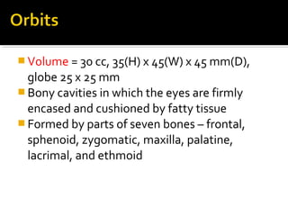
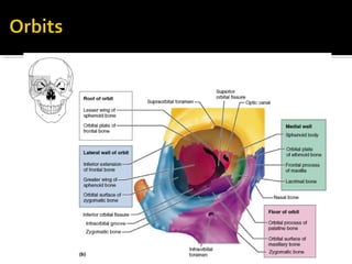
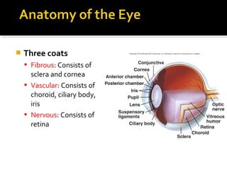
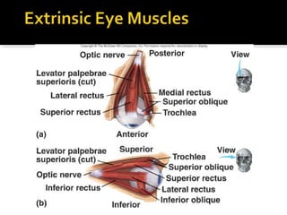
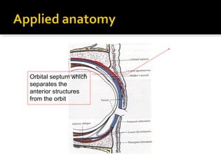
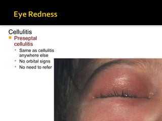
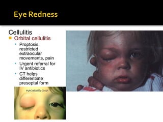
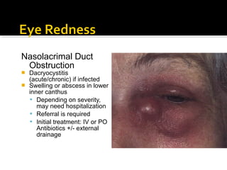
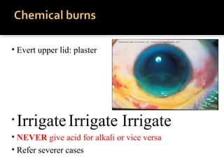
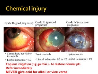
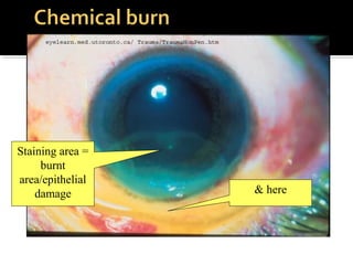
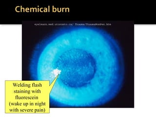
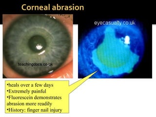
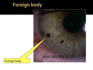
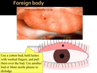
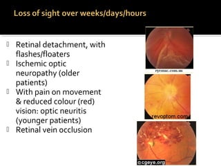
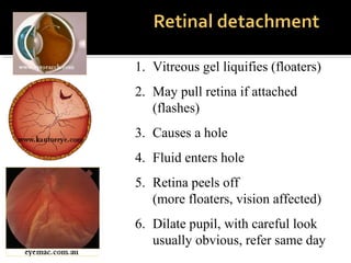
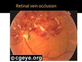
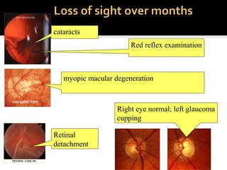
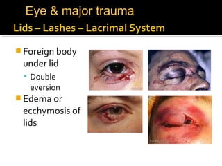
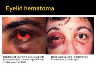
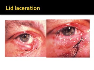
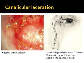
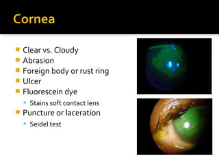
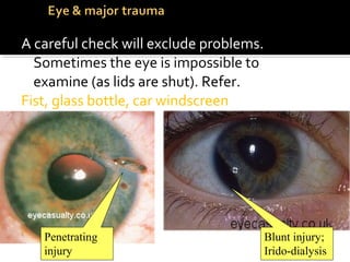
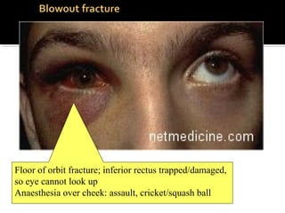
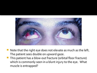
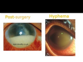
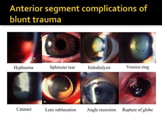
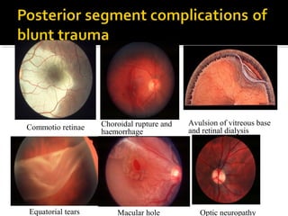
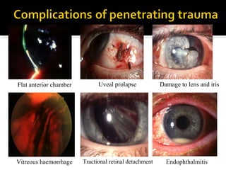
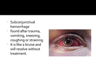
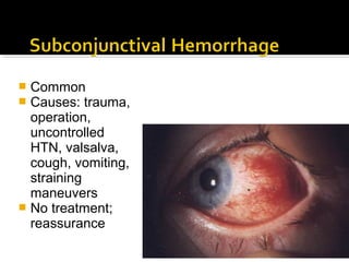
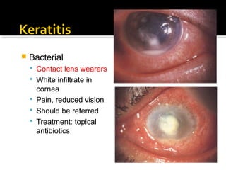
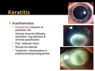
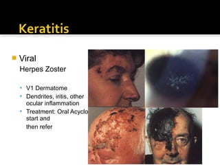
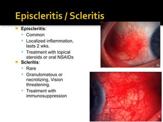
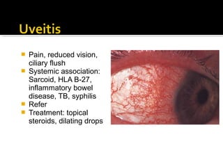
Dr. Riyad Banayot
 Eyelid
 Hematoma
 Marginal laceration
 Canalicular laceration
 Orbital blow-out fracture
 Complications of blunt trauma
 Anterior segment
 Posterior segment
 Chemical injuries
 Volume = 30 cc, 35(H) x 45(W) x 45 mm(D),
globe 25 x 25 mm
 Bony cavities in which the eyes are firmly
encased and cushioned by fatty tissue
 Formed by parts of seven bones – frontal,
sphenoid, zygomatic, maxilla, palatine,
lacrimal, and ethmoid
 Three coats
 Fibrous: Consists of
sclera and cornea
 Vascular: Consists of
choroid, ciliary body,
iris
 Nervous: Consists of
retina
•Red eye
•Loss of vision
•Medical problems
•Trauma
› Lid/orbit infections
› Chemical burns
› Conjunctivitis
› Corneal abrasion
› Foreign body
› Blunt eye injury
› Corneal ulcers
› Acute uveitis
› Acute glaucoma
Orbital septum which
separates the
anterior structures
from the orbit
Cellulitis
 Preseptal
cellulitis
 Same as cellulitis
anywhere else
 No orbital signs
 No need to refer
Cellulitis
 Orbital cellulitis
 Proptosis,
restricted
extraocular
movements, pain
 Urgent referral for
IV antibiotics
 CT helps
differentiate
preseptal form
Nasolacrimal Duct
Obstruction
 Dacryocystitis
(acute/chronic) if infected
 Swelling or abscess in lower
inner canthus
 Depending on severity,
may need hospitalization
 Referral is required
 Initial treatment: IV or PO
Antibiotics +/- external
drainage
• Evert upper lid: plaster
• IrrigateIrrigate Irrigate
• NEVER give acid for alkali or vice versa
• Refer severer cases
• Cornea hazy but visible
iris details
Grade II (good prognosis)
• Limbal ischaemia < 1/3
• No iris details
Grade III (guarded
prognosis)
• Limbal ischaemia - 1/3 to 1/2
• Opaque cornea
Grade IV (very poor
prognosis)
• Limbal ischaemia > 1/2
Copious irrigation ( 15-30 min ) - to restore normal pH.
Refer immediately
NEVER give acid for alkali or vice versa
Staining area =
burnt
area/epithelial
damage & here
Welding flash
staining with
fluorescein
(wake up in night
with severe pain)
•heals over a few days
•Extremely painful
•Fluorescein demonstrates
abrasion more readily
•History: finger nail injury
Foreign body
Use a cotton bud; hold lashes
with washed fingers, and pull
them over the bud. Use another
bud or blunt sterile plastic to
dislodge
 If metal striking-metal is the mechanism
of injury always get an X-Ray/CT scan
of skull (This is mandatory if there is an
open globe injury or suspicion of entry
wound)
 Superficial corneal FB can be removed
with Q-tip or needle tip, otherwise refer
 Rust rings develop after initial removal
 Achy eye, misty
vision
 Previous mild
episodes with haloes
 Pupil fixed (sluggish),
semi-dilated
 Eye feels hard
Press eye with 2
fingers..Try this on your
own eye
n o r m a l s h a llo w
a n t e r io r
c h a m b e r
TI Artery occlusion
Retinal arteriole occlusion:
If within 3 hours, can dislodge clot
(massage, IV diamox, AC paracentesis)
Refer ASAP, aspirin
(diabetes/high cholesterol/smoke/hypertension)
 Retinal detachment, with
flashes/floaters
 Ischemic optic
neuropathy (older
patients)
 With pain on movement
& reduced colour (red)
vision: optic neuritis
(younger patients)
 Retinal vein occlusion
1. Vitreous gel liquifies (floaters)
2. May pull retina if attached
(flashes)
3. Causes a hole
4. Fluid enters hole
5. Retina peels off
(more floaters, vision affected)
6. Dilate pupil, with careful look
usually obvious, refer same day
Retinal vein occlusion
cataracts
Red reflex examination
myopic macular degeneration
Retinal
detachment
Right eye normal; left glaucoma
cupping
 Foreign body
under lid
 Double
eversion
 Edema or
ecchymosis of
lids
Eye & major trauma
Orbital roof fracture if associated with
subconjunctival haemorrhage without
visible posterior limit
Basal skull fracture - bilateral ring
haematomas (‘panda eyes’)
• Repair within 24 hours • Locate and approximate ends of laceration
• Bridge defect with silicone tubing
• Leave in situ for about 3 months
 Clear vs. Cloudy
 Abrasion
 Foreign body or rust ring
 Ulcer
 Fluorescein dye
 Stains soft contact lens
 Puncture or laceration
 Seidel test
A careful check will exclude problems.
Sometimes the eye is impossible to
examine (as lids are shut). Refer.
Fist, glass bottle, car windscreen
Blunt injury;
Irido-dialysis
Penetrating
injury
• Periocular ecchymosis
and oedema
• Infraorbital nerve
anaesthesia
• Ophthalmoplegia -
typically in up- and down-
gaze (double diplopia)
• Enophthalmos - if severe
Floor of orbit fracture; inferior rectus trapped/damaged,
so eye cannot look up
Anaesthesia over cheek: assault, cricket/squash ball
 Note that the right eye does not elevate as much as the left.
The patient sees double on upward gaze.
 This patient has a blow-out fracture (orbital floor fracture)
which is commonly seen in a blunt injury to the eye. What
muscle is entrapped?
Sphincter tear
Cataract Angle recession
Hyphaema
Lens subluxation
Iridodialysis Vossius ring
Rupture of globe
Macular hole Optic neuropathyEquatorial tears
Choroidal rupture and
haemorrhage
Commotio retinae
Avulsion of vitreous base
and retinal dialysis
Flat anterior chamber
Vitreous haemorrhage
Damage to lens and iris
EndophthalmitisTractional retinal detachment
Uveal prolapse
• Subconjunctival
hemorrhage
• found after trauma,
vomiting, sneezing,
coughing or straining.
• It is like a bruise and
will resolve without
treatment.
 Common
 Causes: trauma,
operation,
uncontrolled
HTN, valsalva,
cough, vomiting,
straining
maneuvers
 No treatment;
reassurance
 Bacterial
 Contact lens wearers
 White infiltrate in
cornea
 Pain, reduced vision
 Should be referred
 Treatment: topical
antibiotics
 Fungal
 Frequently preceded by
ocular trauma with
organic matter
 Grayish white infiltrate
surrounded by feathery
infiltrate in cornea
 Pain, reduced vision
 Should be referred
 Treatment: topical
antifungal agents &
systemic therapy if
severe
 Acanthamoeba
 Contact lens wearers at
particular risk
 Anterior stromal infiltrates,
ulceration, ring abscess &
stromal opacification
 Pain, reduced vision
 Should be referred
 Treatment: chlorhexidine or
polyhexamethylenebiguanide
 Viral
Herpes Simplex
 Recurrent dendrites,
corneal edema, iritis
 Refer
 Treatment: Acyclovir
ointment
 Viral
Herpes Zoster
 V1 Dermatome
 Dendrites, iritis, other
ocular inflammation
 Treatment: Oral Acyclovir;
start and
then refer
 Episcleritis:
 Common
 Localized inflammation,
lasts 2 wks.
 Treatment with topical
steroids or oral NSAIDs
 Scleritis:
 Rare
 Granulomatous or
necrotizing, Vision
threatening.
 Treatment with
immunosuppression
 Pain, reduced vision,
ciliary flush
 Systemic association:
Sarcoid, HLA B-27,
inflammatory bowel
disease, TB, syphilis
 Refer
 Treatment: topical
steroids, dilating drops

Emergency eye conditions & trauma

  • 1.
  • 2.
     Eyelid  Hematoma Marginal laceration  Canalicular laceration  Orbital blow-out fracture  Complications of blunt trauma  Anterior segment  Posterior segment  Chemical injuries
  • 3.
     Volume =30 cc, 35(H) x 45(W) x 45 mm(D), globe 25 x 25 mm  Bony cavities in which the eyes are firmly encased and cushioned by fatty tissue  Formed by parts of seven bones – frontal, sphenoid, zygomatic, maxilla, palatine, lacrimal, and ethmoid
  • 5.
     Three coats Fibrous: Consists of sclera and cornea  Vascular: Consists of choroid, ciliary body, iris  Nervous: Consists of retina
  • 7.
    •Red eye •Loss ofvision •Medical problems •Trauma
  • 8.
    › Lid/orbit infections ›Chemical burns › Conjunctivitis › Corneal abrasion › Foreign body › Blunt eye injury › Corneal ulcers › Acute uveitis › Acute glaucoma
  • 9.
    Orbital septum which separatesthe anterior structures from the orbit
  • 10.
    Cellulitis  Preseptal cellulitis  Sameas cellulitis anywhere else  No orbital signs  No need to refer
  • 11.
    Cellulitis  Orbital cellulitis Proptosis, restricted extraocular movements, pain  Urgent referral for IV antibiotics  CT helps differentiate preseptal form
  • 12.
    Nasolacrimal Duct Obstruction  Dacryocystitis (acute/chronic)if infected  Swelling or abscess in lower inner canthus  Depending on severity, may need hospitalization  Referral is required  Initial treatment: IV or PO Antibiotics +/- external drainage
  • 13.
    • Evert upperlid: plaster • IrrigateIrrigate Irrigate • NEVER give acid for alkali or vice versa • Refer severer cases
  • 14.
    • Cornea hazybut visible iris details Grade II (good prognosis) • Limbal ischaemia < 1/3 • No iris details Grade III (guarded prognosis) • Limbal ischaemia - 1/3 to 1/2 • Opaque cornea Grade IV (very poor prognosis) • Limbal ischaemia > 1/2 Copious irrigation ( 15-30 min ) - to restore normal pH. Refer immediately NEVER give acid for alkali or vice versa
  • 15.
  • 16.
    Welding flash staining with fluorescein (wakeup in night with severe pain)
  • 17.
    •heals over afew days •Extremely painful •Fluorescein demonstrates abrasion more readily •History: finger nail injury
  • 18.
  • 19.
    Use a cottonbud; hold lashes with washed fingers, and pull them over the bud. Use another bud or blunt sterile plastic to dislodge
  • 20.
     If metalstriking-metal is the mechanism of injury always get an X-Ray/CT scan of skull (This is mandatory if there is an open globe injury or suspicion of entry wound)  Superficial corneal FB can be removed with Q-tip or needle tip, otherwise refer  Rust rings develop after initial removal
  • 21.
     Achy eye,misty vision  Previous mild episodes with haloes  Pupil fixed (sluggish), semi-dilated  Eye feels hard Press eye with 2 fingers..Try this on your own eye
  • 22.
    n o rm a l s h a llo w a n t e r io r c h a m b e r
  • 23.
  • 24.
    Retinal arteriole occlusion: Ifwithin 3 hours, can dislodge clot (massage, IV diamox, AC paracentesis) Refer ASAP, aspirin (diabetes/high cholesterol/smoke/hypertension)
  • 25.
     Retinal detachment,with flashes/floaters  Ischemic optic neuropathy (older patients)  With pain on movement & reduced colour (red) vision: optic neuritis (younger patients)  Retinal vein occlusion
  • 26.
    1. Vitreous gelliquifies (floaters) 2. May pull retina if attached (flashes) 3. Causes a hole 4. Fluid enters hole 5. Retina peels off (more floaters, vision affected) 6. Dilate pupil, with careful look usually obvious, refer same day
  • 27.
  • 28.
    cataracts Red reflex examination myopicmacular degeneration Retinal detachment Right eye normal; left glaucoma cupping
  • 29.
     Foreign body underlid  Double eversion  Edema or ecchymosis of lids Eye & major trauma
  • 30.
    Orbital roof fractureif associated with subconjunctival haemorrhage without visible posterior limit Basal skull fracture - bilateral ring haematomas (‘panda eyes’)
  • 32.
    • Repair within24 hours • Locate and approximate ends of laceration • Bridge defect with silicone tubing • Leave in situ for about 3 months
  • 33.
     Clear vs.Cloudy  Abrasion  Foreign body or rust ring  Ulcer  Fluorescein dye  Stains soft contact lens  Puncture or laceration  Seidel test
  • 34.
    A careful checkwill exclude problems. Sometimes the eye is impossible to examine (as lids are shut). Refer. Fist, glass bottle, car windscreen Blunt injury; Irido-dialysis Penetrating injury
  • 36.
    • Periocular ecchymosis andoedema • Infraorbital nerve anaesthesia • Ophthalmoplegia - typically in up- and down- gaze (double diplopia) • Enophthalmos - if severe
  • 37.
    Floor of orbitfracture; inferior rectus trapped/damaged, so eye cannot look up Anaesthesia over cheek: assault, cricket/squash ball
  • 38.
     Note thatthe right eye does not elevate as much as the left. The patient sees double on upward gaze.  This patient has a blow-out fracture (orbital floor fracture) which is commonly seen in a blunt injury to the eye. What muscle is entrapped?
  • 40.
    Sphincter tear Cataract Anglerecession Hyphaema Lens subluxation Iridodialysis Vossius ring Rupture of globe
  • 41.
    Macular hole OpticneuropathyEquatorial tears Choroidal rupture and haemorrhage Commotio retinae Avulsion of vitreous base and retinal dialysis
  • 42.
    Flat anterior chamber Vitreoushaemorrhage Damage to lens and iris EndophthalmitisTractional retinal detachment Uveal prolapse
  • 43.
    • Subconjunctival hemorrhage • foundafter trauma, vomiting, sneezing, coughing or straining. • It is like a bruise and will resolve without treatment.
  • 44.
     Common  Causes:trauma, operation, uncontrolled HTN, valsalva, cough, vomiting, straining maneuvers  No treatment; reassurance
  • 45.
     Bacterial  Contactlens wearers  White infiltrate in cornea  Pain, reduced vision  Should be referred  Treatment: topical antibiotics
  • 46.
     Fungal  Frequentlypreceded by ocular trauma with organic matter  Grayish white infiltrate surrounded by feathery infiltrate in cornea  Pain, reduced vision  Should be referred  Treatment: topical antifungal agents & systemic therapy if severe
  • 47.
     Acanthamoeba  Contactlens wearers at particular risk  Anterior stromal infiltrates, ulceration, ring abscess & stromal opacification  Pain, reduced vision  Should be referred  Treatment: chlorhexidine or polyhexamethylenebiguanide
  • 48.
     Viral Herpes Simplex Recurrent dendrites, corneal edema, iritis  Refer  Treatment: Acyclovir ointment
  • 49.
     Viral Herpes Zoster V1 Dermatome  Dendrites, iritis, other ocular inflammation  Treatment: Oral Acyclovir; start and then refer
  • 50.
     Episcleritis:  Common Localized inflammation, lasts 2 wks.  Treatment with topical steroids or oral NSAIDs  Scleritis:  Rare  Granulomatous or necrotizing, Vision threatening.  Treatment with immunosuppression
  • 51.
     Pain, reducedvision, ciliary flush  Systemic association: Sarcoid, HLA B-27, inflammatory bowel disease, TB, syphilis  Refer  Treatment: topical steroids, dilating drops

Editor's Notes

  • #10 The orbital septum separates anterior lid structures and infections from the orbit
  • #11 Preseptal cellulitis typically affects children and is usually secondary to lid infection such as severe acute hordeolum, skin laceration or an insect bite. The infection does not penetrate the orbital septum which separates the anterior structures from the orbit. Examination shows periorbital swelling and tenderness without proptosis (Top picture). Ocular motility, visual acuity and pupillary reactions are all normal. Treatment is with oral antibiotics on an outpatient basis
  • #12 Bacterial orbital cellulitis is an infection of the soft tissues behind the orbital septum. It is much less common but potentially more serious than preseptal cellulitis The following are the main types:1. Sinus-related (ethmoidal sinusitis). It typically affects children2. From adjacent structures such as dental infection. 3. Post-traumatic most commonly develops within 48-72 hours of an injury that penetrates the orbital septum. PoIymicrobial infection is the rule Presentation is with a rapid onset of unilateral chemosis, proptosis and painful diplopia. Examination shows an unwell and pyrexial patient. The proptosis is most frequently lateral and downwards. The eyelids are swollen, erythematous, warm and tender to palpation. Ocular movements are restricted and painful. Orbital cellulitis is an emergency requiring hospital admission
  • #13 In adults NLDO presents as a tender canthal swelling &amp; Mild preseptal cellulitis which may develop into an abscess of the lacrimal sac Treatment consists of systemic antibiotics and warm compresses, and DCR after acute infection is controlled (DCR to create internal fistula from lacrimal sac to nose)
  • #20 Top: A Subtarsal foreign body. Need to evert lid..use a cotton bud. Fluorescein drops show up any scratch/abrasion. Easier to see with a blue light, but an ordinary light is sufficient. Should the foreign body be removed? Yes no What is the treatment? ………………………………………………………………………………………. Does this need referral? Yes no Bottom A corneal foreign body.
  • #21 Treatment consists of: - Removal of FB ASAP - Topical antibiotic ointment - Patching the eye
  • #45 A good history is essential to exclude: trauma, operation, uncontrolled HTN, valsalva, coughing, vomiting or straining maneuvers
  • #46 Predisposing factors include: 1- Contact lens wear is the most common predisposing factor 2- Ocular surface disease post herpetic corneal disease, trauma, corneal exposure and dry eyes Examination reveals acutely painful red eye and a white spot on the cornea. In some cases keratitis can rapidly lead to a corneal ulcer and perforation A bacterial corneal ulcer is a sight-threatening condition which demands urgent identification and eradication of the causative organism. This is best performed with the patient hospitalized
  • #48 Predisposing factors include: 1- Contact lens wear is the most common predisposing factor 2- Ocular surface disease post herpetic corneal disease, trauma, corneal exposure and dry eyes Examination reveals acutely painful red eye and a white spot on the cornea. In some cases keratitis can rapidly lead to a corneal ulcer and perforation A bacterial corneal ulcer is a sight-threatening condition which demands urgent identification and eradication of the causative organism. This is best performed with the patient hospitalized
  • #49 Infection with HSV is extremely common and about 90% of the population are seropositive for HSV antibodies. In spite of this, most infections are subclinical Primary ocular infection typically occurs in children between the ages of 6 months and 5 years. In most cases it is self-limited Clinical picture Blepharoconjunctivitis is usually benign and self-limited. Skin lesions typically involve the lids and periorbital area. Initially, they consist of vesicles which rapidly form superficial crusts and then heal without scarring. Keratitis develops within a few days in about 50% of patients with blepharoconjunctivitis. A fine epithelial punctate keratitis may be a transient finding. A coarse epithelial punctate keratitis may give rise to a variety of epithelial lesions which subsequently progress to dendritic shapes Treatment: topical antiviral ointment should be applied prophylactically to the eye five times a day for about 21 days to prevent keratitis
  • #50 Chickenpox and zoster are different conditions caused by the same virus. Zoster mainly affects elderly patients 15% of all cases of herpes zoster affect the ophthalmic division of the trigeminal nerve (any branch: frontal, lacrimal and nasociliary) Clinical features: Rash is maculopapular and then becomes pustular. The pustules subsequently burst to form crusting ulcers. Initially, the rash is accompanied by periorbital edema. Ocular lesions include conjunctivitis, episcleritis, keratitis, uveitis Keratitis takes the form of small, fine, dendritic or stellate epithelial lesions with tapered ends without bulbs Treatment: Systemic therapy is with acyclovir 800 mg tablets administered five times daily for 7 days as early as possible. Topical therapy consists of antiviral creams and a steroid-antibiotic preparation used three times daily
  • #51 Episcleritis is a common, benign, self-limiting and frequently recurrent disorder which typically affects young adults. It is seldom associated with a systemic disorder and never progresses to scleritis Presentation is with unilateral mild discomfort and tenderness to touch Examination shows sectoral or, rarely, diffuse redness Simple episcleritis usually resolves spontaneously within 1-2 weeks. Mild cases may need topical steroids and/or topical non-steroidal anti-inflammatory drugs (NSAIDs) Scleritis is a granulomatous inflammation of the scleral coat of the eye. It is much less common than episcleritis. About 45% of patients with scleritis, particularly of the necrotizing type, may have one of the following systemic diseases:1. Rheumatoid arthritis (the most frequent)2. Connective tissue vascular disorders Presentation is with a gradual onset of pain and localized redness Early cases show distortion of blood vessels in the affected area, this is followed by the development of scleral necrosis. Eventually, the sclera becomes transparent and the underlying uvea visible Treatment is with oral steroids, Immunosuppressive drugs or combined therapy
  • #52 By strict definition, uveitis is an inflammation of the uveal tract ( iris, ciliary body and retina) The main symptoms of acute anterior uveitis are photophobia, pain, redness, decreased vision and lacrimation The main symptoms of posterior segment inflammation are floaters and impaired vision Uveitis is associated with arthritis: Ankylosing spondylitis, Reiter&amp;apos;s syndrome, Psoriatic arthritis, Juvenile chronic arthritis Uveitis is also associated with chronic systemic infections: AIDS, Acquired syphilis, Tuberculosis, Leprosy Uveitis is also associated with parasitic infections: Toxoplasmosis, Toxocariasis Treatment of uveitis includes: Mydriatics (To give comfort &amp; prevent formation of posterior synechiae); Steroids administered topically in the form of drops or ointment, by periocular injection or systemically. Cytotoxic drugs are used in blinding (usually bilateral), reversible, uveitis which has failed to respond to adequate steroid therapy and intolerance side effects from systemic steroid therapy