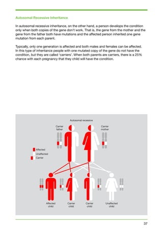
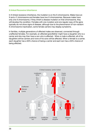
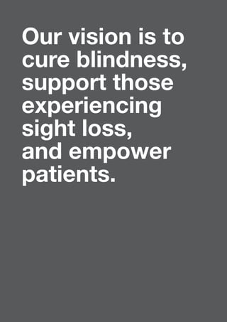
This document provides a summary of conditions that affect the retina. It begins with an introduction from the CEO of Fighting Blindness, noting that over 224,000 people in Ireland are affected by sight loss. It then discusses Fighting Blindness's mission to cure blindness, support those with sight loss, and empower patients. The document provides information on various retinal conditions, including age-related macular degeneration, diabetes-related sight loss, rare inherited conditions like retinitis pigmentosa and Stargardt disease, and Fighting Blindness' research and support services. It aims to be a valuable resource for individuals and families affected by retinal conditions.
To know about definition , causes, types, signs and symptoms, diagnosis, treatment home remedies for floating these can give knowledge by this presentation
Good to know, since we are likely to get cataract sooner or later.
Take care of your precious eyes before cataract starts to form.
Watch this power point presentation.
There are several types of eye problems and visual disturbances. These include blurred vision, halos, blind spots, floaters, and other symptoms.
Changes in vision, blurriness, blind spots, halos around lights, or dimness of vision should always be evaluated by a medical professional. Such changes may represent an eye disease, aging, eye injury, or a condition like diabetes that affects many organs in your body.
For the most severe form of visual loss, see blindness.
http://www.nlm.nih.gov/medlineplus/ency/article/003040.htm
A cataract is a clouding or opacity that
develops in the crystalline lens of the eye or in its envelope, varying in degree from slight opacity to obstructing the passage of light.
Progressive, painless clouding of the natural, internal lens of the eye.
Very important to know about cataract and its symptoms are. It will be able to diagnose it and stop the further damage. Treatment of the cataract is the surgery. The surgery for the cataract is very safe.
Orbis Charity Patron Baroness Uddin Suspended from the House of Lords - 24 Oc...CharityNewsNet
Orbis Charity Patron and quangocrat Baroness Uddin is suspended for a year from parliament in the toughest punishment enacted in the House of Lords in modern times after an inquiry found that each had broken expenses rules to wrongly claim tens of thousands of pounds in expenses. Lady Uddin is ordered to pay back £125,349.10.
Achievements Forum - 2020. TOP 100 Register.HannaOsland
On the pages of this catalogue EBA proudly presents outstanding companies and personalities.
They have all demonstrated remarkable resilience in a year of unprecedented challenges.
Global Medical Cures™ | Living with Low Vision
DISCLAIMER-
Global Medical Cures™ does not offer any medical advice, diagnosis, treatment or recommendations. Only your healthcare provider/physician can offer you information and recommendations for you to decide about your healthcare choices.
Descriptive power point lecture for geriatic carers highlighting basic anatomy and basic facts about the eye and eye problems ;Aging and the eye, common eye disorders, symptoms and signs,
care of the eyes and prevention of blindness and injury for the elderly,
care of the eyes for the blind or visually impaired elderly and mobility concerns for the blind and visually impaired.
We held an improvement collaborative with 19 NHS providers earlier this year to help improve the management of falls in an inpatient setting. As part of the collaborative offer we gave updates on clinical developments.
This resource has been designed to give providers who did not participate in the collaborative access to the clinical update material.
RNIB Scotland and Deaf Action: Innovation in improving outcomes for people wh...Sophie40
RNIB Scotland and Deaf Action are presently undertaking pioneering activity in three key areas which includes: Hidden Sensory Loss (Learning Disability, Stroke and Dementia); Ethnic Minorities; and Early Intervention Family Support. This workshop predominantly explores the Hidden Sensory Loss issues for people with complex needs and introduces the innovative Sensory Champion Training Programme which is being delivered across Edinburgh and the Lothians throughout 2014. Contributed by: RNIB
Similar to A guide-to-conditions-of-the-retina-final (20)
$ATHX @athersys Corporate Overview - Investor Presentation (dated 08-10-18 / 32 slides) - http://www.athersys.com/static-files/df957fcc-48c5-46aa-8ae4-c254e18e05ee As found by "imz72", with growing, insightful shareholder's comments at reddit - https://redd.it/98b693 #MultiStem #Stroke Company Website -http://www.athersys.com/ As posted at Twitter - https://twitter.com/twenty2John/status/1030883310082318337
9/11/2017 Revised Bocce Courts 4 HB (Huntington Beach). This report, given in book form, to each of the seven members of the HB City Council (including, Mayor), and two copies to David Dominguez - Facilities, Development & Concessions Manager (HB).
Special Note: All links should be CLICKABLE (Tap link twice), EXCEPT links on first (3) pages. Links, on first (3) pages of this report are not accessible.
Human embryonic stem cell-derived retinal pigment epithelium in patients with age-related macular degeneration and Stargardt's macular dystrophy: follow-up of two open-label phase 1/2 studies
Published Online: 15 October 2014
Including Comment By, Anthony Atala
Q2 2015 ARM - Alliance for Regenerative Medicine
Quarterly Data Report: Q2 2015 provides an in-depth look at regenerative medicine and advanced therapies sector trends and metrics compiled from more than 580 leading therapeutic companies worldwide.
Part 2 Of 2 Comment/Reply on ACT/Ocata Lancet Report (Oct. 2014)
Reply by, *Steven D Schwartz, Eddy Anglade, Robert Lanza, on behalf of the Ocata
Macular Disease Investigator Group
schwartz@jsei.ucla.edu
Jules Stein Eye Institute Retina Division, and David
Geffen School of Medicine, University of California,
Los Angeles, CA 90095, USA (SDS); and Ocata
Therapeutics Inc, Marlborough, MA, USA (EA, RL)
Source Material: http://www.thelancet.com/pdfs/journals/lancet/PIIS0140-6736(15)61203-X.pdf
March 23, 2015 Sheraton Silver Spring Hotel - Silver Spring, Maryland USA
Bioassays 2015: Scientific Approaches & Regulatory Strategies
Bioassay Development for Human Stem Cell-derived Retinal Pigment Epithelium: Progress and Challenges
Irina Klimanskaya, Ocata Therapeutics, Inc., Marlborough, MA USA
4. The production of this Retinopathies Guide has been made possible
by an unrestricted educational grant from Bayer Healthcare.
This publication is proudly endorsed by:
Written and edited by the Communications Department
at Fighting Blindness:
Dr Maria Meehan, Research Manager
Anna Moran, External Affairs Manager
Caitríona Dunne, Communications Officer
We are very grateful for the expert guidance of Mr David Keegan, Mater
Misericordiae Hospital, Dublin; Dr Paul Kenna, Royal Victoria Eye and
Ear Hospital / Trinity College Dublin; and Prof Jane Farrar, Trinity College
Dublin, in the development of this material.
All information current November 2014
This publication is also available online and in audio format.
Please contact us for a copy.
5. Foreword
On behalf of the Irish College of Ophthalmologists, I am pleased
to introduce this Guide to Conditions of the Retina compiled by
Fighting Blindness.
Many people in Ireland are living with severe vision loss, a
significant proportion of which is due to retinal degenerative
conditions. For individuals and families who have been diagnosed
with a retinopathy, access to clear, relevant, detailed and easily
understandable information is essential. Unfortunately, due to the
rare nature of some retinal conditions, information in this format
has not in the past always been readily available.
The value of providing information and support to people with
conditions of the retina - as well as to the doctors who care for
them - cannot be underestimated. Fighting Blindness has been
doing this successfully for many years through their support
services and events, and has been a real resource to Irish people
who are dealing with this often life changing news. The production
of this Guide to Conditions of the Retina is a shining example of
a strong resource that will be invaluable to eye doctors and their
patients to help them understand the conditions they are facing, as
well as explaining the options that are available.
Miss Marie Hickey-Dwyer
President, Irish College of Ophthalmologists
3
6. Contents
Introduction
Foreword from Miss Marie Hickey-Dwyer, President,
Irish College of Ophthalmologists 3
Welcome Note from Avril Daly, CEO, Fighting Blindness 7
Fighting Blindness
About Fighting Blindness 10
Cure. Support. Empower. 11
Insight Counselling Service
About Insight Counselling Service 14
The Benefit of Psychotherapy 15
Support Services 16
Your Eyes
How Your Eyes Work 20
The Spectrum of Sight Loss 22
What You Can Do to Take Care of Your Eyes 23
Healthcare Professionals Who Look After Your Eyes 24
What Happens at Your Ophthalmology Appointment 24
Questions to Ask Your Doctor 27
Research
Fighting Blindness Research 30
Clinical Trials 32
Human Genetics – A Brief Insight (Prof Jane Farrar, TCD) 34
Inheritance Patterns 36
Target 5000 – Gateway to Vision 41
7. Rare Inherited Conditions
Retinitis Pigmentosa 46
Choroideremia 50
Leber Congenital Amaurosis 52
Usher Syndrome 54
Stargardt Disease 56
Juvenile X-linked Retinoschisis 58
Leber Hereditary Optic Neuropathy 60
Best Disease 62
Cone Rod Dystrophies 64
Achromatopsia 66
Other Retinal Conditions
Age-related Macular Degeneration 70
Diabetes Related Sight Loss 74
Retinal Degeneration as Part of a Syndrome 76
Connect
Connect with Fighting Blindness 80
How to Support Our Work 82
Further Information
Useful Links 84
Irish and International Patient Organisations 85
Glossary of Terms 90
8. Our vision is to
cure blindness,
support those
experiencing
sight loss,
and empower
patients.
6
9. Introduction Note
Welcome to the Fighting Blindness
Guide to Conditions of the Retina
More than 224,000 people in Ireland are affected by sight loss and
this number is expected to rise to 275,000 by 2020. Many forms of
sight loss are caused by degeneration of the layer of tissue at the
back of the eye known as the retina, leading to variable levels of
sight loss specific to each person and depending on the condition
they have. At Fighting Blindness we believe it is vital to understand
the condition that affects you or your family members, so that
you can make informed decisions about your eye care and the
supports that may be needed now and into the future.
The good news is that researchers are investigating the conditions
and identifying the problems, with the intention of developing
treatments and, ultimately, cures. Some conditions, such as
certain types of age-related macular degeneration and diabetes-related
7
sight loss, are already being successfully treated in
Ireland. For other rare, genetic conditions, we are still in the early
investigative stages, but positively moving from the laboratory to
clinical trial stage.
The guide provides information on conditions, from basic research
through to clinical trials, treatment options and relevant support
services. We hope it will be a valuable and useful resource not only
to individuals and families who are affected by retinal conditions,
but also for the allied medical professionals who are an integral
part of diagnosing, treating and caring for them.
While we have provided a substantial volume of information,
we cannot expect to answer all of your questions, but we hope
this resource will point you in the right direction to access
further information. We also welcome you to contact us at info@
fightingblindness.ie if you have any particular queries. We are here
for you.
This resource has been reviewed and sanctioned by world-leading
scientists and the country’s leading ophthalmic specialists, and
we are grateful to them for their input. We are also very grateful
to Bayer Healthcare: without their support we would not be in a
position to publish this important document.
Avril Daly, Chief Executive
10. We believe in the dignity and respect of
all people affected by sight loss, and are
committed to supporting people, whatever
stage they are at on their own journey. We
remain a patient-led organisation and ensure
that the needs of people living with sight
loss are always held as our highest priority.
8
12. About Fighting Blindness
Fighting Blindness is an Irish, patient-led charity that has been funding research into
treatments and cures for conditions that cause sight loss since 1983. We also provide
a professional counselling service for individuals and families who are affected by sight
loss, and are extremely active at national and European level in the areas of advocacy and
patient empowerment. At this moment, we have almost 700 members who are affected
by sight loss as well as thousands of supporters and other stakeholders who are actively
involved in the organisation. We represent the 224,000 people in Ireland who are living
with severe vision loss.
Founded in 1983, Fighting Blindness originally operated as a support group for people
who had been diagnosed with a genetic form of sight loss. The determination of those
involved moved them to act powerfully on their belief that someday treatments and cures
would be available for the conditions affecting them. They knew that the key was to
investigate the disorders, and that meant investing in research. We have come a long way
since those early days. To date, Fighting Blindness has invested more than €15 million
into research in Ireland, covering over 70 projects. We have been at the forefront of some
of the most significant scientific breakthroughs in the genetics of inherited eye disease,
and we are now moving into the realm of clinical trials.
We believe in the dignity and respect of all people affected by sight loss, and are
committed to supporting people, whatever stage they are at on their own journey. We
remain a patient-led organisation and ensure that the needs of people living with sight
loss are always held as our highest priority.
Cure. Support. Empower.
The three-tiered approach of our strategy – Cure. Support. Empower. – informs
everything that we do. We are excited and invested in the future, so we are searching for
treatments and cures for sight loss. However, we know that facing a diagnosis can be
extremely difficult for the individual affected, as well as their wider family and network.
Often, the nature of genetic conditions means that multiple members of a family can be
affected, increasing the impact of the reality of sight loss. We actively support, counsel,
inform and assist people on that journey. It is also crucial that we act positively and
advocate on behalf of people with sight loss to ensure that the patient voice is always
heard in issues of policy, healthcare and services.
10
13. 11
Our Purpose
Cure
Promote and facilitate the
development of treatments
and cures which are
accessible to all patients
affected by sight loss.
Support
Develop our counselling
service into a nationwide
programme, ensuring access
and support of the highest
standard is available to
patients and family members
who are living with sight loss.
Empower
Work in partnership with all
stakeholder groups in the
areas of health, science,
industry and government
to empower patients and to
achieve the greatest impact
in the global fight against
blindness.
14. My daughter rapidly lost a major portion
of her eyesight when she was eleven as
a result of a genetic condition called
Stargardt disease. Since then Fighting
Blindness have been invaluable in terms
of the support, information and hope they
provide through their great counselling and
work towards finding cures.
Ger, Kilkenny
12
16. 14
The Benefits of Psychotherapy
Sight is probably our most treasured sense, and the thought of
losing it naturally gives rise to feelings of fear and uncertainty
about the future. The issues that a person with sight loss faces
are both practical and psychological in nature as one makes the
transition from a sighted to a partially or even non-sighted lifestyle.
The journey of adapting and coping with new and uninvited
circumstance is unique for each individual. That needs to be
understood and respected by all who offer encouragement and
assistance in order to ensure that the process of adjustment is not
impeded.
At such a challenging time engaging in psychotherapy can help.
Working with a psychotherapist in a safe, trusting and non-judgemental
environment allows the individual with sight loss
the time and space to make sense of what is actually happening
to them, away from the pressure of well-meaning but often ill-informed
and anxious family and friends. With the appropriate
support and guidance it is possible to work through and surmount
difficulties to find new and life-enhancing meaning and purpose in
living.
The psychotherapist offers a relationship where the person’s innate
inner wisdom and potential for positive change can be realised
and harnessed. By freeing the natural healing process in the client,
the integrity of personhood is honoured and championed. It takes
time, commitment and courage to undertake such a journey but
with the help and facilitation of a trained psychotherapist it is
possible to reach the desired destination.
Sometimes life can be unfair. We study hard but don’t achieve
the hoped for grade; we train assiduously but never get picked
for the first team; we work enthusiastically but are overlooked for
promotion; we invest cautiously but the stock market crashes;
we experience bereavement and the grief that ensues; and we
risk loving another only to be rejected. But we still survive. The
human being is designed to heal given the right circumstances and
opportunities. We can’t stop bad things happening; we can only
choose how to respond. We take our life experiences and mine
them for the gold of wisdom that lights our path through dark and
alien territory. We grow and evolve so that sight loss informs but
does not have to determine how we live our lives.
17. 15
It is not just the individual with sight loss who is affected but also their partner, children,
extended family and friends. At Insight, we work with all those affected directly and
indirectly by sight loss. We offer a range of services including psychotherapy, support
groups and technological assistance in conjunction with other relevant agencies.
Sight loss is a life changing experience, but not a life ending one. We are here to help.
John Delany, Senior Counselling Manager
Insight Counselling Service
The Insight Counselling Service is a professional counselling service for people and
families who are affected by sight loss.
The centre was established by Fighting Blindness in 2002 to help people cope with the
challenges of being diagnosed with, and living with, conditions that cause sight loss.
Losing your vision can also impact your family and friends, so the service can help those
close to you adjust to new challenges as well.
No matter what your level of sight loss, we are here to help and provide any support you
may need. We offer a variety of services and invite you to get in touch if there is anything
that you feel may be of benefit to you.
The Insight Counselling Service can be contacted using the details below:
Email: insight@fightingblindness.ie
Please contact Fighting Blindness on 01 6789 004 and we can
put you in contact with the Insight Counselling Service.
18. Support Services
Crisis Counselling
Crisis counselling can help individuals deal with an upsetting event or situation by offering
assistance and support while aiming to improve a person’s coping strategies in the here
and now. It is usually for a short period of time.
Long Term Psychotherapy
This involves developing a therapeutic relationship over time with a therapist in order
to address mental health concerns or to deal with a source of life stress. It can help
the person to develop skills for dealing with or overcoming stress, worries, problematic
thoughts or behaviours.
Couples’ Therapy
This type of support is often very useful in providing a safe and private setting for a
couple to work through challenges together whether in their relationship or as a result of
external circumstances. It is important that each partner in the relationship feels like they
have equal say, in a balanced session.
Family Support
Family sessions can involve everyone and can be comprised entirely of adults, or can
involve children too, if suitable. It provides a setting that is respectful of each person’s
position so that when the family group is leaving, they all feel that they’ve had a chance to
be heard and understood.
Support Groups
In support groups, the individuals support each other, while the therapist facilitates
and ensures that nobody is dominating the session. These sessions are about sharing
experiences as opposed to telling another person what to do. They provide a safe
environment which encourages people to interact. These groups are organic and
develop around the needs on that particular day, rather than following a set programme.
We currently hold monthly support groups in Dublin, Cork and Limerick on the first
Wednesday, last Wednesday and third Thursday of the month respectively.
Telephone Support
For people living outside of Dublin or for those who are unable to travel, we offer a
telephone support service. This service can also be helpful for someone who needs an
immediate response.
16
19. Information about Conditions
We endeavour to provide information to people who have been diagnosed with a retinal
condition as well as providing updates on any research that may be developing in that
area. We do this in several ways through reports and updates on our website and in our
published materials, as well as through the public engagement day at our annual Retina
Conference. We are also very happy to discuss this over the phone through the Fighting
Blindness office.
Connect with Other Services
We can help you to identify any other services which may be of benefit to you. These
services include organisations such as the NCBI-the national sight loss agency, the Irish
Guide Dogs, and ChildVision who offer training in mobility, independent living skills, visual
aids and assistive technology amongst other services.
Hospital Supports
We provide support services at the Research Department of the Royal Victoria Eye and
Ear Hospital in Dublin every Wednesday and Thursday morning, and the Mater Hospital
Eye Clinic in Dublin each Thursday morning.
The Exchange Club
This era of fast moving technology offers people who are visually impaired an opportunity
to improve the quality of their communications with others, using such equipment as
mobile phones with Talks, iPhones with voiceover and interface with laptops, etc. The
Exchange Club is a place for people with a visual impairment to meet friends and help
each other to get the best use of such technology. The Exchange Club meets on Monday
mornings from 11am to 1pm. For further information, please contact Fighting Blindness
on 01 6789 004 or email insight@fightingblindness.ie.
How will I provide? I was married with two
children and another one on the way when I
was diagnosed with Ushers in 1999. Fighting
Blindness provided me with invaluable
support in counselling and group meetings.
I met some incredible people. It’s important
to talk.
Dave, Dublin
17
20. The eye is one of the most complex organs
in the body. There are many parts that make
up the eye, all of which are necessary for the
eye to function properly.
18
22. How Your Eyes Work
Light rays enter the eye through the cornea, pupil and lens. These light rays are focused
onto the retina, the tissue lining the back of the eye. The retina converts light rays
into impulses which are then sent through the optic nerve to the brain, where they are
recognised as images.
20
Optic Nerve
Ciliary Body
Vitreous
Rod Cells
Cone
Cells
Retinal Pigment Epithelium (RPE)
23. Parts of the Eye
Cornea: The cornea is the transparent outer layer of the eye that refracts, or redirects,
light to a sharp focus at the retina.
Lens: The lens is a transparent structure inside the eye that works with the cornea to
focus. It fine-tunes images and allows us to see at various distances.
Iris: The iris is the visible outer layer that gives us our eye colour. It adjusts the size of the
pupil, determining how much light reaches the retina.
Pupil: The pupil is the hole in the centre of the eye that lets light in. It gets smaller in
bright conditions to let less light in and bigger in dark conditions to let more light in.
Retina: The retina is a light-sensitive tissue that lines the back of the eye. It consists of
rod and cone cells. These cells collect the light signals directed onto them and send them
as electrical signals to the optic nerve at the back of our eye.
Macula: The macula is the central part of the retina that allows us to achieve high-quality
vision and accounts for our ability to read, to drive safely and to see the world in detail
and colour. The macula is yellow due to the collection of pigment from coloured fruit and
vegetables we eat as part of our daily diet.
Fovea: The fovea is the centre of the macula which provides the sharp vision.
Rod Cells: Rod cells are concentrated around the edge of the retina. They help us to see
things that aren’t directly in front of us, giving us a rough idea of what is around us. They
help us with our mobility and getting around, by stopping us from bumping into things.
They also enable us to see things in dim light and to see movement.
Cone Cells: Cone cells are concentrated in the centre of our retina where the light is
focused by the cornea and lens. This area is called the macula. Cone cells give us our
detailed vision which we use when reading and looking at people’s faces. They are also
responsible for most of our colour vision.
Retinal Pigment Epithelium (RPE): The retinal pigment epithelium is a layer of cells
located just outside the retina and is attached to the choroid.
Choroid: The choroid is a layer containing blood vessels that lines the back of the eye; it
is located between the retina and the sclera.
Sclera: The sclera is the white outer coat of the eye, surrounding the iris.
Optic Nerve: The optic nerve carries information of what you see from the retina to the
brain.
Vitreous: The clear, jelly-like substance found in the middle of the eye that helps to
regulate eye pressure and shape.
21
24. The Spectrum of Sight Loss
Most people who have a visual impairment are able to see something. The level of
vision can vary from being able to distinguish between light and dark, to seeing large
objects and shapes, to seeing everything but as a blur. Visual impairment is a term used
to describe all levels of sight loss. It covers moderate sight loss, severe sight loss and
blindness. People are not either sighted or blind; there are different degrees of sight loss
and different ways in which sight loss can affect a person’s vision. For example, some
conditions can affect the peripheral vision, and others can affect the central vision. Sight
loss can affect people of all ages and can have an impact on all aspects of a person’s life:
social, school and work, and everyday tasks.
22
25. What You Can Do
to Take Care of Your Eyes
No matter what your level of vision, it is important to look after your eye health
and protect whatever sight you do have. There are a number of factors that can
exacerbate or contribute to certain conditions, so it is important to be aware of
them. The list below explains what you can do to take care of your eyes.
1. Have Regular Eye Tests
It is recommended that people have an eye test every two years. A regular eye test can
identify any early indications of diseases, some of which are treatable if caught early.
It is important to maximise any useful vision you have by wearing the best possible
prescription, your optometrist can help you with this.
2. Don’t Smoke
Your eye is a complex organ that needs oxygen to survive; smoking reduces the amount
of oxygen in your bloodstream, so less oxygen reaches the eye. This causes oxidative
stress and damages the retina and also causes cell death to retinal pigment epithelium
(RPE) cells. Smoking is a risk factor for developing age-related macular degeneration
and diabetic retinopathy. Stopping smoking can stop or reverse damage to the eyes,
depending on the severity of the condition. Passive or second-hand smoke also causes
damage to the eye and should be avoided. You can find information about how to quit
smoking at www.quit.ie.
3. Wear Sunglasses
Ultraviolet (UV) light from the sun’s rays can cause damage to your eyes. To reduce risks
always wear sunglasses when in the sun. Check your shades have a UV factor rating and
block 100 percent of UV rays. Your sunglasses should carry the CE mark, which indicates
that they meet European safety standards. Close-fitting wraparound glasses will block
more light and offer better protection. Be aware that UV rays can still cause damage when
it is cloudy and overcast.
4. Eat the Right Food
Some foods can help protect against certain eye conditions, like cataracts and age-related
23
macular degeneration due to the specific nutrients they contain. These nutrients
are called lutein or zeaxanthin, and are found in many fruits and vegetables including
mango, squash, broccoli, green beans, and spinach.
5. Take Regular Screen Breaks
If you use a computer, take frequent breaks from your screen – at least once an hour.
Resting your eyes can help you avoid headaches, eyestrain and soreness.
26. Healthcare Professionals
Who Look After Your Eyes
There are a number of different healthcare professionals that you may interact with when
it comes to looking after your eyes, including opticians, optometrists, ophthalmologists
and orthoptists. Each of these professionals has a different role in caring for eye health,
explained below.
What is an optician?
Opticians are technicians trained to design, verify and fit lenses and frames, contact
lenses, and other devices to correct eyesight.
What is an optometrist?
Optometrists are healthcare professionals who provide primary vision care ranging from
sight testing and correction to the diagnosis, treatment, and management of vision
changes. An optometrist is not a medical doctor.
What is an ophthalmologist?
An ophthalmologist, also called an eye doctor, is a medically trained doctor who has
undertaken further specialist training and study in the human eye. In the Republic of
Ireland there are two types of Eye Specialists; Medical Eye doctors who undergo 11
years of clinical medical training, and Eye Surgeons who undergo on average 14 years of
clinical medical training.
What is an orthoptist?
An orthoptist is an allied health professional involved in the assessment, diagnosis, and
management of disorders of the eyes, extra-ocular muscles and vision. Orthoptists are an
important part of the eye care team and work in close association with ophthalmologists,
usually in a hospital based setting. They are involved in many areas of care, including
paediatrics, neurology, community services, rehabilitation, geriatrics, neonatology and
ophthalmic technology.
What Happens at Your
Ophthalmology Appointment?
When you visit your ophthalmologist, you will be asked to perform a number of tests
that measure different parts of your vision and look at different parts of your eyes. Some
of the tests are very quick and straightforward while others will take more time as they
require drops in your eyes to take affect before they can be performed. Some of the tests
require you to look at something and respond about what you see. Your ability to perform
these tests will depend on your level of vision, and some people may find them difficult
or frustrating. Other tests involve the measurement of involuntary responses of the eye
or taking photographs of the eye. Some of the tests that are used are explained below.
If you have any questions about any of the tests or are unsure of anything taking place
during your appointment, don’t be afraid to ask your ophthalmologist or test technician to
explain it you.
24
27. Visual Acuity Testing
Most people are familiar with visual acuity testing, which involves reading letters from
a chart while sitting or standing a certain distance away. The purpose of this test is to
measure your central vision which is the ability to see fine detail.
Colour Testing
This test measures your colour perception. You will be asked to look at a series of
images composed of many small circles. Someone with normal colour vision can
recognize numbers within the images.
Visual Field Testing
This test measures the scope or range of vision, including peripheral (side) vision and
central vision. A light is brought in from the side on a screen, and slowly moved to the
centre of vision. You are asked to press a button as soon as you see the light.
Electroretinogram (ERG)
This measures the electrical response of the light-sensitive cells in the eye, the rods
and cones. It also measures retinal function. Prior to this test, you will be given drops in
your eyes to dilate the pupils. You will also be given an anaesthetic eye drop to numb
your eyes. A special type of recording contact lens will then be placed over your eye
and electrodes will be placed on the skin near the eye. You will be asked to watch
some flashing lights; these are used to stimulate the retina. The electrodes measure the
electrical response of the retina to the flashing lights. The test will be performed first in
a dark room and then again when the lights are turned on. The test does not cause any
pain, but some people may find it uncomfortable. Because of the drops used, you may
notice that your vision is blurred for quite some time afterwards.
Fundus Photographs
This test takes a photograph of the retina fundus at the back of the eye using a special
camera. The images will show any changes or abnormalities in the back of the eye. You
will be given drops to dilate your eyes before this test.
Fluorescein Angiogram
During this test a special dye, called fluorescein, is injected into the bloodstream. The
dye is injected through the arm. Its purpose is to highlight the blood vessels in the
back of the eye so they can be photographed. This makes it easier for a doctor to see
abnormalities in the back of the eye.
25
28. If you have any questions about anything
that happens at your appointment or
are unsure about any of the information
you receive, don’t be afraid to ask your
ophthalmologist or test technician to
explain it you.
26
29. Questions to Ask your Doctor
You may feel overwhelmed by the information you receive at your appointment so it
can be useful to make a list of questions beforehand to take with you. Here are some
suggestions that might be helpful;
• What is my diagnosis?
• How will my vision be affected?
A doctor will not be able to give you an exact answer to this question as everyone is
different, and conditions can progress at different rates in different people. They will be
able to tell you which parts of your vision are most likely to be affected, for example,
your central vision or peripheral vision.
• How will it affect my home and work life?
The doctor may be able to give you an example of how the condition will affect an
everyday task such as reading or driving.
• What is the short-term and long-term prognosis for my disease or condition?
Many conditions will cause your vision to change over time. A doctor will not be able to
give you an exact prognosis or time line but may able to give you an idea of what can
happen in the short and long term in most cases of your condition.
• What caused the disease or condition?
Is your condition genetic or can it be affected by lifestyle or environmental factors.
• Are there local agencies or organisations that can help me learn more about my
condition and vision loss?
There may be an organisation or support group that deals specifically with your
condition, or that may be able to give you more information.
• Has my vision changed since my last visit?
• Could medication or health supplements I take have any effect on my vision?
If this is something you are unsure about, bring a list of any medications or
supplements to your appointment and ask your doctor about them.
27
30. The importance of Fighting Blindness in
the development of the genetics lab in TCD
and the progress of our research into retinal
conditions cannot be over-emphasised.
Without the initial funding provided by the
group in the 80’s, we would not be at the
stage we are at now where we are moving
towards potentially the first gene therapy for
the most common cause of RP.
Prof Jane Farrar, Trinity College Dublin
28
32. Fighting Blindness Research
Since it was founded in 1983, Fighting Blindness has invested over €15 million in more
than 70 research projects. In 1989, researchers in Trinity College Dublin, funded by
Fighting Blindness, were the first in the world to identify the Rhodopsin gene, the first
gene implicated in the condition retinitis pigmentosa (RP). Since then we have relentlessly
pursued solutions to different causes of sight loss. Our work focuses on rare inherited and
age-related retinal degenerative diseases.
Fighting Blindness investigates every realistic avenue of medical research that has a
clear potential to result in therapeutic interventions for people living with all forms of
sight loss. Over the past number of years, we have broadened our scope from funding
genetic research to looking at new and emerging technologies, particularly cell-based
technologies and population studies. Basic and translational research remains key to our
research strategy through our five pillars of research.
30
33. Genes and Gene Therapy
The theory behind gene therapy is to treat the disease by repairing the abnormal gene.
This is achieved by replacing the disease-causing faulty gene with a “normal” copy into
an individual’s cells. The most successful method to deliver the gene to the cells is by
using a harmless virus that has been genetically modified to carry human DNA. The eye
has proved to be an ideal organ for gene therapy as it is well protected from the body’s
immune response. These early successes are paving the way in the near future for
treatments for both inherited and non-inherited forms of blindness.
Cell Therapy and Regenerative Medicine
Stem cell technology holds great potential for improving the sight of people with a
visual impairment, particularly to replace photoreceptors that have been lost due to
degeneration. A number of studies are currently being undertaken in order to develop
new therapies to treat, or prevent a loss of vision. Central to this research is the
development of our understanding of how different types of stem cells behave, and how
best to harness their potential in the eye. It is important to realise that stem cells are not
a one-stop, generic cure, but they do hold exciting potential for vision repair.
Retina Implant Technology
Retina implant technology involves the use of microelectronics and microchip
electrodes surgically implanted into the back of the eye (retina) to restore the function
of the damaged light-activated cells found there. These photoreceptor cells respond to
light and convert it to an electrical signal which is passed to nerve cells in the eye, and
then ultimately to the brain where it is perceived as vision.
Novel Drug Therapy
Sustained scientific research has led to a better understanding of the underlying cause
of disease for a large number of retinal degenerative conditions. These efforts have led
to the exploration of novel drug therapies to “compensate” for the defective part of how
vision is processed. Another approach involves using drugs to target pathways that may
slow the death of photoreceptor cells, thus preserving vision for longer. Many of these
drugs are repurposed drugs; they may already be approved for use in a completely
different disease and are being tested for their effectiveness in the eye. These therapies
are attractive as they may have the potential to be administered to many different
genetic subtypes independent of the causative gene for the degeneration.
Population Studies
Population studies, also known as epidemiology, is the study of a group of individuals
taken from the general population who share a common characteristic, such as a health
condition. Our Target 5000 research project is a good example of this type of study. You
can read more about Target 5000 on page 41 of this booklet.
31
Our Five Pillars of Research
34. Clinical Trials
What is a Clinical Trial?
Clinical trials are medical research studies in which people volunteer to participate.
Carefully conducted clinical trials are the safest and fastest way to find treatments that
work in people. A clinical trial is used to evaluate the safety and effectiveness of a new
procedure, medication or device to prevent, diagnose or treat an eye disease or disorder.
There are four different phases to a clinical trial. The time from a phase one to a phase
four trial can take many years.
Phase 1 Trial
These are the earliest trials in the life of a new drug or treatment. They are usually small
trials, recruiting anything up to about 30 patients. Phase 1 trials are undertaken to
determine the safety of a potential treatment. People recruited to Phase 1 trials often
have advanced eye disease.
Phase 2 Trial
A phase two trial tests the potential new treatment in a larger number of volunteers,
to learn more about how the body responds to the treatment, the optimal dose of the
treatment and how the treatment affects a certain eye condition. If the results of a phase
two trial show that a new treatment may be as good as an existing treatment or better,
they move to phase three.
Phase 3 Trial
Phase three trials are usually much larger than phase one or two, sometimes involving
hundreds or thousands of patients in many different settings. Phase three trials are
usually randomised. This means the researchers put the people taking part into two or
more groups at random. One group gets the new treatment and the other the standard
treatment or a placebo (non-active) treatment.
Phase 4 Trial
Phase four trials are performed after a drug has been shown to work and has been
granted a license. They are performed in order to understand more about the treatment,
by evaluating its safety and effectiveness in larger numbers of patients, subgroups of
patients, and to compare and/or combine it with other available treatments.
32
35. 33
Fighting Blindness is committed to aiding
the discovery of new treatments for eye
diseases through research and ensuring
they are implemented in Ireland through
advocacy. This unified vision of Fighting
Blindness Ireland is integral to all we
wish to achieve.
David Keegan,
Consultant Ophthalmic Surgeon
Mater Hospital Dublin
36. Human Genetics – A Brief Insight
Each human individual is composed of approximately ten to one hundred trillion cells.
Each one of these cells has a control centre called a nucleus where vital information
is stored. The nucleus enables the correct components of the cell to be formed so
that the cell can function efficiently. Our genetic material, called deoxyribonucleic acid
(DNA), is found in the nucleus of the cell. In humans, the DNA is divided among the
chromosomes in the nucleus. Almost all cells in a human have 23 pairs of chromosomes.
It is just the cells involved in reproduction, that is, ova and sperm that have a single set of
chromosomes – egg and sperm cells simply have 23 chromosomes. In this way when an
egg is fertilised, the correct number of 23 pairs of chromosomes is reconstituted.
The chromosome material is made of DNA, and this DNA is wound around proteins called
histone proteins. The DNA found in human chromosomes contains the genetic code
that enables cells to function properly. The human genetic code has three billion letters
(akin to a book with a million pages). These letters are called nucleotides or bases; the
whole code is termed the human genome. Approximately one in 1,000 letters of code
varies between each human, which means that we are 99.9% the same as each other.
Although the human genome comprises three billion letters of code found in 23 pairs of
chromosome in each cell, this vital material is highly compacted and tightly packaged into
the nucleus. Indeed there are about two meters of DNA extremely tightly packaged into
every nucleus of every cell. As each person has ten to one hundred trillion cells, they have
hundreds of thousands of kilometres of DNA in their body.
What does the genetic code / your genome do?
The genome codes for the key components that enable each of your ten to one
hundred trillion cells to survive and to function properly. Major building blocks in your
cells are the various proteins the cells contain. The genetic code has sections within it
(involving approximately 2% of the total genetic code) that are dedicated to supplying
the information that will allow the correct proteins with the correct sequence to be
generated in cells. Each section of DNA that encodes a protein is given the term “gene”.
Humans have approximately 20,000-25,000 genes, found in the DNA of the 23 pairs
of chromosomes in the nucleus. The letters of code in the DNA are translated into a
language of proteins, so that the sequence that the protein has is directly related to the
sequence that the section of DNA or gene has.
So what about genetic disorders?
Given that the DNA code is directly related to protein sequences, it is therefore not
surprising that when the DNA code has a mistake or a mutation in it, if this occurs in a
region of the genetic code that is dedicated to making a protein, then an incorrect protein
with a mistake in it will be generated. If this protein in turn has a vital function in the cell,
the cell may not function properly and indeed may possibly die. This is the basis of many
genetic disorders. There are over 3,000 genetic diseases in humans that are caused
by a mutation in one of the 20,000 - 25,000 genes found in the human genome. These
disorders, involving a mistake in just a single gene, are called Mendelian disorders.
34
37. So what is the relevance of this to patients and human health?
We are now in the era of “genomics” and what is termed the era of “big data” where it is
possible to obtain the DNA sequence of someone’s genome quite rapidly and generate
large amounts of data. The genomics revolution enables scientists to begin to understand
which genes are causative of which disorders. In essence, this means that one can begin
to decipher for a given patient what may be the underlying genetic cause of their disorder.
Next generation sequencing (NGS) technologies can be used to rapidly sequence parts of
a patient’s genome, which is what Fighting Blindness is doing as part of the Target 5000
project which you can read about on page 41 of this booklet. Importantly it also enables
scientists and clinicians to develop therapies that are directed towards the primary cause
of a disorder – this is termed rational drug design where, for example, genetic information
is used to develop innovative therapies that are targeted to correcting the precise problem
and hence notably may represent more effective treatments. For example for a patient
who does not have a normal copy of a gene (a copy without a mutation), delivering a
normal gene represents a logical solution for many genetic disorders. There are to date
approximately 1,900 gene therapy trials in humans reported; encouragingly some of these
therapies in trial have provided real benefit to patients (www.clinicaltrials.gov).
Mitochondrial disorders and the mitochondrial genome
While the DNA found in 23 pairs of chromosomes in the nucleus of human cells
represents the majority of DNA present in every cell of an individual’s body, there is one
notable exception. Surrounding the nucleus is what is termed the cell cytoplasm. The cell
cytoplasm is enclosed from other cells by the cell membrane. Much of the machinery of
the cell is found in the cell cytoplasm. A really vital part of this cellular machinery is found
in structures termed ‘mitochondria’. These are structures (or organelles) that are central
to generating the energy that enables a cell to function. This energy is in the form of ATP
and is essential for the many functions that any cell undertakes – the mitochondria are
called the ‘power houses’ of the cell. The mitochondria in the cell cytoplasm have their
own DNA or genome. The mitochondrial DNA genome is much smaller than the DNA
genome in the nucleus, just approximately 16,600 letters of code versus three billion.
In each cell there are hundreds of mitochondria, and in each mitochondria there are
between two to ten copies of the mitochondrial DNA genome. It turns out that mistakes
or mutations in the code of the mitochondrial genome can also give rise to disorders
– these are called mitochondrial disorders. One such mitochondrial disorder is Leber
hereditary optic neuropathy (LHON) which causes significant visual loss and for which a
number of gene therapies are under development. See page 60 for more information on
mitochondrial disorders.
Future perspective
There are so many questions that are still to be answered, however, the technologies
that allow scientists to address such questions are improving rapidly. Undoubtedly there
are exciting times ahead with significant scientific discoveries and the development of
effective therapies within our grasp; such advances should improve the quality of life for
patients with both genetic and acquired disorders.
Prof Jane Farrar, TCD
35
38. Inheritance Patterns
What are the different ways a genetic disease may be inherited?
The diagrams of family trees, also known as pedigrees, are shown below and these
illustrate the different ways in which inherited retinal degenerations can run in families. An
affected person is shown in red and an unaffected person is shown in white.
Autosomal Dominant Inheritance
Autosomal dominant inheritance occurs when just one copy of a gene mutation is enough
to cause an individual to become affected by the condition. The mutation can lead to
development of the condition even when the second copy of the gene is normal. Each
affected person usually has one affected parent, and these disorders tend to occur in
every generation of an affected family.
Both males and females can be affected, however some may be affected so mildly that
they may not even be aware that they have signs of the disease. In rare cases, someone
with a dominant mutation may not show any signs of the condition. In autosomal
dominant inheritance, each affected individual has a 50%chance with each pregnancy of
having a child affected by the condition.
36
Autosomal dominant
Affected
Unaffected
Affected
father
Unaffected
mother
Affected
child
Unaffected
child
Unaffected
child
Affected
child
39. Autosomal Recessive Inheritance
In autosomal recessive inheritance, on the other hand, a person develops the condition
only when both copies of the gene don’t work. That is, the gene from the mother and the
gene from the father both have mutations and the affected person inherited one gene
mutation from each parent.
Typically, only one generation is affected and both males and females can be affected.
In this type of inheritance people with one mutated copy of the gene do not have the
condition, but they are called ‘carriers’. When both parents are carriers, there is a 25%
chance with each pregnancy that they child will have the condition.
37
Autosomal recessive
Carrier
father
Carrier
mother
Affected
Unaffected
Carrier
Affected
child
Carrier
child
Carrier
child
Unaffected
child
Affected Unaffected Carrier
40. X-linked Recessive Inheritance
In X-linked recessive inheritance, the mutation is on the X chromosome. Males have an
X and a Y chromosome and females have two X chromosomes. Because males have
only one X chromosome, if they inherit a disease mutation on that chromosome, they
will develop the condition. Females with one mutation and one regular copy of the gene
typically do not show signs of disease, although due to the phenomenon of non-random
X chromosome-inactivation, some female carriers may display symptoms.
In families, multiple generations of affected males are observed, connected through
unaffected females. For example, an affected grandfather might have a daughter who is a
carrier and she may then have a son who is affected. When a male is affected, all of his
daughters will be carriers and none of his sons will be affected. When a female is a carrier,
each daughter has a 50% chance of being a carrier and each son has a 50% chance of
being affected.
38
X-linked recessive, carrier mother
Unaffected
X Y X X
X Y X X
X X X Y
Affected
Carrier
Unaffected
father
Carrier
mother
Unaffected
son
Unaffected
daughter
Carrier
daughter
Affected
son
41. Mitochondrial Inheritance
Mitochondrial inheritance, also known as maternal inheritance, applies to genes
contained within mitochondrial DNA. Mitochondria are structures within each cell that
convert molecules into energy. Because only egg cells and not sperm cells contribute
mitochondria to a developing embryo, only females pass on mitochondrial mutations
to their children. These conditions can affect both males and females and can appear
in every generation of a family, but fathers do not pass down these conditions to their
children. The severity of symptoms may vary from one affected individual to another; even
within the same family due to a ‘dosage’ effect. This is due to the fact that we have many
mitochondria in each cell. In one individual, if only a small proportion of mitochondria
in each cell have the mutation, symptoms will be mild. In another individual, if a higher
proportion of mitochondria in each cell carry the mutation, symptoms will be more severe.
Uncertain Inheritance
Very often, a person diagnosed with an inherited condition has no other family members
with the disease. There are several possible reasons for only finding one person affected
in the family. The mutation may be a new event in that person, or other family members
may have the same mutation, but may have not yet been diagnosed with the condition.
They may have a later age of onset, or have milder signs of the disease. The mutation
might have been in the family for a long time, but by chance, no other family members
have been affected. For autosomal recessive disease, carriers may have been present
in the mother’s and father’s side of the family for several generations, but a child won’t
develop a condition unless both parents are carriers and both pass on a mutation to their
child.
39
42. The Research Foundation at the Eye and
Ear Hospital, Dublin has been working
with Fighting Blindness and the Genetics
Department at Trinity College Dublin from
the very beginning to spearhead world-class
research into inherited retinal degenerations.
This work, initiated by Fighting Blindness,
has made a major contribution to the
worldwide effort to identify the genetic
basis for many of these conditions and pave
the way for future treatments. While past
achievements have been significant,
I firmly believe that, working together,
the best is yet to come.
Dr Paul Kenna, Trinity College Dublin and
Royal Victoria Eye and Ear Hospital
40
43. Target 5000 – Gateway to Vision
Our Target 5000 research project aims to provide genetic testing for the estimated 5,000
people in Ireland who have a genetic retinal condition. These conditions include, among
others;
• Retinitis Pigmentosa (RP)
• Usher Syndrome
• Stargardt Disease
• Leber Hereditary Optic Neuropathy (LHON)
• Leber Congenital Amaurosis (LCA)
• Choroideremia
• Retinoschisis
Why is genetic testing important for me?
Until now, one of the challenges confronted by someone who has one of these conditions
was the lack of a precise diagnosis. It’s often impossible to tell which specific type of
retinal disease a patient has based on just looking at the eye, because inherited retinal
diseases are some of the most complicated of all genetic conditions, involving more than
200 genes. Receiving a complete diagnosis requires genetic testing. Through genetic
screening of the person affected and their family members, Target 5000 will provide more
detailed information about the nature and inheritance pattern of the condition.
How does it work?
The researchers will use DNA sequencing to try to find the gene that is mutated and
causing the condition. This can be looked at like peeling an onion: First they look at the
outside layer which contains the known genes associated with retinal degenerations.
There are approximately 200 genes known so far, and these are estimated to account
for conditions in about 60% of people affected. If your gene is in this first outside layer
then finding the gene is relatively straightforward. However if the gene is not found in this
outside layer then further work is needed to look at your entire genome. This will take
a longer time, but the technology is constantly being refined and so this may happen
sooner than we expect.
What happens when they find the gene mutation causing my condition?
When a causative mutation is discovered, you will be contacted by the hospital and
an appointment will be set up so that you can receive the results of your genetic tests.
Information about your gene and condition will then be added to a national patient
registry. This registry will enable us to identify patients who are eligible for clinical trials.
Research projects have now advanced through basic research to first stage clinical
trials. Many of these therapies will rely on repairing the specific gene abnormality for
each individual. That is why it is vital that we identify the exact genetic profile of people
affected by a retinal degenerative condition in Ireland.
41
44. How can I get involved?
You can register for Target 5000 by contacting our research manager on 01 6789 004 or
target5000@fightingblindness.ie. You will be asked to provide some basic information
including personal details and details about your condition. This information is
confidential and stored in a secure file accessed only by the research manager.
What happens next?
There are three sites involved in the Target 5000 project. They are based at the Royal
Victoria Eye and Ear Hospital, Dublin, the Mater Hospital, Dublin, and the Royal Victoria
Hospital in Belfast. The research teams will receive the requests for genetic sequencing
from us and will call you when they have an upcoming clinic. When you attend your
appointment, there are a couple of different things that will happen. The appointment may
take up to three or four hours.
• You will be asked to sign a consent form indicating that you are a voluntary participant
in the project.
• You will be asked some questions about your condition, including onset of symptoms.
• You will also be asked about any family history of sight loss and if any family members
are affected by similar or other sight loss conditions. This information is hugely
important so that clinicians can try to determine inheritance patterns of conditions.
• You will be asked to give a blood sample. The blood samples will be sent directly to
Trinity College Dublin (TCD) and held securely. DNA will be extracted from the blood
samples in TCD. A portion of the DNA sample is kept in secure storage in TCD, and a
portion is sent for next generation sequencing to an accredited laboratory. A raw, DNA
sequence file will then be sent back to TCD for analysis.
• The clinician will perform a series of visual tests. This is very important because as
well as finding out the genetic cause of the condition, we also want to compare this
with what your current vision is like. You will find a list of these tests on page 25 of this
booklet.
How long will it take to get my results?
It is impossible to predict how long it will take to find your causative mutation as this will
differ greatly from person to person, depending on the gene involved. When the research
team finds your mutation, they will contact you to set up an appointment. The causative
mutation will be added to the patient registry in order to match the clinical assessment
data with a molecular diagnosis. This data may be used in the future to identify potential
eligible participants for clinical trials.
If you have any questions about any of the information above or would like to find out
more about the Target 5000 project, please contact the Fighting Blindness research
manager on 01 6789 004 or target5000@fightingblindness.ie.
42
45. I was really excited when I heard about the
Target 5000 project. I often feel very helpless
when it comes to my condition because
there is no treatment and nothing that I can
do to improve the outcome or even get an
idea of how it will progress. Target 5000
is something proactive that I can do and
that my family can get involved in as well.
It makes me feel like I’m doing something
useful that will help me find out more about
my condition, but also help research that
may lead to some form of treatment in
the future.
Triona, Offaly
43
46. Retinal degeneration research is one of
the most exciting and dynamic areas of
current medical and scientific investigation.
Until recently, there were no treatment
options available for most of these
conditions, but that has begun to change.
I hope this guide gives an overview of
how far we have come, as the number
of therapies currently in research and
development continues to grow.
Dr Maria Meehan
Research Manager, Fighting Blindness
44
48. Retinitis Pigmentosa
Retinitis pigmentosa (RP) refers to a group of inherited diseases that affect the
photoreceptor (light sensing) cells that are responsible for capturing images from the
visual field. These cells line the back of the eye in the region known as the retina.
People with RP experience a gradual decline in their vision because the two types of
photoreceptor cells - rod and cone cells - die. Rod cells are present throughout the retina,
except for the very centre, and they help with night vision. Cone cells are also present
throughout the retina, but are concentrated in the central region of the retina (the macula).
They are useful for central (reading) vision, for colour vision and for side vision. In RP, the
rod cells, and eventually the cone cells stop working, causing vision loss; however, many
people with RP retain useful central vision well into middle age.
What are the symptoms of RP?
Rod cells are usually initially involved as previously mentioned and difficulty seeing in
dim light, including transitioning from light to dark and vice versa, is one of the earliest
symptoms experienced. There can be a very variable range in the onset of RP. Some
people are diagnosed in childhood while others are not affected until they are adults. The
condition is slowly degenerative, but the rate of progression and degree of visual loss can
vary from person to person and even among affected members of the same family. It is
therefore very difficult to predict what an individual’s vision will be like at a specific time in
the future. Both eyes are usually affected in a similar way.
What is the cause of RP and how is it inherited?
In Ireland, it is estimated that 1 in 4000 people have RP, which equates to approximately
1500 individuals. RP is one of the most complicated genetic conditions of all, and over
50 different genes have been identified to be causative for various forms of RP. There are
various inheritance patterns for RP, including autosomal dominant (30-40%), autosomal
recessive (50-60%) and X-linked (5-15%). Approximately 50% of RP patients will have
a history of at least one other family members also being affected. 50% of patients will
not have a positive family history. While their RP is still caused by a gene alteration, it
might not be possible to determine the inheritance pattern in these patients. See previous
section on inheritance patterns here on page 36.
The autosomal dominant form of disease tends to follow a milder course with
maintenance of preserved vision well into late middle age. The X-linked form is the most
severe and central vision may be lost by the third decade. RP is a genetic disease, but
cases with no family history also commonly occur. If a family member is diagnosed with
RP, it is strongly advised that other members of the family also have an eye exam by an
eye doctor (ophthalmologist) who is specially trained to detect retinal diseases.
What treatments are available?
Maximising the remaining vision that an individual has is a crucial first step to take, and
there are many new low vision aids including telescopic and magnifying lenses. The wide
range of assistive technologies for people with visual impairments provides plenty of
choice for users at all stages of sight loss, and this technology has also removed many
barriers to education and employment.
46
49. There are, as of yet, no proven or effective cures for RP although research in this area has
recently accelerated. The term RP represents an extremely varied number of diseases,
as scientists have now identified more than 50 genes that can have mutations that cause
RP and a number of these were discovered in Irish individuals by research performed
in Trinity College, Dublin. It is likely that mutations in more than 100 different genes will
eventually be identified in coming years.
Typically, each person with RP only has damage in one pair of genes, and gene therapy to
replace defective genes by inserting healthy genes into the retina via harmless viruses has
begun to be explored in clinical trials for a small number of RP genes. Scientists have also
begun to treat animals with many other forms of RP with this approach and several more
treatment trials in humans are expected to begin in the near future. Each of these hoped-for,
47
new treatments will be specific for the gene responsible for the individual patient’s
form of RP. This is the reason why Fighting Blindness considers Target 5000, designed
to identify the genes responsible for inherited retinal degenerations in Irish patients,
including those with RP, to be so important.
Another research area currently being explored is the area of promising drug treatments
that aim to preserve the function of the rod and cone photoreceptor cells, thereby
keeping them alive for longer. Many of these drugs are “repurposed” drugs, which may
have already been approved for a different disease, and are now being tested for their
effectiveness in RP. It is estimated that as few as 5% of cone cells need to be preserved
by such a treatment in order to have a huge impact on quality of life by the maintenance
of a small but significant amount of central vision.
Gene therapy and drug therapies hold huge promise to treat individuals at an early to
mid-stage of disease progression where there are still some viable rod and cone cells
present. For individuals who may have lost a significant portion or all of their vision, there
are other technologies that are being investigated, such as stem cell therapies and retinal
implant technologies.
Stem cell technology holds great potential to replace retinal cells that have already died
due to degeneration. Scientists are currently working on replacing two different cell
types by stem cell therapy - retinal pigment epithelium (RPE) cells and photoreceptor
cells. RPE cells are a special type of cell that support the photoreceptor cells, but are
not responsible for “seeing”, therefore it is hoped that replacement of RPE cells will help
the retina function better, prevent further vision loss, and help nourish surviving retinal
cells. This cell type would need to be replaced in time to help support a retina that is still
working. Efforts at transplanting stem cell-derived photoreceptor cells are at an even
earlier stage of research, but a number of recent animal studies have shown the potential
to restore function in the eye, which may pave the way for human studies in the future.
Retinal implants are a form of biomedical technology currently being developed for
retinitis pigmentosa. A number of these implants have shown success in delivering a
form of artificial vision to individuals with total vision loss due to RP. When all or most of
the photoreceptor cells have died, they can theoretically be replaced by an electronic
microchip that brings a visual image to the remaining cells of the retina. These microchips
electronically signal the remaining retinal cells which pass the signal down the optic
50. nerve for processing as a visual image by the brain. At the moment, these devices do
not restore natural vision, but can help to restore mobility, by allowing an individual to
see a difference in light and dark to the point where they can tell how to walk through a
doorway.
Despite the lack of current treatments for RP, it is still very important to continue having
regular general eye check-ups. This is because people with RP are still at risk for other
kinds of eye problems that can affect the general population, and may be treatable. RP
patients tend to develop cataracts at an earlier age than the non-RP population and can
do very well from cataract surgery, although the visual outcome obviously depends on the
severity of the retinal degeneration. Regular visits to your eye doctor can also make you
aware of current advances as we learn more about RP.
Useful Links
RP Fighting Blindness www.rpfightingblindness.org.uk
48
51. 49
Fighting Blindness is a key driver of vision
research in Ireland. They have not only
funded postgraduate and postdoctoral
researchers in my group to advance vision
research, but they have also enabled us
to directly interact with patients, clinicians
and pharma, thereby providing a multi-dimensional
perspective on the vision
research needs and potential in Ireland.
Dr Breandán Kennedy,
University College Dublin
52. Choroideremia
Choroideremia (alternative spelling: Choroideraemia) is a genetic condition that
causes progressive vision loss mostly in men and is due to a degeneration of the
specialised light-sensing photoreceptor cells that line the back of the eye. The vision
loss due to choroideremia gets worse over time, eventually leading to blindness, but
the rate of progression can vary between two individuals. Choroideremia is likely to be
underdiagnosed as its symptoms are quite similar to a number of other retinal conditions
such as retinitis pigmentosa. The distinctive appearances at the back of the eye and
X-linked inheritance pattern help eye doctors to make the diagnosis.
What are the symptoms of Choroideremia?
Choroideremia causes damage to the network of blood vessels behind the retina that
are known as the choroid. The choroid supplies oxygen and nutrients to support and
nourish the retinal pigment epithelial (RPE) cells and the photoreceptor (rod and cone)
cells. One of the earliest symptoms of the condition is difficulty seeing at night time, due
to rod cell death because of a lack of nourishment from the choroid. Over time, the visual
field narrows and progresses to tunnel vision and blindness commonly occurs in late
adulthood.
What is the cause of Choroideremia and how is it inherited?
Choroideremia is a rare disease, estimated to affect approximately 1 in 50,000 people,
although the exact prevalence in Ireland is currently unknown. Unlike some other retinal
degenerations, such as retinitis pigmentosa, cases of choroideremia are due to mutations
in just one gene, known as CHM. This gene makes an essential protein called REP-1,
which is involved in escorting essential nutrients between cells in the back of the eye.
However, about 20% of patients with a clinical diagnosis of Choroideremia have been
found not to have a mutation in the CHM gene.
Choroideremia is genetically passed through families by an X-linked pattern of
inheritance. The CHM gene is located on the X chromosome. Females have two X
chromosomes, but generally only one of the chromosomes will carry a faulty copy of the
gene and the other functioning copy will compensate. Therefore, females are carriers
of the condition, but do not generally display the severe symptoms of the disease. In
women, one or other of the two X chromosomes is randomly inactivated in every cell.
Usually, in female carriers of X-linked disease genes, including CHM, this results in 50%
of the retinal cells working on the altered CHM gene and the other 50% working on the
normal copy of the CHM gene. These women will have very subtle, if any, symptoms of
the disease. Inactivation, however, in some women may be skewed in favour of either
the normal or the altered CHM gene copy. If greater than 50% of the normal CHM copy
is inactivated the carrier female will have more in the way of symptoms. In rare, extreme
cases of skewed inactivation the carrier female might be almost as severely affected as
a male. Males only have one X chromosome and will become affected by the condition if
he receives a faulty copy of the gene. Affected males cannot pass on the disease to their
sons, because they pass on their Y chromosome. Men affected with Choroideremia must
pass on the disease gene to all of their daughters, who then themselves become carriers
of the gene.
50
53. See previous section on inheritance patterns here on page 36.
If a family member is diagnosed with choroideremia, it is strongly advised that other
members of the family also have an eye exam by an eye doctor (ophthalmologist) who is
specially trained to detect retinal diseases.
What treatments are available?
Maximising the remaining vision that an individual has is a crucial first step to take, and
there are many new low vision aids including telescopic and magnifying lenses. The wide
range of assistive technologies for people with visual impairments provides plenty of
choice for users at all stages of sight loss, and this technology has also removed many
barriers to education and employment.
There are no proven treatments for choroideremia although in recent years there have
been momentous leaps made in clinical research and development. Over the past twenty
years, researchers have identified the causative CHM gene, explored gene therapy in
mouse models of disease and performed necessary safety tests of the treatment. This
has culminated in the authorisation of a small gene therapy clinical trial, and more trials
are expected in the near future. In these gene therapy trials, the researchers engineer
a small, safe virus to deliver the correct version of the CHM gene into the light-sensing
photoreceptor cells in the retina. The patient’s retina is first detached and then the
virus is injected underneath using a very fine needle. Early results from this trial have
been positive, no safety issues have been reported and in some cases, gains in vision
have been observed. However, it is still early days, and monitoring of the safety and
effectiveness of this treatment will be a priority for the researchers over the coming years.
Useful links
Choroideremia Research Foundation www.choroideremia.org
51
54. Leber Congenital Amaurosis
Leber Congenital Amaurosis (LCA) is a rare genetic eye disease that appears at birth or
in the first few months of life. The extent of vision loss varies from patient to patient, but
can be quite severe (with little to no light perception). LCA is actually a term given to a
group of diseases that are caused by mutations in at least 14 genes. The gene mutations
lead to failure in function of the photoreceptor cells (rod and cones cells that receive light),
ultimately causing cell degeneration. Given the severity of the condition, it is one of the
most extensively researched inherited retinal disorders, and a number of clinical trials
have begun recently.
What are the symptoms of LCA?
There are many different types of LCA, and the disease can present differently in different
children. However, there are some basic symptoms that are often associated with LCA.
These include nystagmus (involuntary jerky rhythmic eye movement), photophobia
(sensitivity to light) and slow pupillary response to light. Eye-pressing and rubbing the
eyes with a knuckle or finger can be common with babies and children who have very
little vision. This can cause damage to the cornea (keratoconus) and lens and may result
in a loss of fatty tissue around the eyes causing the eyes to look deep-set. The extent of
degeneration depends on the type of LCA the child has and for some types of LCA the
vision (or lack of vision) remains stable.
What is the cause of LCA and how is it inherited?
LCA is a very rare condition which is estimated to affect around 1 in 80,000 in the
population. Forms of LCA are caused by a mutation in one of a number of genes that are
important for retinal function and it is usually inherited in an autosomal recessive manner.
However, there are rare incidences where the inheritance pattern may be autosomal
dominant. See previous section on inheritance patterns here on page 36.
What treatments are available?
At present, there are no proven effective treatments for LCA. However a number of clinical
trials have begun or are due to begin for some subtypes of the condition.
Gene therapy for a form of LCA known as LCA2 has begun to gather momentum in recent
years, and a number of small clinical trials are currently underway. In the LCA2 clinical
trials, gene replacement therapy involves engineering a “normal” copy of the RPE65 gene
in the laboratory and injecting harmless viral particles containing the gene into the back of
the eye to allowing integration of the gene into the DNA. Encouragingly, there have been
no safety issues reported with these clinical trials thus far and improvements in vision
have been observed. These trials are on-going, and more trials for other subtypes of LCA
are planned to begin recruitment in the near future.
52
55. Promising human clinical trials investigating an oral drug are also underway in forms
of LCA due to alterations in the RPE65 gene or the LRAT gene. These therapies aim to
improve vision by bypassing the biochemical effects of lack of RPE65 or LRAT and thus
“reawakening” dormant or sleeping retinal cells in order to allow the patient to see.
It is extraordinary that this very uncommon, and severe, inherited retinal degeneration
is the subject of two different clinical trials, both of which have shown early evidence of
success. This gives justifiable hope that less severe inherited retinal degenerations might
also treatable in the foreseeable future.
53
56. Usher Syndrome
Usher syndrome refers to a group of genetic conditions that have both hearing loss
and progressive deterioration in vision due to retinitis pigmentosa. While patients with
retinitis pigmentosa do have a greater tendency to develop hearing difficulties as they age
compared to the general population, the term Usher syndrome is reserved for patients
where the hearing loss becomes obvious at a very early age. The vision loss aspect of the
syndrome is due to a form of retinitis pigmentosa which affects the photoreceptor cells,
and which are responsible for capturing images from the visual field. Usher syndrome
causes a gradual decline in vision because two types of photoreceptor cells known as
the rod and cone cells begin to degenerate and die. Usher syndrome is responsible for
the majority of cases of deaf-blindness, but the rate of progression of vision loss can vary
widely from person to person and even within members of the same family. Some people
with Usher syndrome may also have problems with their balance.
What are the symptoms of Usher syndrome?
The major symptoms of Usher syndrome are hearing loss and an eye disorder called
retinitis pigmentosa, or RP. The deafness is the first symptom to become apparent,
usually from birth. There are broadly three different clinical types of Usher syndrome, type
1, type 2, and type 3. In Ireland, types 1 and 2 are the most common types. People with
Usher type 1 develop profound deafness from birth and early childhood symptoms of
RP. The deafness is generally so early in onset and so severe that hearing aids may not
be of value, although cochlear implants may be beneficial, and patients fail to develop
intelligible speech. Patients with Usher type 2 also have an early onset hearing loss but
the deafness is less severe than in type 1 and thus the child will benefit from hearing aids
and will develop intelligible speech. In type 2 Usher syndrome the features of RP usually
become obvious during their teenage years. Usher type 3 is a very rare form of Usher and
is generally found in people with their family origins in Finland.
What is the cause of Usher syndrome and how is it inherited?
The prevalence of Usher syndrome varies from country to country, but it is a rare
condition affecting approximately 1 in 10,000 people. Usher syndrome is a genetic
disease that occurs when there are mutations (defects) in genes that are important for the
function of both photoreceptors in the retina and hair cells in the cochlea, or inner ear. So
far, researchers have found 11 genes that are associated with the three main subtypes of
the syndrome. Usher syndrome is always inherited in a recessive pattern. Patients with a
combination of early-onset partial deafness and retinitis pigmentosa but due to alterations
in the mitochondrial DNA do not fall into the category of Usher syndrome. See previous
section on inheritance patterns here on page 36.
If a family member is diagnosed with Usher syndrome, it is strongly advised that other
members of the family also have an eye exam by an eye doctor (ophthalmologist) who is
specially trained to detect retinal diseases. As the deafness becomes obvious at a much
earlier age than the RP in patients with Usher syndrome, it is particularly important that
younger siblings with a hearing problem have a careful eye examination.
54
57. What treatments are available?
Current treatment focuses on helping an individual adapt to hearing and vision loss and
maximising the vision and hearing that an individual has. This includes the use of hearing
aids, assistive listening devices, cochlear implants, mobility training, and low vision
services. The wide range of assistive technologies available provides plenty of choice for
users at all stages of sight and hearing loss and this technology has also removed many
barriers to education and employment.
Prospects for the development of effective Usher syndrome treatments have never been
brighter. The syndrome is passed down through a recessive pattern of inheritance with
only one pair of genes affected and is therefore a prime candidate for gene therapy. Gene
therapy involves using harmless viruses to deliver and insert a healthy copy of the gene
into the retina, thereby restoring function. This approach has led to exciting initial results
in a number of other retinal diseases. Usher syndrome gene therapy is quite challenging
as the genes affected tend to be extremely large in size and difficult to deliver. However,
scientists are exploring other harmless viruses that have the ability to safely deliver these
large genes. One trial is currently in the early stages of recruitment in the USA and France,
and more trials are planned in the future.
Some forms of Usher syndrome are due to mutations known as “nonsense” mutations
that lead to the incomplete and premature production of essential proteins. There are now
new drugs in the very early stages of testing for a number of other rare genetic conditions
due to nonsense gene mutations that may have the potential to stop the defective protein
synthesis and produce the correct form of the protein. Neuroprotective agents that aim to
preserve the function of the rod and cone photoreceptor cells for longer also hold huge
potential for Usher syndrome, by maintaining a small, but significant amount of central
vision.
Many of the therapies that are in development will not bring back the lost vision that
occurs due to Usher syndrome, but they do have the potential to slow down or halt
further degeneration, until other therapies, such as the exciting developments that have
occurred in stem cell research, become available. Efforts at transplanting stem cell-derived
photoreceptor cells are still at an early stage of research, but a number of recent
animal studies have shown the potential to restore function in the eye, which may pave
the way for human studies in the future.
Despite the lack of current treatments for Usher syndrome, general eye check-ups are
important. This is because people with Usher syndrome are still at risk for other kinds
of eye problems that can affect the general population, such as cataracts, and may be
treatable. Regular visits to your eye doctor can also make you aware of current advances
as we learn more about these conditions.
Useful Links
Deafblind Ireland www.deafblindireland.ie
The Anne Sullivan Foundation www.annesullivan.ie
Irish Deaf Society www.irishdeafsociety.ie
Usher Syndrome Coalition www.usher-syndrome.org
Sense www.sense.org.uk
55
58. Stargardt Disease
Stargardt disease is the most common form of inherited juvenile macular degeneration
and affects the portion of the retina known as the macula. The macula is the central
portion of the retina that contains photoreceptor cells known as cone cells that are
responsible for central (reading) vision and for colour vision. Stargardt disease, also
known as fundus flavimaculatus, is usually diagnosed in individuals under the age
of 20 when decreased central vision is first noticed; for example a person may have
difficulty seeing the board in class. The progression of visual loss varies hugely between
individuals, but side or peripheral vision is usually preserved. This means that patients
with Stargardt disease rarely have issues with independent mobility.
What are the symptoms of Stargardt disease?
Central vision is usually the first affected, and the first symptom is often difficulty reading.
Blind spots can occur, and these may increase in size over time. The condition can be
slowly degenerative and progressive, but it is very uncommon for someone with Stargardt
disease to become completely blind. Both eyes are usually affected in a similar manner
and colour vision may be affected in the later stages of disease. The rate of progression
and degree of visual loss can vary from person to person and even among affected
members of the same family. It is, therefore, very difficult to predict what an individual’s
vision will be like at a specific time in the future.
What is the cause of Stargardt disease and how is it inherited?
The prevalence of Stargardt disease worldwide is estimated to be 1 in 8,000 to 1 in
10,000 and in Ireland this equates to approximately 600-800 affected individuals. The
majority of people with Stargardt disease have the recessive form of disease, involving
mutations in the ABCA4 gene, which provides instructions to make the ABCA4 protein.
If there is a faulty ABCA4 gene it leads to the build-up of a toxic waste product known
as A2E in the retinal pigment epithelium and can lead to macular degeneration and
progressive loss of vision.
A very rare form of Stargardt disease may be caused by mutations in the ELOVL4 gene
and follows an autosomal dominant form of inheritance. Mutations in the ELOVL4 gene
can make dysfunctional ELOVL4 protein clumps that can interfere with photoreceptor cell
functions leading to cell death. See previous section on inheritance patterns here on page
36.
If a family member is diagnosed with Stargardt disease, it is strongly advised that other
members of the family also have an eye exam by an eye doctor (ophthalmologist) who is
specially trained to detect retinal diseases.
56
59. What treatments are available?
At present, there are no effective treatments for Stargardt disease, but there is research
to suggest that UV sunlight can increase the toxicity of the waste products accumulating
in the retina. Therefore, it is recommended that people with Stargardt disease wear UV-screening
57
sunglasses when out in direct sunlight. Recent evidence also suggests that
taking extra vitamin A, such as in a vitamin supplement, may have a negative effect on the
condition and should be avoided.
Gene therapy is currently being explored for the recessive form of Stargardt disease
caused by ABCA4 mutations. The idea behind gene therapy is that a “normal” copy of
the ABCA4 gene can be engineered in the lab. This can then be injected into the eye of
an affected individual, delivering the correct copy of the gene and therefore stopping the
degeneration of the sight of the individual. This approach is currently in the early stages
of a clinical trial in the USA and France in order to find out if this is a safe approach. If this
gene therapy is found to be safe, the trial will move on to the next stage which will be to
test how effective this experimental treatment is.
Stem cells are amazing cells that are produced in the body and have the remarkable
potential to develop into many different cell types in the body during early life and growth.
A number of groups worldwide are currently manipulating stem cells from various origins
in order to produce retinal pigment epithelial cells (RPE) and these cells have been
injected into a number of patients. Thus far, these cells have proven to be safe and well
tolerated. The transplantation of these functional cells into patients may prevent the
death of the remaining photoreceptors and may, therefore, help Stargardt patients avoid
blindness. These early stage clinical trials are currently on-going. Efforts at transplanting
stem cell-derived photoreceptor cells are at an even earlier stage of research, but a
number of recent animal studies have shown the potential to restore function in the eye,
which may pave the way for human studies in the future.
Another interesting approach that is currently being studied as a therapy involves
research into a modified form of vitamin A called deuterated vitamin A. The researchers
hope that this may help slow the accumulation of A2E by blocking its formation
downstream in the visual cycle. They are currently testing the safety of this approach.
Please note however that recent evidence suggests that taking extra “normal” vitamin A,
such as in a vitamin supplement from a health shop, may have a negative effect on the
condition and should be avoided as this can increase toxic levels of A2E.
Despite the lack of current treatments for Stargardt disease, general eye check-ups are
important. This is because people with Stargardt disease are still at risk for other kinds of
eye problems that can affect the general population and may be treatable. Regular visits
to your eye doctor can also make you aware of current advances and new treatments as
we learn more about the condition.
60. Juvenile X-linked Retinoschisis
Juvenile X-linked retinoschisis is a rare genetic disease of the retina and affects primarily
boys and young men. Retinoschisis is a condition in which an area of the retina (the
tissue lining the inside of the back of the eye that transmits visual signals to the optic
nerve and brain) has separated into two layers. The part of the retina that is affected by
retinoschisis will have poorer vision, however very few people with retinoschisis lose all of
their vision.
What are the symptoms of retinoschisis?
Affected boys are usually identified in primary school, but occasionally are identified as
young infants. Most boys with retinoschisis present with a mild decrease in central vision
in primary school that may be subtle and not perceptible. They may continue to lose
vision into their teens. Once they are adults, their vision often stabilises until they are in
their 50s or 60s. Retinoschisis patients are more susceptible to retinal detachment and
eye haemorrhage (bleeding) than other people and they should have regular examinations
with an eye doctor. When detected early, a complicating retinal detachment can be
treated surgically.
What is the cause of retinoschisis and how is it inherited?
The exact prevalence of retinoschisis is currently unknown, but it is thought to affect
between one in 5,000 to 25,000 people, and is therefore a rare condition. X-linked
retinoschisis is usually caused by mutations (defects) in the RS1 (retinoschisin 1) gene
located as the name suggests on the X chromosome. Men have only one X chromosome,
while women have two. Therefore, because women almost always have another
functioning X chromosome, they typically retain normal vision, even as carriers. However,
in rare circumstances, due to the phenomenon of ‘non-random X chromosme inactivation’
some female carriers may have symptoms. Men, on the other hand, will develop
suboptimal vision if they have an affected X chromosome. Affected males cannot pass on
the disease to their sons, because they pass on their Y chromosome. Men affected with
retinoschisis must pass on the disease gene to all of their daughters who become carriers
of the condition.
What treatments are available?
Maximising the vision that an individual has remaining is a crucial first step to take, and
there are many new low vision aids including telescopic and magnifying lenses.
Currently, there are no medical or surgical treatments available for retinoschisis.
Glasses may improve the overall quality of vision in a patient with retinoschisis who
is also nearsighted or farsighted, but will not repair the nerve tissue damage from the
retinoschisis.
58
61. In 1997, researchers identified mutations in the RS1 gene on the X chromosome that
cause retinoschisis. Scientists then studied the gene to determine its function in the
retina and this work has greatly enhanced efforts to develop treatments for this condition.
A number of groups worldwide are now working on developing gene therapies for
retinoschisis. These innovative therapies aim to deliver healthy copies of the RS1 gene by
a non-toxic virus, therefore replacing defective copies at the back of the eye. This type of
gene therapy has been shown to be safe in other conditions, and it is hoped that small
clinical trials in order to determine safety of this approach in retinoschisis will begin soon.
General eye check-ups are important for people with retinoschisis as these men and
boys are still at risk for other kinds of eye problems that can affect the general population
and may be treatable. They are also more vulnerable to retinal detachments and
haemorrhages. Regular visits to your eye doctor can also make you aware of current
advances as we learn more about retinoschisis.
59
62. Leber Hereditary Optic Neuropathy
Leber hereditary optic neuropathy (LHON) is a genetic disease that leads to sudden vision
loss during young adult life. Men are more likely to be affected than women. LHON is
a disorder caused by mutations in the genetic code of the mitochondria which are little
subunits that reside within the cell. Mitochondria are also known as the “powerhouses of
the cell” as they constantly convert energy locked in our food into energy that the cell can
use.
Our eyes are our most energy hungry organs and a lack of energy production can lead
to degeneration and death of retinal ganglion cells (RGCs) which are the nerve cells that
communicate visual information to the brain. Loss of these cells leads to subsequent
degeneration of the optic nerve and visual loss. However, it is worth noting that a
significant percentage of people who possess a mutation that causes LHON do not
develop any features of the disorder.
Loss of vision due to LHON does not cause any eye pain, but it is quite an alarming
experience as the loss of central vision presents suddenly and can progress quite quickly,
leaving only peripheral vision. This means that the majority of people with LHON retain
independent mobility but cannot focus on anything straight ahead or see fine detail.
What are the symptoms of LHON?
Affected individuals do not usually present any symptoms until they develop visual
blurring affecting their central vision. These vision problems may begin in one eye or both
simultaneously. If one eye is affected, then similar symptoms appear in the other eye on
average eight weeks later.
Over time, the vision in both eyes worsens with a severe loss of sharpness and a fading
of colour vision. The vision loss mainly affects central vision, which is needed for tasks
such as reading, driving and recognising faces. In a small percentage of cases, the central
vision loss can improve; but in most cases loss of vision is permanent.
The severity of symptoms may vary from one affected individual to another, even within
the same family due to a ‘dosage’ effect. This is due to the fact that we have many
mitochondria in each cell. In one individual, if only a small proportion of mitochondria
in each cell have the mutation, symptoms will be mild. In another individual, if a higher
proportion of mitochondria in each cell carry the mutation, symptoms will be more severe.
What is the cause of LHON and how is it inherited?
LHON follows a mitochondrial pattern of inheritance, which is also known as maternal
inheritance. Only egg cells and not sperm cells contribute mitochondria to a developing
embryo, therefore only females can pass mitochondrial conditions to their children.
Fathers affected by LHON or carrying LHON mutations do not pass the condition to their
children.
60
63. Often, people who develop LHON have no family history of the condition. However, it
is currently impossible to predict which members of a family who carry a mutation will
eventually develop vision loss. More than 50% of men and more than 85% of women with
a mitochondrial mutation will never experience vision loss. LHON is a very rare condition.
Exact prevalence is unknown in Ireland, but it is thought to affect between 1 in 30,000 to
1 in 50,000 individuals worldwide. Research has revealed that three particular mutations
in mitochondrial genes account for between 85% – 90% of cases of LHON.
What treatments are available?
Currently, there are no approved treatments or cures for LHON. There is strong evidence
that environmental factors such as smoking and excessive alcohol consumption are
linked to why some susceptible people develop the condition and others do not.
Therefore many actions that contribute to a healthy lifestyle should be adopted by those
at risk. A number of new therapies are also in development for the treatment of LHON,
including pharmaceutical compounds and gene therapies.
There is a growing body of evidence to suggest that idebenone, an antioxidant
compound initially developed for the treatment of Alzheimer’s disease, may be effective
in the treatment of LHON. Studies have found that idebenone is effective during the
earliest phase of symptoms and protects from further retinal ganglion cell loss. Other
antioxidants are currently being examined for their protective effects on the retina.
A number of research teams are investigating gene based therapies for the treatment of
LHON including our own dedicated researchers in Trinity College, Dublin. Gene therapies
that target the cell nucleus have been successfully employed in other retinal conditions
but progress in LHON has been slower as there is a need to deliver such a therapy
targeted to the mitochondria, of which there may be hundreds or thousands within a
single cell. However, many of these barriers have now been overcome and progress in
LHON gene therapy will be eagerly monitored over the coming years.
Useful Links
LHON Society www.lhonsociety.org
61
64. Best Disease
Best disease, also known as vitelliform macular dystrophy, is an inherited form of retinal
degeneration affecting the portion of the retina known as the macula. The macula is the
central part of the retina containing the photoreceptor cells known as cone cells that are
responsible for fine visual detail and colour perception. Best disease is usually diagnosed
during the teenage years, but vision does not generally deteriorate until later in life. The
progression of visual loss varies between individuals but side or peripheral vision usually
remains unaffected. Therefore, people with Best disease do not generally have issues
with independent mobility.
What are the symptoms of Best disease?
The first symptoms of Best disease can vary from person to person but always involves
the central vision. In the initial stages, a fatty yellow pigment builds up in the cells
underneath the macula. Overtime, this abnormal accumulation of this substance can
damage the cone cells located in the macula, leading to a blurring or distortion of central
vision. Best disease generally doesn’t affect peripheral or side vision. It does not always
affect both eyes equally and clearer vision is sometimes retained in one eye.
What is the cause of Best disease and how is it inherited?
Best disease is a genetic disease and is caused by mutations in the BEST1 gene which
produces the protein Bestrophin-1. Best disease is passed down through families by
an autosomal dominant pattern of inheritance. In this pattern of inheritance, an affected
person has a mutated BEST1 gene paired with a normal copy of the BEST1 gene. When
the affected person has children with an unaffected partner, there is a 50 percent chance
that the affected parent will pass the mutated BEST1 gene to each child. The unaffected
partner will only pass normal copies of BEST1 genes. A child who does not have a
mutated BEST1 gene will not have the disease and cannot then pass the disease to his or
her children. Owing to the variable expression of the BEST1 mutation some people who
have the mutation may have very mild, or in rare cases no, symptoms. More information
about patterns of inheritance can be found on page 36.
What treatments are available?
Currently, there is no treatment for Best’s disease. Genetic research identified the
BEST1 gene as causative for Best disease in 1998 and researchers are now working
on understanding the function of this gene in the retina. Recently, there have been
encouraging gene therapy studies in preclinical models of Best disease and these
findings mark the first clear steps to developing a therapy that could prevent vision
loss. Using this approach, which has proven safe in other retinal conditions, researchers
engineer small, safe viruses to deliver the correct version of the BEST1 gene to the retina.
62
65. No matter what your level of sight loss, we
are here to help and provide any support you
or your family may need. We offer a variety
of services and invite you to get in touch
if there is anything that you feel may be of
benefit to you.
63