Downloaded 223 times


![Nerves
Motor and most sensory innervation (except for the nasal region) of the
pharynx is mainly through branches of the vagus [X] and glossopharyngeal
[IX] nerves, which form a plexus in the outer fascia of the pharyngeal wall.](https://image.slidesharecdn.com/pharynx-170528153500/85/Pharynx-60-320.jpg)
![ The pharyngeal plexus i s formed by:
the pharyngeal branch of the vagus nerve [X] ,
branches from the external laryngeal nerve from the superior laryngeal
branch of the vagus nerve [X] , and
pharyngeal branches of the glossopharyngeal nerve [IX] .](https://image.slidesharecdn.com/pharynx-170528153500/85/Pharynx-61-320.jpg)
![ The pharyngeal branch of the vagus nerve [X] originates from the upper
part of its inferior ganglion above the origin of the superior laryngeal
nerve and is the major motor nerve of the pharynx.
All muscles of the pharynx are innervated by the vagus nerve [X] mainly
through the pharyngeal plexus, except for the stylopharyngeus , which is
innervated directly by a branch of the glossopharyngeal nerve [IX]](https://image.slidesharecdn.com/pharynx-170528153500/85/Pharynx-62-320.jpg)
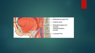
![ Each subdivision o f the pharynx has a different sensory innervation:
The nasopharynx is innervated by a pharyngeal branch of the maxillary nerve
[V2] that originates in the pterygopalatine fossa and passes through the
palatovaginal canal in the sphenoid bone to reach the roof of the pharynx.
The oropharynx is innervated by the glossopharyngeal nerve [IX] via the
pharyngeal plexus .
The laryngopharynx is innervated by the vagus nerve [X] via the internal branch
of the superior laryngeal artery.](https://image.slidesharecdn.com/pharynx-170528153500/85/Pharynx-64-320.jpg)


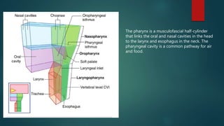
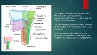
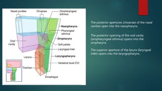
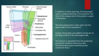
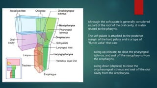
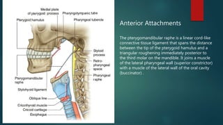
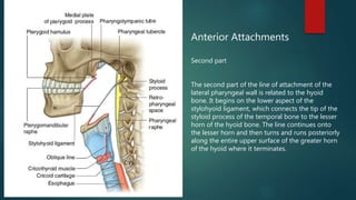
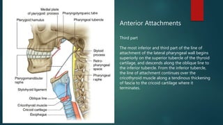
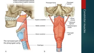
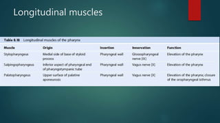
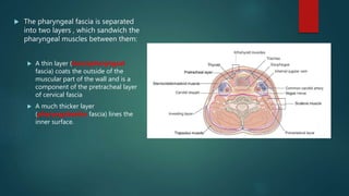
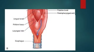
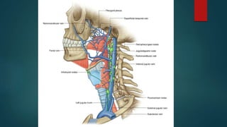
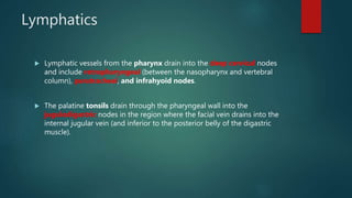
The pharynx is a musculofascial tube that connects the oral and nasal cavities to the larynx and esophagus. It is divided into three regions - the nasopharynx, oropharynx, and laryngopharynx. Various structures open into the pharynx, including the choanae, oral cavity, and larynx. The pharynx contains muscles that allow it to constrict during swallowing to push food into the esophagus. It is supplied by arteries, veins, lymphatics and nerves including the vagus and glossopharyngeal nerves.


