
More Related Content
What's hot
What's hot (20)
Similar to Agarose electrophoresis
Similar to Agarose electrophoresis (20)
Recently uploaded
Recently uploaded (20)
Agarose electrophoresis
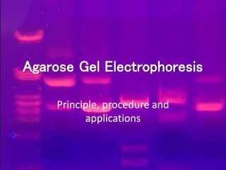
- 1. Agarose Gel Electrophoresis Principle, procedure and applications
- 2. Why electrophoresis? • To separate DNA fragments from each other • To determine the sizes of DNA fragments • To determine the presence or amount of DNA • To analyze restriction digestion products
- 3. Introduction Of Agarose Gel Electrophoresis • Agarose gel electrophorresis is a method to separate DNA or RNA molecules by size. • This is achieved by moving negatively charged nucleic acid molecules through an agarose matrix with an electric field (electrophoresis). • Shorter molecules move faster and migrate faster than longer ones .
- 4. Principle of electrophoresis • powerful separation method frequently used to analyze DNA fragments generated by restriction enzymes • convenient analytical method for determining the size of DNA molecules in the range of 500 to 30,000 base pairs. • employs electromotive force to move molecules through a porous gel
- 5. Principle (cont.) • separates molecules from each other on the basis of –size and/or –charge and/or –shape • basis of separation depends on how the sample and gel are prepared
- 7. *Gel Electrophoresis Materials: Hardware Casting tray Gel combs Power supply Gel tank Cover Electrical leads Gel Electrophoresis Materials: Hardware
- 8. Agarose • A linear carbohydrate polymer extracted from seaweed , agarobiose • forms a porous matrix as it gels – shifts from random coil in solution to structure in which chains are bundled into double helices
- 9. agarose •Basic unit of agar which is a cell wall and intercellular component of some red marine algae, usually Gelidium and Gracillaria. •Linear polysaccharide that contains double helices stabilized by water molecules. •Exterior hydroxyl groups allow helices to aggregate into suprafibers that branch off to form a matrix.
- 10. *AGAROSE GEL A highly purified uncharged polysaccharide derived from agar. Used to separate macromolecules such as nucleic acids, large proteins and protein complexes. It is prepared by dissolving 0.5% agarose in boiling water and allowing it to cool to 40°C. It is fragile because of the formation of weak hydrogen bonds and hydrophobic bonds.
- 11. TYPES OF AGAROSE • Standard Agarose - LE Gels at 35-38oC; Melts at 90-95oC Becomes opaque at high concentrations • Low Melting Agarose (NuSieve) Gels at 35oC; Melts at 65oC Often used to isolate DNA fragments from gel Intermediate forms or combinations of LE and NuSieve can provide sturdy, translucent gels at high agarose concentrations .
- 12. Concentrations of agarose used % Agarose (w/v) Size Range (kb pairs)for Optimal Separation • 0.5 2-30 • 0.75 0.7-20 • 1.0 0.5-10 • 1.5 0.2-3 • 2.0 0.1-2 • 3.0 (Nu-Sieve) 0.07-1.5 • 4.0 (N-S) 0.04-0.9 • 5.0 (N-S) 0.03-0.6 • 6.0 (N-S) 0.01-0.4
- 13. Gel Casting Trays • available in a variety of sizes and composed of UV-transparent plastic. • The open ends of the trays are closed with tape while the gel is being cast, then removed prior to electrophoresis.
- 14. Applied voltage • voltage, rate of migration • The higher the voltage, the more quickly the gel runs • But if voltage is too high, gel melts • The best separation will apply voltage at no more than 5V/cm of gel length.
- 15. Buffers • During electrophoresis water undergoes hydrolysis : H2O H + OH- • Buffers prevent the pH from changing by reacting with the H+ or OH- products • Most common buffer used is called TRIS – [tris(hydroxymethyl)aminomethane]
- 16. Buffers (cont.) • Another compound is added to make Tris an effective buffer — either boric or acetic acid • Another compound is added to bind metals EDTA • The buffer is either TBE or TAE TBE is made with Tris/Boric Acid/EDTA TAE is made with Tris/Acetic Acid/ EDTA
- 17. Staining of DNA • To make DNA fragments visible after electrophoresis, the DNA must be stained • The favorite—ethidium bromide • When bound to DNA it fluoresces under ultraviolet light (reddish –orange colour) • Convenient because it can be added directly to the gel • Sensitive—detects 0.1ug of DNA
- 18. *Dye DNA and place into gel The gel is made out of agarose, which is similar to jello. The gel is made with wells at one end so that the DNA can be loaded into the gel.
- 19. Ethidium bromide • The standard concentration used in staining DNA in gels is 0.5-1ug/mL • Ethidium bromide is a fluorescent dye that intercalates between bases of nucleic acids and allows very convenient detection of DNA fragments in gels. • Inserting itself between the base pairs in the double helix
- 20. Staining of DNA (cont.) • UV absorbance maxima at 300 and 360 nm and emission maxima at 590 nm. • Detection limit of bound DNA is 0.5-5 ng/band. • ethidium bromide is mutagenic so care must be taken while handling the dye. • Othe alternatives for ethidium bromide : Methylene blue Syber safe xylene cyanol bromphenol blue
- 21. A Comb • A comb is placed in the liquid agarose after it has been poured • Removing the comb from the hardened gel produces a series of wells used to load the DNA
- 22. DNA ladder • It is a solution of DNA molecules of different length • DNA Ladder consists of known DNA sizes used to determine the size of an unknown DNA sample. • The DNA ladder usually contains regularly spaced sized samples which when run on an agarose gel looks like a "ladder".
- 24. Method For Electrophoresis Add running buffer, load samples and marker Run gel at constant voltage until band separation occurs Pour into casting tray with comb and allow to solidify View DNA on UV light box and show results Prepare agarose gel Melt, cool and add Ethidium Bromide. Mix thoroughly.
- 25. • DNA is negatively charged. +- Power DNA • When placed in an electrical field, DNA will migrate toward the positive pole (anode). H O2 • An agarose gel is used to slow the movement of DNA and separate by size.
- 26. +- Power DNA How fast will the DNA migrate? strength of the electrical field, buffer, density of agarose gel… Size of the DNA! *Small DNA move faster than large DNA …gel electrophoresis separates DNA according to size small large
- 27. • Within an agarose gel, linear DNA migrate inversely proportional to the log10 of their molecular weight.
- 28. *Smaller pieces of DNA travel farther than Larger pieces of DNA End result!
- 29. applications • Solve criminal cases • Solve paternity cases • Diagnose genetic diseases • Determine genetic kinship among species
- 30. applications • Gel electrophoresis is commonly used in plant breeding and genomics for genotyping with molecular markers, • specific DNA fragments used as markers and isolated from individual plants are amplified by the polymerase chain reaction (PCR) and the resulting DNA fragments are subsequently loaded on a gel.
Editor's Notes
- https://s10.lite.msu.edu/res/msu/botonl/b_online/e26/26d.htm