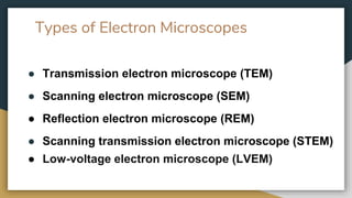
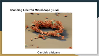
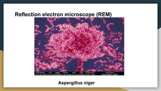
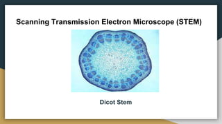
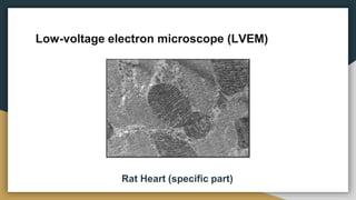
Electron Microscopy to
Study the Micro
Organisms
Electron Microscope
● The electron microscope is a type of microscope that uses
electrons to create an image of the target.
● It has much higher magnification or resolving power than a
normal light microscope.
● As the wavelength of an electron can be up to 100,000
times shorter than that of visible light photons.
Principle: An electron
microscope uses an 'electron
beam' to produce the image of the
object and magnification is obtained
by 'electromagnetic fields'; unlike
light or optical microscopes, in
which 'light waves' are used to
produce the image and
magnification is obtained by a
system of 'optical lenses'.
Types of Electron Microscopes
● Transmission electron microscope (TEM)
● Scanning electron microscope (SEM)
● Reflection electron microscope (REM)
● Scanning transmission electron microscope (STEM)
● Low-voltage electron microscope (LVEM)
Transmission electron microscope (TEM)
● Beam of electrons is transmitted through the specimen
which can be less than 100 nm ultrathin section or a
specimen grid. An image is formed due to interaction of
electrons with the specimen.
● The image is magnified on a imaging device like
fluorescent screen or a photographic film.
● Maximum resolution of ×5,00,000.
● To study Biological tissue specimens, Biological Sections,
organic polymers, Etc.
● The bacterium Escherichia coli (E. coli for short) is crucial
in modern biotechnology. Scientists use it to store DNA
sequences from other organisms, to produce proteins and to
test protein function.
Transmission electron microscope (TEM)
Escherichia coli
Scanning electron microscope (SEM)
● Works on the principle of raster scanning and helps in
studying the specimens.
● The image is formed due to the conversion of energy in
the electrons into heat, low-energy secondary electron,
High energy backscattered electrons and light emission or
X-Ray emission.
● Specimen surface, such as its topography and
composition can be obtained. The yeast Candida
albicans may cause infections of the mucosa in mouth.
● Maximum resolution of X30,000.
Scanning Electron Microscope (SEM)
Candida albicans
Reflection Electron Microscope (REM)
● REM is a combination of imaging, diffraction, and
spectroscopy techniques for characterization of topography,
crystal structure, and composition of surfaces of single crystals
● Scattered electrons are being used for the image formation.
● Aspergillus niger is a haploid filamentous fungus which is
used for waste management and biotransformations in
addition to its industrial uses, such as production of citric acid
and extracellular enzymes.
Reflection electron microscope (REM)
Aspergillus niger
Scanning Transmission Electron Microscope (STEM)
● It is known as the advanced version of SEM
● It focuses on a narrow spot and image of the specimen will
be created by raster scanning technique.
● It also picks up the electron that passes through the
specimen, technique of TEM and helps in creating the image
of a better resolution.
● Maximum resolution of X10,000,000
Scanning Transmission Electron Microscope (STEM)
Dicot Stem
Low-voltage electron microscope (LVEM)
● This electronic microscope works with low amount of energy
that is as low as 6–25 kV.
● This electronic microscope works on a similar technique as
that of TEM.
● It overcomes the higher resolution, possibility of X-ray
microanalysis of TEM.
● Magnification 1.0–3 nanometers
Low-voltage electron microscope (LVEM)
Rat Heart (specific part)
Applications of Electron Microscopy to
study Microorganisms
● Helps us to view the organisms that are not visible to naked
eyes nor to that of high microscope.
● Gives us the idea of the morphology of an organism and also
its surface texture.
● Helps us to study the individual cell structure and its
structure.
● Helps us to understand and differentiate with the types of
cells present in various organs.
Thank You

Electron microscopy to study the micro organisms

  • 1.
    Electron Microscopy to Studythe Micro Organisms
  • 2.
    Electron Microscope ● Theelectron microscope is a type of microscope that uses electrons to create an image of the target. ● It has much higher magnification or resolving power than a normal light microscope. ● As the wavelength of an electron can be up to 100,000 times shorter than that of visible light photons.
  • 3.
    Principle: An electron microscopeuses an 'electron beam' to produce the image of the object and magnification is obtained by 'electromagnetic fields'; unlike light or optical microscopes, in which 'light waves' are used to produce the image and magnification is obtained by a system of 'optical lenses'.
  • 4.
    Types of ElectronMicroscopes ● Transmission electron microscope (TEM) ● Scanning electron microscope (SEM) ● Reflection electron microscope (REM) ● Scanning transmission electron microscope (STEM) ● Low-voltage electron microscope (LVEM)
  • 5.
    Transmission electron microscope(TEM) ● Beam of electrons is transmitted through the specimen which can be less than 100 nm ultrathin section or a specimen grid. An image is formed due to interaction of electrons with the specimen. ● The image is magnified on a imaging device like fluorescent screen or a photographic film. ● Maximum resolution of ×5,00,000.
  • 6.
    ● To studyBiological tissue specimens, Biological Sections, organic polymers, Etc. ● The bacterium Escherichia coli (E. coli for short) is crucial in modern biotechnology. Scientists use it to store DNA sequences from other organisms, to produce proteins and to test protein function. Transmission electron microscope (TEM)
  • 7.
    Transmission electron microscope(TEM) Escherichia coli
  • 8.
    Scanning electron microscope(SEM) ● Works on the principle of raster scanning and helps in studying the specimens. ● The image is formed due to the conversion of energy in the electrons into heat, low-energy secondary electron, High energy backscattered electrons and light emission or X-Ray emission. ● Specimen surface, such as its topography and composition can be obtained. The yeast Candida albicans may cause infections of the mucosa in mouth. ● Maximum resolution of X30,000.
  • 9.
    Scanning Electron Microscope(SEM) Candida albicans
  • 10.
    Reflection Electron Microscope(REM) ● REM is a combination of imaging, diffraction, and spectroscopy techniques for characterization of topography, crystal structure, and composition of surfaces of single crystals ● Scattered electrons are being used for the image formation. ● Aspergillus niger is a haploid filamentous fungus which is used for waste management and biotransformations in addition to its industrial uses, such as production of citric acid and extracellular enzymes.
  • 11.
    Reflection electron microscope(REM) Aspergillus niger
  • 12.
    Scanning Transmission ElectronMicroscope (STEM) ● It is known as the advanced version of SEM ● It focuses on a narrow spot and image of the specimen will be created by raster scanning technique. ● It also picks up the electron that passes through the specimen, technique of TEM and helps in creating the image of a better resolution. ● Maximum resolution of X10,000,000
  • 13.
    Scanning Transmission ElectronMicroscope (STEM) Dicot Stem
  • 14.
    Low-voltage electron microscope(LVEM) ● This electronic microscope works with low amount of energy that is as low as 6–25 kV. ● This electronic microscope works on a similar technique as that of TEM. ● It overcomes the higher resolution, possibility of X-ray microanalysis of TEM. ● Magnification 1.0–3 nanometers
  • 15.
    Low-voltage electron microscope(LVEM) Rat Heart (specific part)
  • 16.
    Applications of ElectronMicroscopy to study Microorganisms ● Helps us to view the organisms that are not visible to naked eyes nor to that of high microscope. ● Gives us the idea of the morphology of an organism and also its surface texture. ● Helps us to study the individual cell structure and its structure. ● Helps us to understand and differentiate with the types of cells present in various organs.
  • 17.