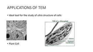
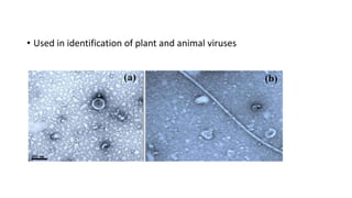
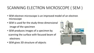
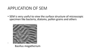
ELECTRON MICROSCOPY
Bhalerao Sainath
Sub. T.- Tiwari S.
WHAT ARE ELECTRON MICROSCOPES ?
• Electron microscope are scientific instruments that use highly
energetic electron to examine objects on a very fine scale as,
• Topography ( Surface features)
• Morphology ( Size and Shape )
• Crystallographic Information (arrangements of atoms)
INVENTION OF EMs
• To study objects of < 0.2 micro meter
• For analysis of sub cellular structure
• Study of cell organelles
• Cell metabolism
• An Ems can magnify structure from 100-250000 Times than light
microscope.
TYPES OF ELECTION MICROSCOPE
• There are two types of Ems,
• Transmission Electron Microscope (TEM) – Allows one to study inner structure
of objects
• Scanning Electron Microscope (SEM) – Used for visualise the surface of
objects
TRANSMISSION ELECTRON MICROSCOPE
• TEM was discovered by Max Knoll and Ernst Ruska in 1931.
• TEM is a Microscopy technique where a beam of electrons is transmitted
through an ultra thin specimen.
• An image is formed from the interaction of the electrons transmitted through
vthe specimen
• TEM provides morphological and crystalline info. The images allows
researchers to view specimen on a molecular level by making it possible to
analyse structure and texture.
APPLICATIONS OF TEM
• Ideal tool for the study of ultra structure of cells
• Plant Cell
• Used in identification of plant and animal viruses
ADVANTAGES OF TEM
• TEM offer the most powerful magnification, potentially over 1 million
times or more
• TEMs provides information on element and compound structure
• Images are high quality and detailed
• They are easy to operate with proper training
SCANNING ELECTRON MICROSCOPE ( SEM )
• SEM electron microscope is an improved model of an electron
microscope
• SEM is used for the study three dimensional
image of the specimen
• SEM produces images of a specimen by
scanning the surface with focused beam of
electrons
• SEM gives 3D structure of objects
APPLICATION OF SEM
• SEM is very useful to view the surface structure of microscopic
specimen like bacteria, diatoms, pollen grains and others
.
Basillus megatherium
• SEM is employed in the analysis of structural features of compound
eyes of insects
ADVANTAGES OF SEM
• Better resolution
• Work with low voltage
• Fast imaging
• Easy to operate

Electron Microscopy and its type Transmission electron microscopy

  • 1.
  • 2.
    WHAT ARE ELECTRONMICROSCOPES ? • Electron microscope are scientific instruments that use highly energetic electron to examine objects on a very fine scale as, • Topography ( Surface features) • Morphology ( Size and Shape ) • Crystallographic Information (arrangements of atoms)
  • 3.
    INVENTION OF EMs •To study objects of < 0.2 micro meter • For analysis of sub cellular structure • Study of cell organelles • Cell metabolism • An Ems can magnify structure from 100-250000 Times than light microscope.
  • 4.
    TYPES OF ELECTIONMICROSCOPE • There are two types of Ems, • Transmission Electron Microscope (TEM) – Allows one to study inner structure of objects • Scanning Electron Microscope (SEM) – Used for visualise the surface of objects
  • 5.
    TRANSMISSION ELECTRON MICROSCOPE •TEM was discovered by Max Knoll and Ernst Ruska in 1931. • TEM is a Microscopy technique where a beam of electrons is transmitted through an ultra thin specimen. • An image is formed from the interaction of the electrons transmitted through vthe specimen • TEM provides morphological and crystalline info. The images allows researchers to view specimen on a molecular level by making it possible to analyse structure and texture.
  • 6.
    APPLICATIONS OF TEM •Ideal tool for the study of ultra structure of cells • Plant Cell
  • 7.
    • Used inidentification of plant and animal viruses
  • 8.
    ADVANTAGES OF TEM •TEM offer the most powerful magnification, potentially over 1 million times or more • TEMs provides information on element and compound structure • Images are high quality and detailed • They are easy to operate with proper training
  • 9.
    SCANNING ELECTRON MICROSCOPE( SEM ) • SEM electron microscope is an improved model of an electron microscope • SEM is used for the study three dimensional image of the specimen • SEM produces images of a specimen by scanning the surface with focused beam of electrons • SEM gives 3D structure of objects
  • 10.
    APPLICATION OF SEM •SEM is very useful to view the surface structure of microscopic specimen like bacteria, diatoms, pollen grains and others . Basillus megatherium
  • 11.
    • SEM isemployed in the analysis of structural features of compound eyes of insects
  • 12.
    ADVANTAGES OF SEM •Better resolution • Work with low voltage • Fast imaging • Easy to operate