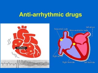
2. Introduction
o a - ‘without’; rhuthmos – ‘rhythm’ i.e. it means
‘without rhythm’
o Also known as ‘Cardiac dysrhythmia’
o Cardiac arrhythmias are a group of conditions in
which the heart beats with an irregular or
abnormal rhythm.
o Arrhythmias/ dysrhythmia: abnormality in the site
of origin of impulse, rate, or conduction.
3.
4. Physiology of cardiac rate
and rhythm
• Cardiac myocytes are electrically excitable
• Resting intracellular voltage of myocardial
cells is negative -90mV (SA node is -40mV)
• Resting state - K+ inside and Na+ outside cell
(Na+/K+ pump)
• Action potential occurs when Na+ enters the
cell and sets up a depolarising current
• Stimulation of a single muscle fibre causes
electrical activity to spread across the
myocardium
5. Phases of action potential of
cardiac cells
• Phase 0 rapid depolarisation
(inflow of Na+)
• Phase 1 partial repolarisation
(inward Na+ current deactivated,
outflow of K+)
• Phase 2 plateau (slow inward
calcium current)
• Phase 3 repolarisation (calcium
current inactivates, K+ outflow)
• Phase 4 pacemaker potential
(Slow Na+ inflow, slowing of K+
outflow) ‘autorhythmicity’
• Refractory period (phases 1-3)
Phase 4
Phase 0
Phase 1
Phase 2
Phase 3
0 mV
-80mV
Phase 4
8. Cardiac Action Potential
• Divided into five phases (0,1,2,3,4)
– Phase 4 - resting phase (resting membrane potential)
• Phase cardiac cells remain in until stimulated
• Associated with diastole portion of heart cycle
• Addition of current into cardiac muscle (stimulation)
causes
– Phase 0 – opening of fast Na channels and rapid depolarization
• Drives Na+ into cell (inward current), changing membrane potential
• Transient outward current due to movement of Cl- and K+
– Phase 1 – initial rapid repolarization
• Closure of the fast Na+ channels
• Phase 0 and 1 together correspond to the R and S waves of the
ECG
9. Cardiac Action Potential (con’t)
• Phase 2 - plateau phase
– sustained by the balance between the inward movement of Ca+ and
outward movement of K +
– Has a long duration compared to other nerve and muscle tissue
– Normally blocks any premature stimulator signals (other muscle tissue
can accept additional stimulation and increase contractility in a
summation effect)
– Corresponds to ST segment of the ECG.
• Phase 3 – repolarization
– K+ channels remain open,
– Allows K+ to build up outside the cell, causing the cell to repolarize
– K + channels finally close when membrane potential reaches certain
level
– Corresponds to T wave on the ECG
11. Differences between nonpacemaker and
pacemaker cell action potentials
• PCs - Slow, continuous depolarization during rest
• Continuously moves potential towards threshold for a
new action potential (called a phase 4 depolarization)
12. Sinus rhythm
• Sinoatrial node is cardiac
pacemaker
• Normal sinus rhythm 60-100
beats/min
• Depolarisation triggers
depolarisation of atrial
myocardium
• Conducts more slowly
through AV node
• Conducts rapidly through His
bundles and Purkinje fibres
13.
14. Sinus rhythm
• Sinoatrial rate controlled by autonomic
nervous system
• Parasympathetic system predominates (M2
muscarinic receptors)
• Sympathetic system (ß1 receptors)
– Increased heart rate (positive chronotropic
effect)
– Increased automaticity
– Facilitation of conduction of AV node
15. ECG
• Recording of electrical activity of the heart
• Net sum of depolarisation and repolarisation
potentials of all myocardial cells
• P-QRS-T pattern
• P - atrial depolarisation
• QRS - ventricular depolarisation
• T - ventricular repolarisation
16. ECG (EKG) showing wave
segments
Contraction
of atria
Contraction
of ventricles
Repolarization
of ventricles
17.
18. Conventional locations for the chest electrodes are illustrated below. The arrow
indicates the direction of polarity from negative to positive.
Lead I: In lead I the positive electrode is below
the left clavicle and the negative below the
right.
Lead II: In lead II the positive electrode is
below the left pectoral muscle and the
negative below the right clavicle .
19. Lead III: Lead III is displayed by attaching
the positive electrode beneath
the left pectoral muscle and the negative
below the left clavicle. Although these
simulate or approximate the I, II, and III
leads of the standard EKG, they are not
identical
MCL1: Another popular monitoring lead is
the modified precordial leads (MCL1 lead).
To connect this lead, the negative electrode
is placed near the left shoulder, usually
under the outer third of the left clavicle, and
the positive is placed to the right of the
sternum in the fourth intercostal space.
20. Definition of arrhythmia
• Cardiac arrhythmia is an abnormality of
the heart rhythm
• Bradycardia – heart rate slow (<60
beats/min)
• Tachycardia – heart rate fast (>100
beats/min)
21. Clinical classification of
arrhythmias
• Heart rate (increased/decreased)
• Heart rhythm (regular/irregular)
• Site of origin (supraventricular /
ventricular)
• Complexes on ECG (narrow/broad)
22.
23. Mechanisms of Cardiac Arrhythmias
• Result from disorders of impulse
formation, conduction, or both
• Causes of arrhythmias
– Cardiac ischemia
– Excessive discharge or sensitivity to
autonomic transmitters
– Exposure to toxic substances
– Genetic
– Unknown etiology
24.
25.
26.
27.
28. Mechanisms of arrhythmia
production
• Re-entry (refractory tissue reactivated due to
conduction block, causes abnormal
continuous circuit. eg accessory pathways
linking atria and ventricles in Wolff-Parkinson-
White syndrome)
• Abnormal pacemaker activity in non-
conducting/conducting tissue (eg ischaemia)
• Delayed after-depolarisation (automatic
depolarisation of cardiac cell triggers ectopic
beats, can be caused by drugs eg digoxin)
29. Disorders of impulse formation
• No signal from the pacemaker site
• Development of an ectopic pacemaker
– May arise from conduction cells (most are capable of
spontaneous activity)
– Usually under control of SA node à if it slows down too
much conduction cells could become dominant
– Often a result of other injury (ischemia, hypoxia)
• Development of oscillatory afterdepolariztions
– Can initiate spontaneous activity in nonpacemaker tissue
– May be result of drugs (digitalis, norepinephrine) used to
treat other cardiopathologies
30.
31.
32. Disorders of impulse conduction
• May result in
– Bradycardia (if have AV block)
– Tachycardia (if reentrant circuit occurs)
Reentrant
circuit
34. Atrial flutter is related to atrial fibrillation, but the atrial
frequency - counted from the P-waves - is much lower -
usually around 300 bpm and the AV-conduction is more
regular. AV-blocks is 2:1, but the ratio can also be 3:1, 4:1 etc.
Atrial flutter is recognized in the ECG as sawtooth-like P-
waves
Atrial Flutter
35. Atrial fibrillation:
Sinus node no longer controls the rhythm An excitation wave
with 400-600 cycles per min, courses continuously through the
atrial wall over a circular pathway about the origin of the great
veins (the circus motion theory). There is a continuous
activation with more than 400 P-waves per min, where regular
atrial contraction is impossible. Untreated atrial fibrillation has a
QRS-frequency of 150-180 bpm
36. Tachycardia occurs in paroxysms and
is either of atrial or ventricular origin.
1) Atrial tachycardia is elicited in the
atrial tissue outside the SN as an atrial
frequency around 200 bpm. Often only
every second impulse passes the AV-
node to the ventricles, so a 2:1 AV-
block is found in the ECG.
2) Ventricular tachycardia is elicited
from one focus in the ventricular tissue
with a frequency around 200 bpm
(more than 120 bpm) and abnormal
intraventricular impulse conduction
(disturbed QRS complexes). There are
no P-waves in the ECG, and the QRS-
complexes are broad and irregular.
Atrial & Ventricular Tachycardia
37. Paroxysmal Supraventricular Tachycardia (PSVT)
PSVT is a sudden onset of atrial tachycardia (150-200/min)
mostly due to circus movement type of re-entry occurring within
or around the AV node.
38. Atrial ectopic beats appear as early
(premature extrasystoles) and
abnormal P-waves in the ECG; they
are usually followed by normal QRS-
complexes
Ventricular ectopic beats
(extrasystoles) are recognized in the
ECG by their wide QRS-complex
(above 0.12 s), since they originate in
the ventricular tissue and slowly
spread throughout the two ventricles
without passing the Purkinje system.
The ventricular ectopic beat is
recognized by a double R-wave
Ectopic Beats
39. The first-degree AV block is a
prolongation of the PQ (PR)-interval
(above 0.2 s) implying a delay of the
conduction - not a real block.
The second-degree AV block
occurs when some signals are not
conducted to the AV-node, so some
of the P-waves are not followed by
QRS-complexes.
The third degree AV block
(complete AV-block) is a total block
of the conduction between the SN
and the ventricles. Also blocked His
bundle conduction results in an AV-
block.
AV Block
Atrioventricular block is blockage of the conduction from the atria to the AV-node.
40. This ECG is a classic example of torsades de pointes,
Torsades is a form of ventricular tachycardia that can most often be due to
medications. The QRS complexes during this rhythm tend to show a series
of "points up" followed by "points down" often with a narrow waist between.
Torsades de pointes
41. Antiarrhythmic drugs
• Biggest problem – antiarrhythmics can
cause arrhythmia!
– Example: Treatment of a non-life
threatening tachycardia may cause fatal
ventricular arrhythmia
– Must be vigilant in determining dosing,
blood levels, and in follow-up when
prescribing antiarrhythmics
42. Management of arrhythmias
• Acute management (clinical
assessment of patient and diagnosis)
• Prophylaxis
• Non-pharmacological
• Pharmacological (some antiarrhythmics
are also proarrhythmic)
47. Vaughan Williams classification
of antiarrhythmic drugs
• Class I: block sodium channels
– Ia (quinidine, procainamide,
disopyramide) AP
– Ib (lignocaine) ¯AP
– Ic (flecainide) «AP
• Class II: ß-adrenoceptor
antagonists (atenolol, sotalol)
• Class III: prolong action
potential and prolong refractory
period (suppress re-entrant
rhythms) (amiodarone, sotalol)
• Class IV: Calcium channel
antagonists. Impair impulse
propagation in nodal and
damaged areas (verapamil)
Phase 4
Phase 0
Phase 1
Phase 2
Phase 3
0 mV
-80mV
II
I
III
IV
48. Class I
• Class I – blocker’s of fast Na+ channels
Subclass IA
• Cause moderate Phase 0 depression
• Prolong repolarization
• Increased duration of action potential
• Includes
– Quinidine – 1st antiarrhythmic used, treat both atrial
and ventricular arrhythmias, increases refractory
period
– Procainamide - increases refractory period but side
effects
– Disopyramide – extended duration of action, used
only for treating ventricular arrthymias
49. Subclass IB
• Weak Phase 0 depression
• Shortened depolarization
• Decreased action potential duration
• Includes
– Lidocane/Lignocaine (also acts as local anesthetic) –
blocks Na+ channels mostly in ventricular cells, also
good for digitalis-associated arrhythmias
– Mexiletine - oral lidocaine derivative, similar activity
– Phenytoin – anticonvulsant that also works as
antiarrhythmic similar to lidocane
50. Lidocaine
• Class Ib (blocks Na+ channels, reduces AP
duration)
• Ventricular arrhythmias (acute treatment)
• IV infusion only (2 hour half life, high first
pass metabolism)
• Hepatic metabolism (inhibited by cimetidine,
propranolol)
• SE mainly CNS - drowsiness, disorientation,
convulsions, hypotension
51. Subclass IC
– Subclass IC
• Strong Phase 0 depression
• No effect of depolarization
• No effect on action potential duration
• Includes
– Flecainide (initially developed as a local anesthetic)
» Slows conduction in all parts of heart,
» Also inhibits abnormal automaticity
– Propafenone
» Also slows conduction
» Weak β – blocker
» Also some Ca2+ channel blockade
52. Flecainide
• Class Ic (block Na+ channels, no change to
AP)
• Slows conduction in all cardiac cells
• Acute treatment /prophylaxis
• Supraventricular tachycardias
• Paroxysmal atrial fibrillation
• Ventricular tachycardias
• Oral/IV
• Long acting (T1/2 14 hours)
• Hepatic metabolism, urinary elimination
54. Class II
• Class II – β–adrenergic blockers
– Based on two major actions
1) blockade of myocardial β–adrenergic receptors
2) Direct membrane-stabilizing effects related to Na+ channel blockade
– Includes
• Propranolol
– causes both myocardial β–adrenergic blockade and membrane-
stabilizing effects
– Slows SA node and ectopic pacemaking
– Can block arrhythmias induced by exercise or apprehension
– Other β–adrenergic blockers have similar therapeutic effect
• Metoprolol
• Nadolol
• Atenolol
• Acebutolol
• Pindolol
• Sotalol
• Timolol
• Esmolol
55. Class III
• Class III – K+ channel blockers
– Developed because some patients negatively
sensitive to Na channel blockers (they died!)
– Cause delay in repolarization and prolonged
refractory period
– Includes
• Amiodarone – prolongs action potential by delaying K+ efflux
but many other effects characteristic of other classes
• Ibutilide – slows inward movement of Na+ in addition to
delaying K + influx.
• Bretylium – first developed to treat hypertension but found to
also suppress ventricular fibrillation associated with
myocardial infarction
• Dofetilide - prolongs action potential by delaying K+ efflux
with no other effects
56. Amiodarone
• Class III - increases refractory period and AP
• Major effect acutely is depression of AV node
• Acute treatment/prophylaxis
• Atrial and ventricular arrhythmias
• Oral or IV (central line)
• Loading and maintenance doses
• T1/2 54 days
• Large volume of distribution
• Accumulates
• Hepatic metabolism- biliary and intestinal
excretion
58. Class IV
• Class IV – Ca2+ channel blockers
– slow rate of AV-conduction in patients
with atrial fibrillation
– Includes
• Verapamil – blocks Na+ channels in addition to
Ca2+; also slows SA node in tachycardia
• Diltiazem
59. Verapamil
• Class IV (calcium channel blocker)
• Prolongs conduction and refractoriness in AV
node, slows rate of conduction of SA node
• Acute treatment/prophylaxis
• Used IV/oral
• SUPRAVENTRICULAR NOT VENTRICULAR
ARRHYTHMIAS
• Do not use IV verapamil with ß- blocker (heart
block)
• T1/2 6-8 hours
61. Adenosine
• Not in Vaughan Williams class
• Purine nucleotide (activates adenosine
receptors)
• Slows AV nodal conduction
• Acute treatment
• Termination of SVT/ diagnosis of VT
• Given IV only (rapid bolus)
• T1/2 < 2 seconds
63. Digoxin
• Not in Vaughan Williams class
• Cardiac glycoside (digitalis, foxglove)
• Act on Na/K-ATPase of cell membrane
(inhibits Na+/K+ pump, increases intracellular
Na+ and calcium)/ increases vagal activity
• Increase cardiac contraction and slows AV
conduction by increasing AV node refractory
period
64. Digoxin
• Atrial fibrillation or flutter (controls ventricular
rate)
• Acute treatment/prophylaxis
• Oral/IV
• Loading and maintenance doses
• T1/2 36 hours
• Excreted by kidneys
• Narrow therapeutic index
• Therapeutic drug monitoring
• Reduce dose in elderly/renal impairment
66. Pacemakers
• Surgical implantation of electrical leads attached to a
pulse generator
• Over 175,000 implanted per year
1) Leads are inserted via subclavicle vein and advanced to the
chambers on the vena cava (right) side of the heart
2) Two leads used, one for right atrium, other for right ventricle
3) Pulse generator containing microcircuitry and battery are
attached to leads and placed into a “pocket” under the skin
near the clavicle
4) Pulse generator sends signal down leads in programmed
sequence to contract atria, then ventricles
• Pulse generator can sense electrical activity generated
by the heart and only deliver electrical impulses when
needed.
• Pacemakers can only speed up a heart experiencing
bradycardia, they cannot alter a condition of
tachycardia