The document describes eicosanoid synthesis derived from 20-carbon fatty acids, primarily focusing on arachidonic acid as a major precursor. It details the different types of eicosanoids, such as prostaglandins and leukotrienes, their synthesis pathways, and the enzymatic processes involved in their formation. Additionally, the document discusses heme synthesis and respiratory burst mechanisms in immune cells, emphasizing the production of reactive oxygen species and their roles in defense mechanisms.
Introduction to eicosanoids, their sources, synthesis routes, and types including prostaglandins, leukotrienes, thromboxanes. Arachidonic acid as a major precursor.
Biosynthesis pathways of eicosanoids: cyclic and linear pathways through COX for prostaglandins and LOX for leukotrienes.
Details on lipid numbers, structures of prostaglandins, thromboxanes, leukotrienes, and lipoxins, and their synthesis mechanisms.
Structure and significance of porphyrins, with detailed synthesis processes of heme, involving various enzymes and reactions.
Mechanism of respiratory burst in phagocytes, including oxygen-dependent processes for pathogen elimination, involving reactive oxygen species.
Eicosanoids
• The term"eicosanoids" is used as a collective name for
molecules derived from 20-carbon fatty acids
• Fatty acids of the n-6 family deriving from linoleic acid
are the main source of eicosanoids
• Arachidonic acid (20:4n-6) being the major precursor
• Eicosanoids are synthesized in vivo through several
routes
• some compounds being formed by more than one
mechanism
• In the plant kingdom, several potent derivatives from
linolenic acid (octadecanoid-derived compounds) are
found and have hormone-like functions
(phytohormones)
3.
Lipid number
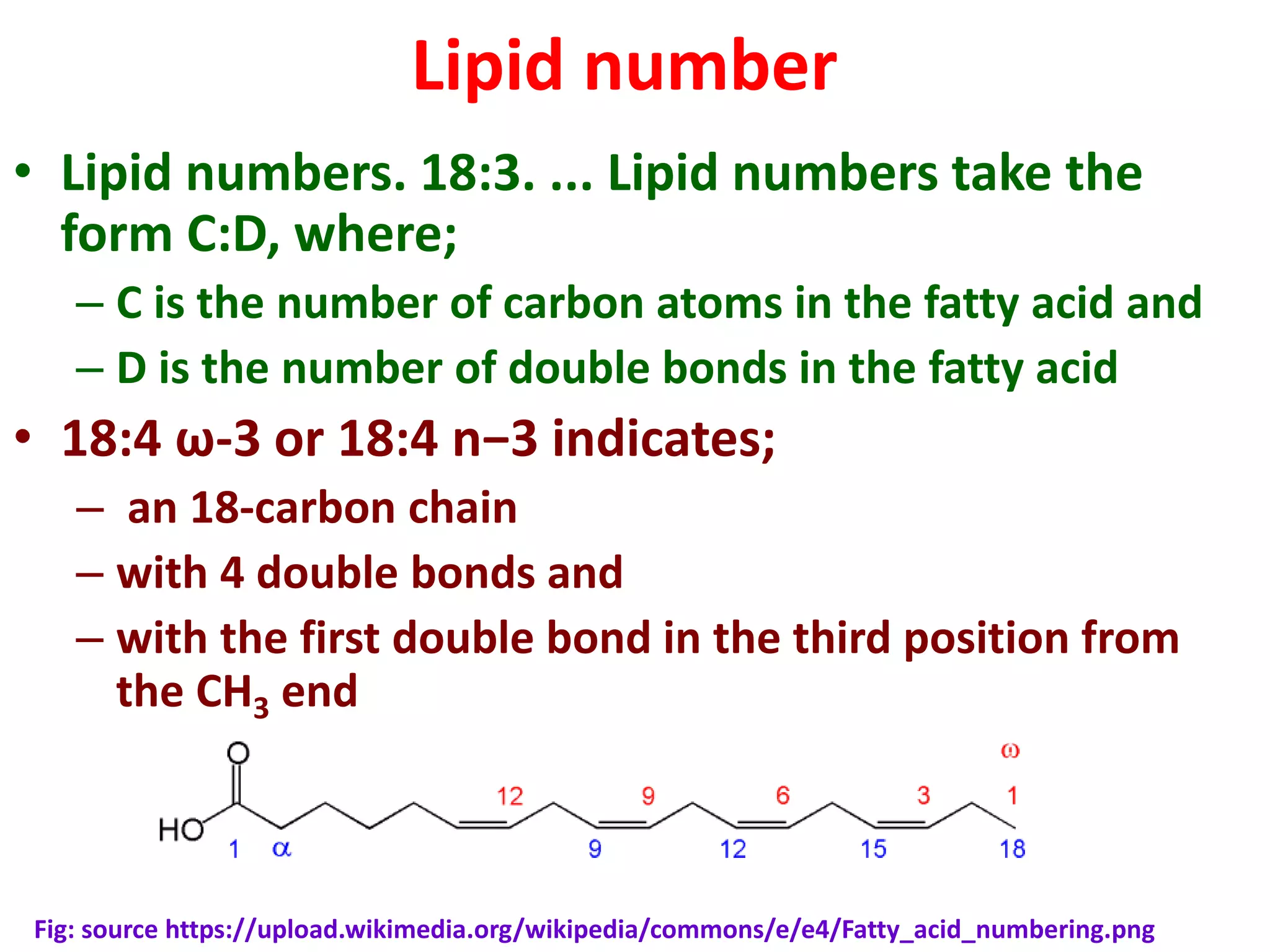
• Lipidnumbers. 18:3. ... Lipid numbers take the
form C:D, where;
– C is the number of carbon atoms in the fatty acid and
– D is the number of double bonds in the fatty acid
• 18:4 ω-3 or 18:4 n−3 indicates;
– an 18-carbon chain
– with 4 double bonds and
– with the first double bond in the third position from
the CH3 end
Fig: source https://upload.wikimedia.org/wikipedia/commons/e/e4/Fatty_acid_numbering.png
4.
Eicosanoid synthesis
• Eicosanoidsconsists of prostaglandins, thromboxanes, leukotrienes
and lipoxins
• Prostaglandins and thromboxanes are identified as prostanoids
• The number of C-C double bonds are used as subscripts with the
name of the prostanoids
• Majority of biologically active prostaglandins and thromboxanes are
referred as series 2 molecules – presence of 2 C-C double bonds
• Predominant Leukotrienes are series 4 molecules -4 C-C double
bonds
• Important series 1 prostaglandins and thromboxanes are also
present
• Prostaglandins are originally shown to be synthesised in the
prostate gland
• Thromboxanes from platelets (thrombocyte)
• Leucotrenes from leucocytes
• Lipoxins are synthesised through lipoxygenase interactions
5.
Names of Prostaglandins
•Prostaglandins are originally shown to be
synthesised in the prostate gland
• Thromboxanes from platelets and
• Leukotrienes from leukocytes
• Their names are derived from the place of synthesis
https://www.google.co.in/search?q=p
latelets&rlz=1C1VFKB_enIN599IN599
&source=lnms&tbm=isch&sa=X&ved=
0ahUKEwjS45uNr53QAhUIvo8KHVv1A
JUQ_AUICCgB&biw=1280&bih=621#i
mgrc=O0EbkI9EC2Sl8M%3A
Source of Fig
https://www.google.co.in/search?q=leuk
ocytes&rlz=1C1VFKB_enIN599IN599&sou
rce=lnms&tbm=isch&sa=X&ved=0ahUKE
wjP2a_Mr53QAhXFo48KHeeZAeYQ_AUIC
CgB&biw=1280&bih=621#imgrc=NswWA
woMbeBGIM%3A
6.
Eicosanoid synthesis
• Twomain pathways involved in the biosynthesis of
eicosanoids
– PG and TX are synthesised by the cyclic pathway
– The leukotrenes by the linear pathway
• Cyclic pathway initiated by Prostaglanding G/H
synthase
– This enzyme has 2 activities – COX and peroxidase
• Two forms of COX- COX1 an dCOX2
– COX-1 expressed constitutively – gastric mucosa, kidney,
platelets and vascular endothelial cells
– COX-2 is inducible-in macrophages and monocytes
express in response to inflammation
• COX-1 and COX-2 catalyse – arachidonic acid to
PGG2 and PGH2
7.
Cyclic pathway
• Numerousstimuli- epinephrine, thrombin and
bradykinin activates phospholipase A2-(PLA2)
• PLA2 hydrolyses arachidonic acid from cell
membrane phospholipids
• Bradykinin receptor coupled with G-protein
activation
• Increased intracellular calcium ions
• Protein kinase C activated
• PKC phosphorylation and Ca2+ ions activate ER
membrnane- associated cPLA2 isoform
8.
Synthesis of prostaglandins
•Hydrolysis of arachidonic acid form
phosphatidylinositol bisphosphate (PIP2)
• Arachidonic acid is converted to PGH2 –action
of COX-1 and COX-2
• Prostaglandin is synthesised from PGH2
• Action of different PGE enzymes catalyse the
synthesis of different PGs
• Thromboxanes are synthesised from PGH2 by
thromboxane synthase
10.
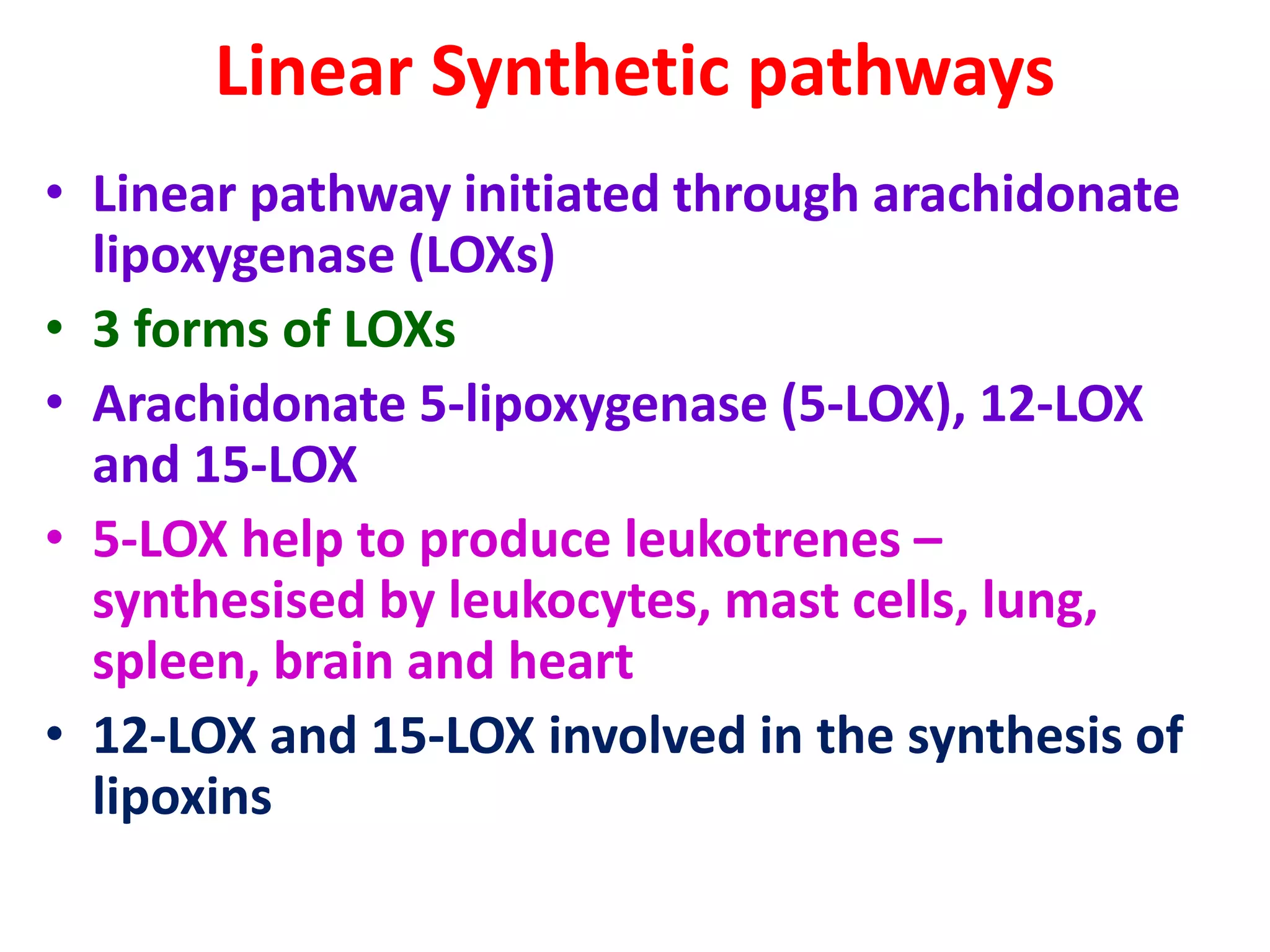
Linear Synthetic pathways
•Linear pathway initiated through arachidonate
lipoxygenase (LOXs)
• 3 forms of LOXs
• Arachidonate 5-lipoxygenase (5-LOX), 12-LOX
and 15-LOX
• 5-LOX help to produce leukotrenes –
synthesised by leukocytes, mast cells, lung,
spleen, brain and heart
• 12-LOX and 15-LOX involved in the synthesis of
lipoxins
11.
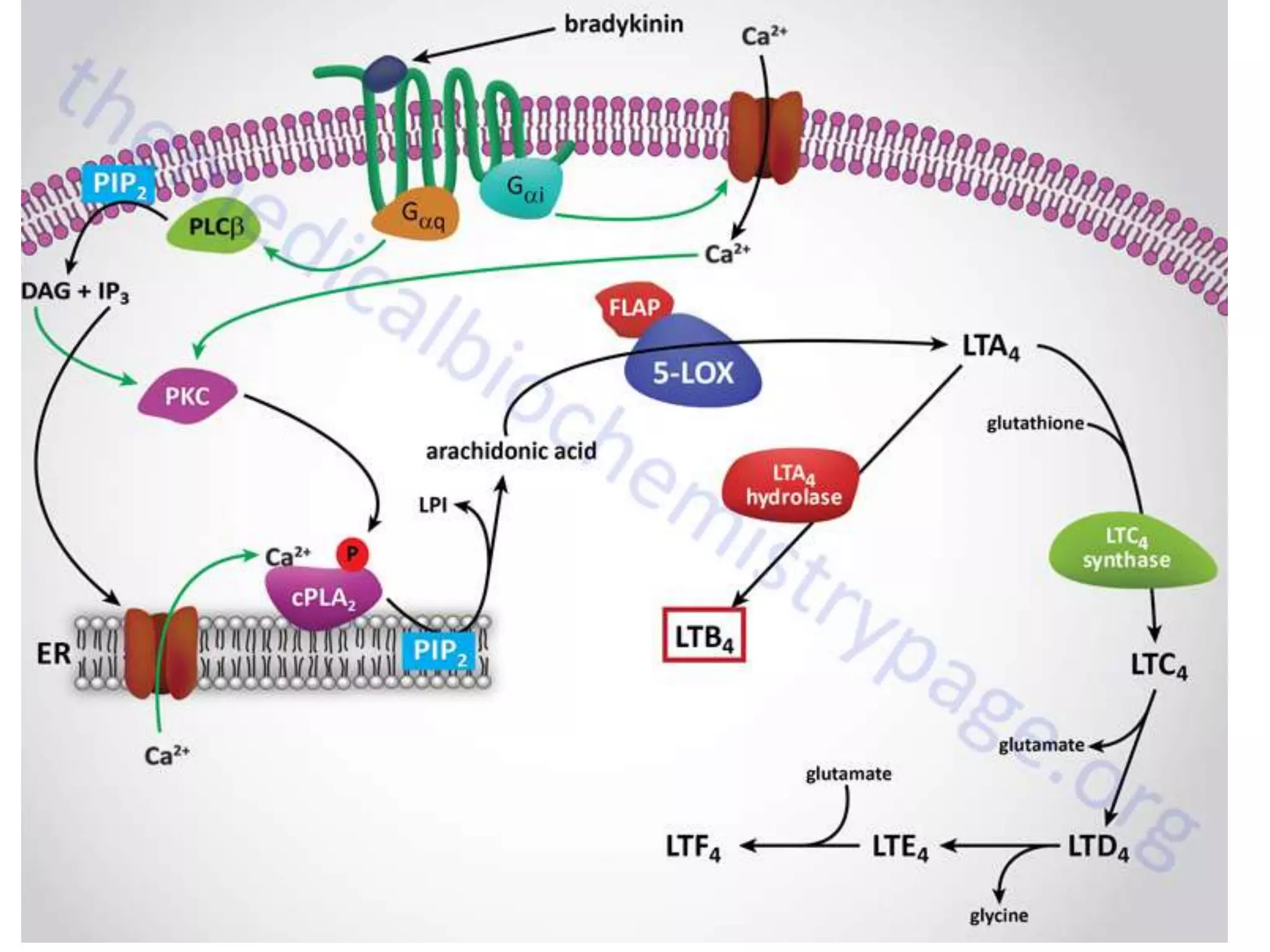
Synthesis of Leukotrienes
•Numerous stimuli- epinephrine, thrombin,
bradykinin activates phospholipase A2-(PLA2)
• PLA2 hydrolyses arachidonic acid from cell
membrane phospholipids
• Bradykinin receptor coupled with G-protein
activation
• Increased intracellular calcium ions
• Protein kinase C activated
• PKC phosphorylation and Ca2+ ions activate ER
membrnane- associated cPLA2 isoform
12.
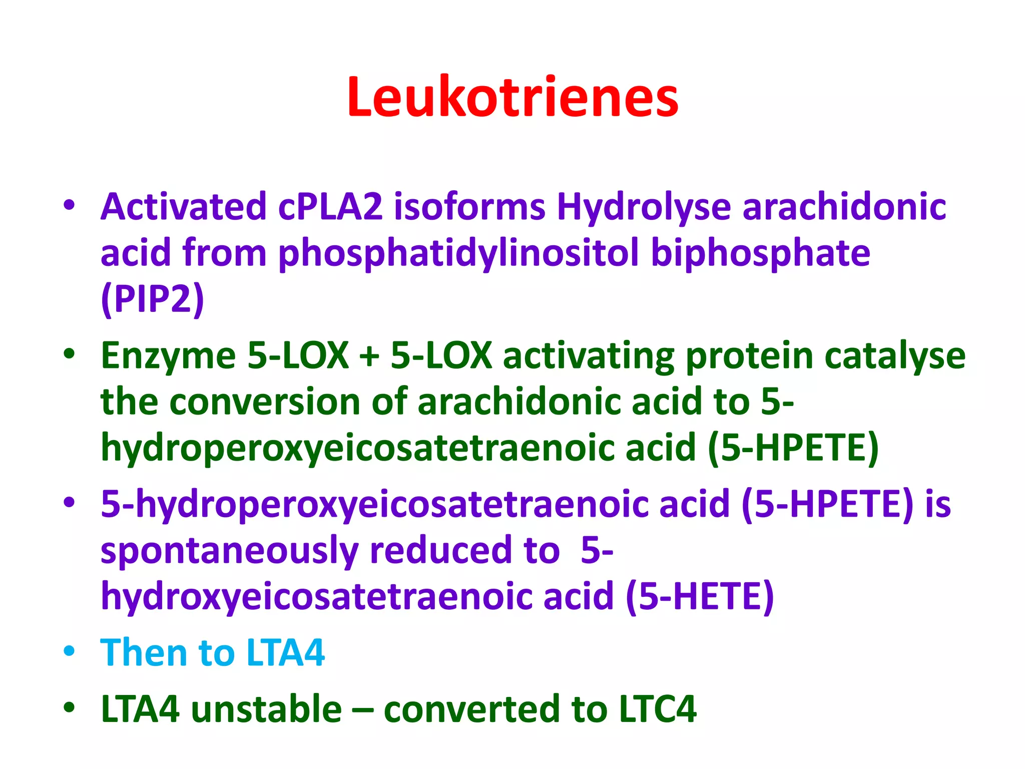
Leukotrienes
• Activated cPLA2isoforms Hydrolyse arachidonic
acid from phosphatidylinositol biphosphate
(PIP2)
• Enzyme 5-LOX + 5-LOX activating protein catalyse
the conversion of arachidonic acid to 5-
hydroperoxyeicosatetraenoic acid (5-HPETE)
• 5-hydroperoxyeicosatetraenoic acid (5-HPETE) is
spontaneously reduced to 5-
hydroxyeicosatetraenoic acid (5-HETE)
• Then to LTA4
• LTA4 unstable – converted to LTC4
14.
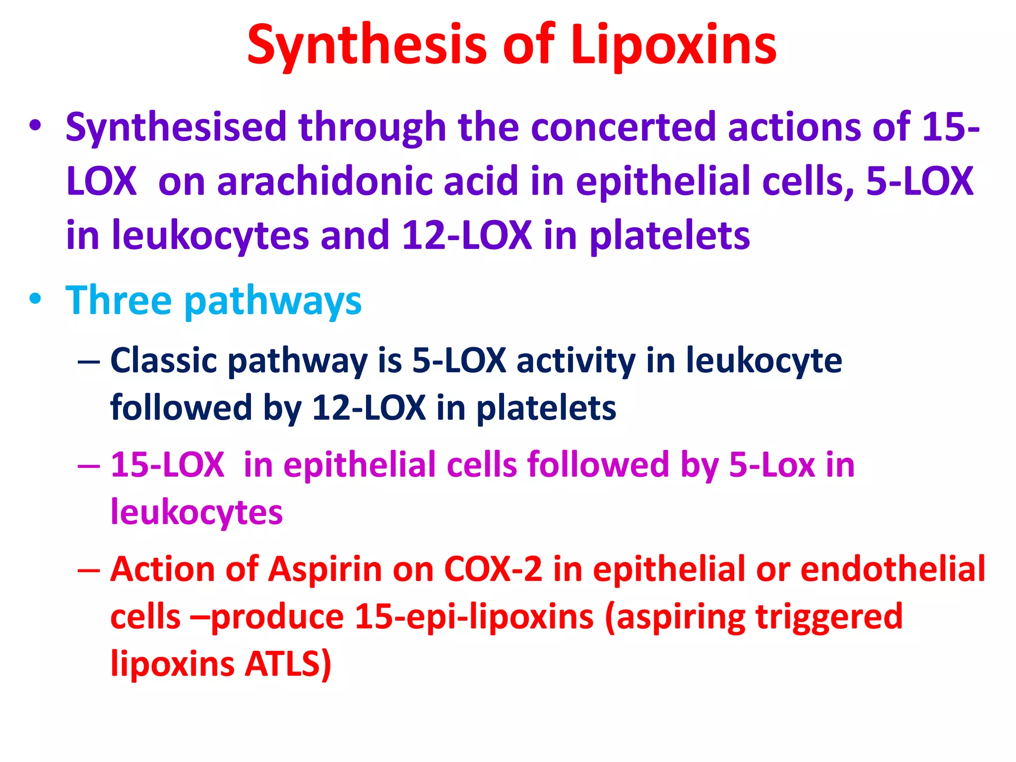
Synthesis of Lipoxins
•Synthesised through the concerted actions of 15-
LOX on arachidonic acid in epithelial cells, 5-LOX
in leukocytes and 12-LOX in platelets
• Three pathways
– Classic pathway is 5-LOX activity in leukocyte
followed by 12-LOX in platelets
– 15-LOX in epithelial cells followed by 5-Lox in
leukocytes
– Action of Aspirin on COX-2 in epithelial or endothelial
cells –produce 15-epi-lipoxins (aspiring triggered
lipoxins ATLS)
16.
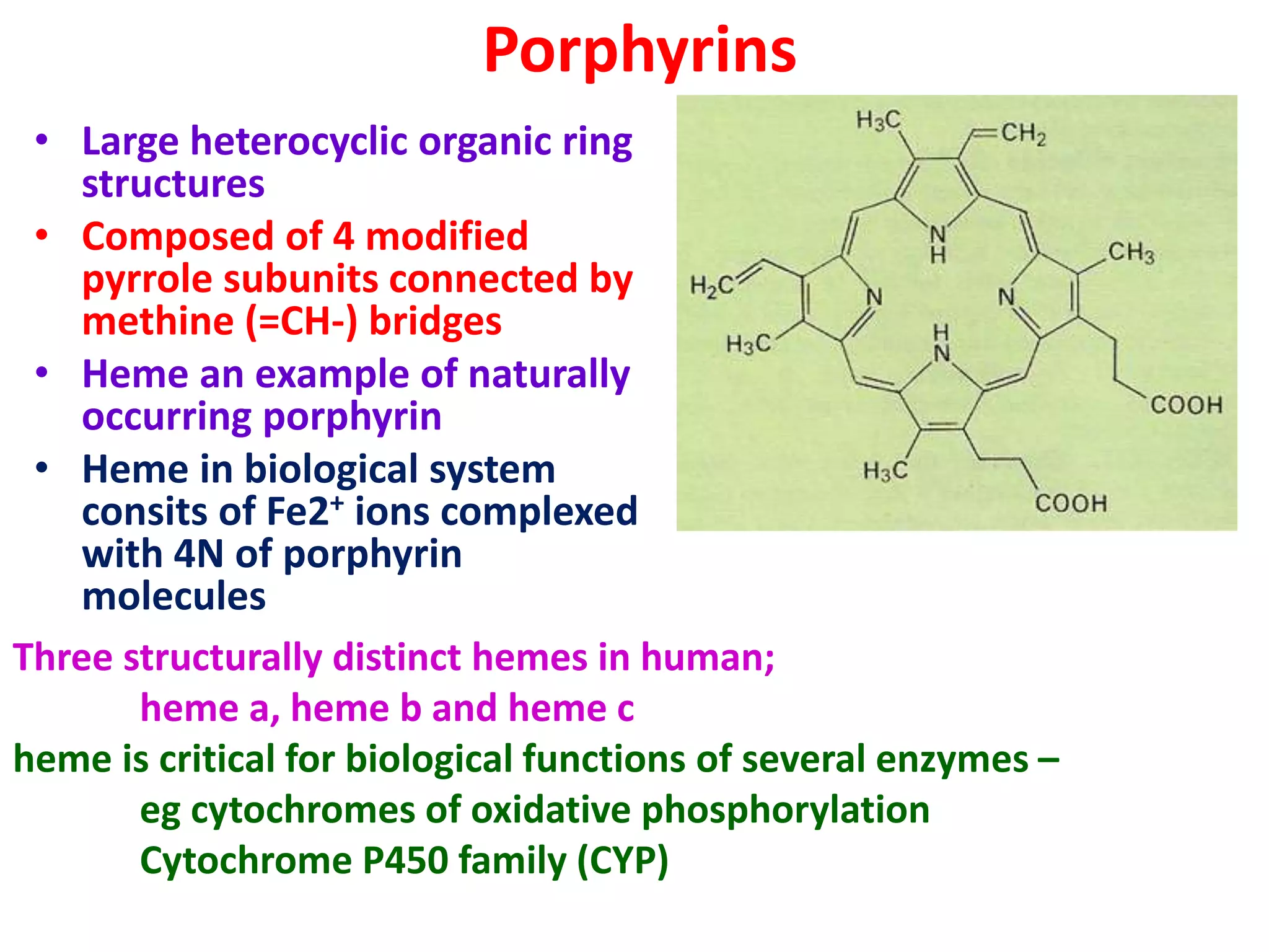
Porphyrins
• Large heterocyclicorganic ring
structures
• Composed of 4 modified
pyrrole subunits connected by
methine (=CH-) bridges
• Heme an example of naturally
occurring porphyrin
• Heme in biological system
consits of Fe2+ ions complexed
with 4N of porphyrin
molecules
Three structurally distinct hemes in human;
heme a, heme b and heme c
heme is critical for biological functions of several enzymes –
eg cytochromes of oxidative phosphorylation
Cytochrome P450 family (CYP)
17.
Heme synthesis
• Firstreaction in mitochondria
• Condensation of one succinyl-coA by pyridoxal phosphate
– requiring enzyme (vitamin B6) – δaminolevulinic acid synthase
(ALAS)
– forming δ aminolevulinic acid (5 aminoleuvinic acid)
• This is the rate limiting reaction in heme synthesis
• ALA is transported to cytosol
• ALA dehydratase (porphobilinogen synthetase) dimerises
2molcules of ALA
• Forms Porphobilinogen
• Head-to-tail condensation of 4 molecules
of porphobilinogen -form linear
tetrapyrrole intermediate –
hydroxymethylbilane
18.
Heme synthesis
• Enzymefor Head-to-tail condensation of 4 molecules of
porphobilinogen is porphobilinogen deaminase (PBG
deaminase)
• Hydroxymethylbilane has two fates
• One due to enzymatic action
• Other due to non-enzymatic action
• Hydroxymethylbilane is enzymatically converted to
uroporphyrinogen III
• Mediated by the enzyme uroporphyrinogen III synthase
• uroporphyrinogen III is decarboxylated by uroporphyrinogen
decarboxylase
• Forms coproporphyrinogens
• Coproporphyrinogen III is most important in heme synthesis
• Coproporphyrinogen III transported to the interior of the
mitochondria
19.
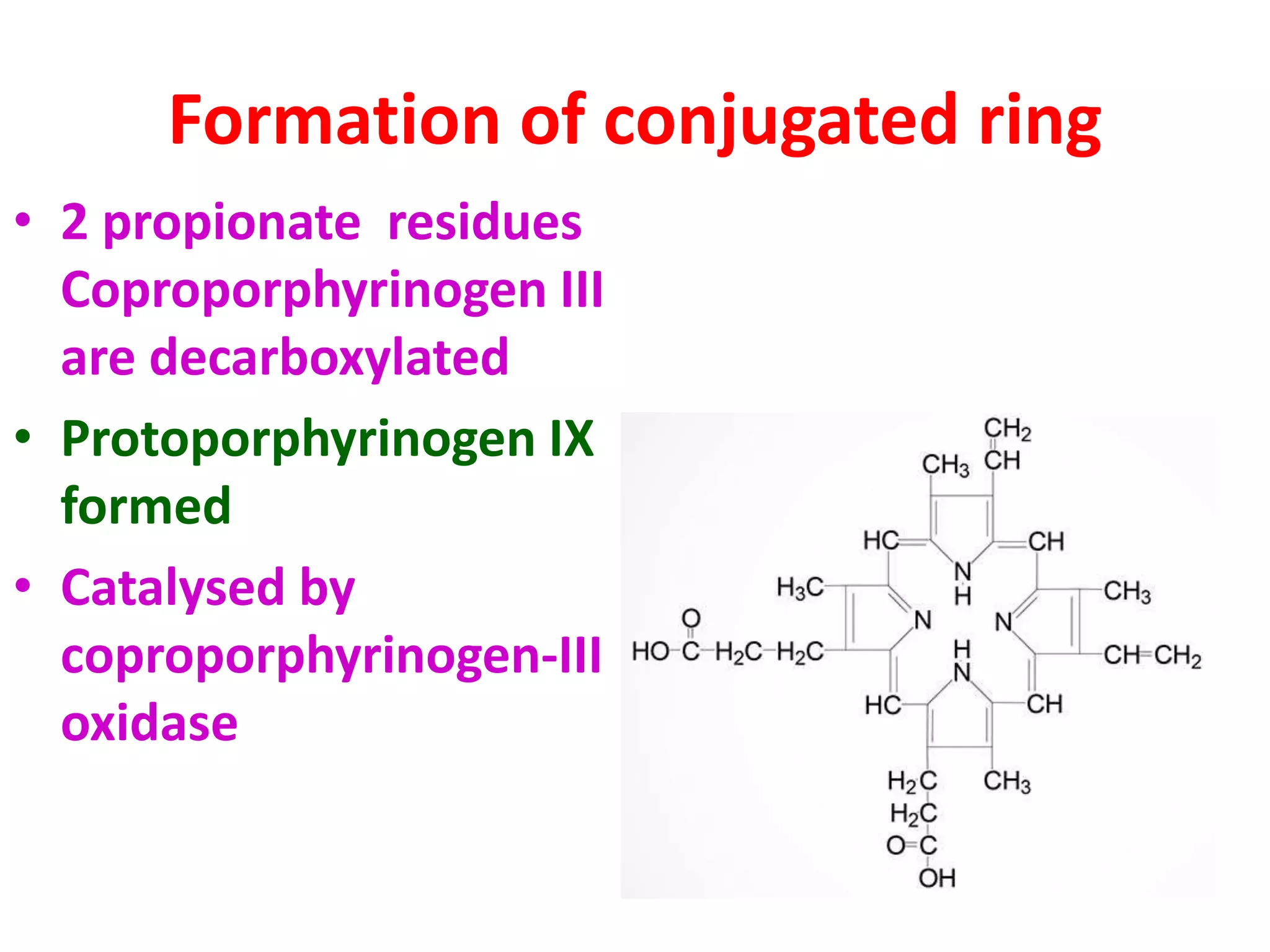
Formation of conjugatedring
• 2 propionate residues
Coproporphyrinogen III
are decarboxylated
• Protoporphyrinogen IX
formed
• Catalysed by
coproporphyrinogen-III
oxidase
20.
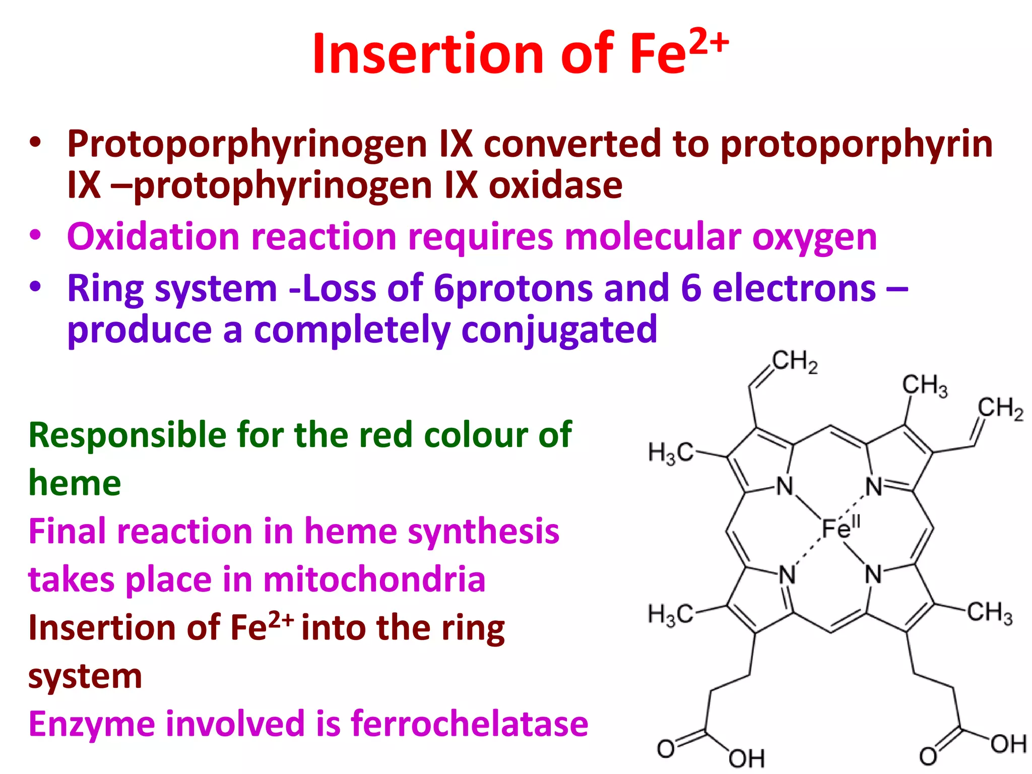
Insertion of Fe2+
•Protoporphyrinogen IX converted to protoporphyrin
IX –protophyrinogen IX oxidase
• Oxidation reaction requires molecular oxygen
• Ring system -Loss of 6protons and 6 electrons –
produce a completely conjugated
Responsible for the red colour of
heme
Final reaction in heme synthesis
takes place in mitochondria
Insertion of Fe2+ into the ring
system
Enzyme involved is ferrochelatase
21.
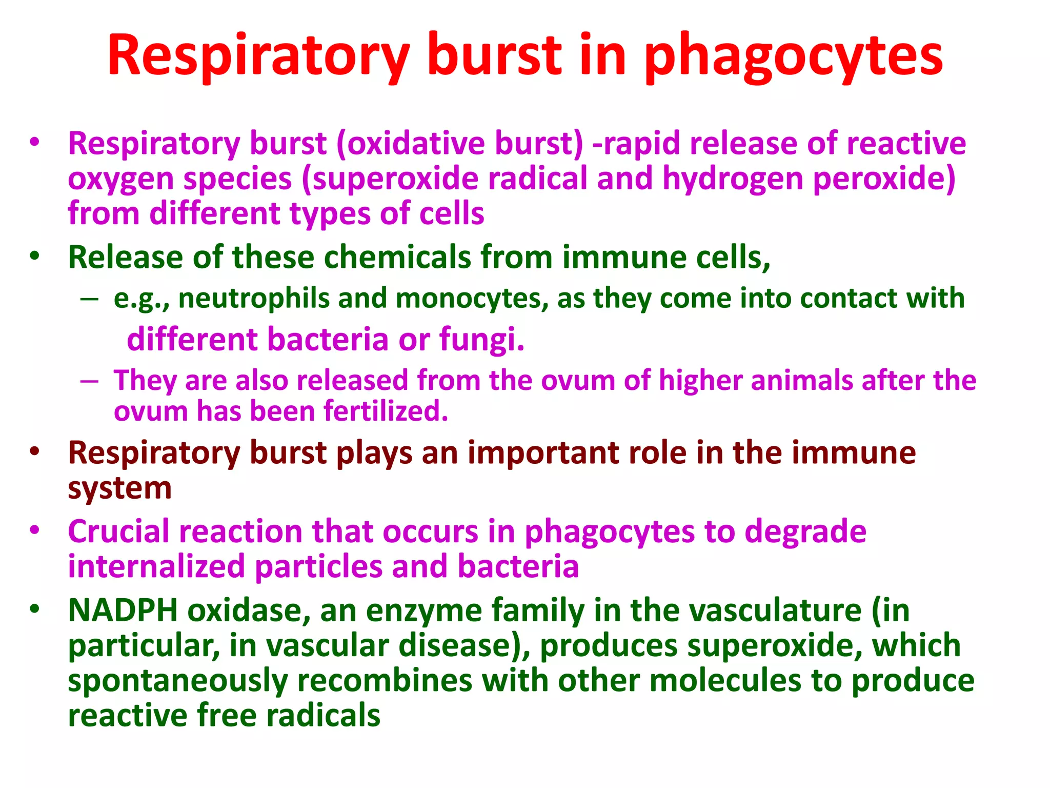
Respiratory burst inphagocytes
• Respiratory burst (oxidative burst) -rapid release of reactive
oxygen species (superoxide radical and hydrogen peroxide)
from different types of cells
• Release of these chemicals from immune cells,
– e.g., neutrophils and monocytes, as they come into contact with
different bacteria or fungi.
– They are also released from the ovum of higher animals after the
ovum has been fertilized.
• Respiratory burst plays an important role in the immune
system
• Crucial reaction that occurs in phagocytes to degrade
internalized particles and bacteria
• NADPH oxidase, an enzyme family in the vasculature (in
particular, in vascular disease), produces superoxide, which
spontaneously recombines with other molecules to produce
reactive free radicals
22.
Phagocytic killing
• Oxygendependent killing
• Oxygen independent killing
• Oxygen dependent phagocytic killing
– Activated phagocytes produce a number of reactive
oxygen intermediates and
– Nitrogen intermediates
• When exposed to certain stimuli- phagocytes
(Neutrophils, oesinophils, and macrophages)
increase oxygen uptake- upto 50 fold
• Oxygen burst
23.
Superoxide radical
• Eliminationof invading micro-organisms by neutrophils,
monocytes, and macrophages
• depends heavily on the generation of reactive oxygen species
during the phagocytosis-associated respiratory burst
• NADPD oxidase also called respiratory burst oxidase
• Present in phagocyte membrane
• toxic oxidants are released to the inside and outside of the cell
• Catalyse reduction of O2
- by adding electron
• 2O2 + NADPH 2O2
- + NADP+ + H+
• 2O2
- + H+ H2O2
- + O2
• the oxidants superoxide anion (O2), hydrogen peroxide (H2O2),
hypochlorous acid, and hydroxyl radical, created in this process-
• carry the potential to damage the phagocytes themselves as
well as other cells at sites of inflammation
24.
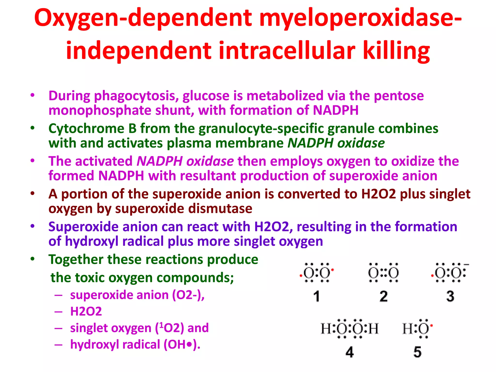
Oxygen-dependent myeloperoxidase-
independent intracellularkilling
• During phagocytosis, glucose is metabolized via the pentose
monophosphate shunt, with formation of NADPH
• Cytochrome B from the granulocyte-specific granule combines
with and activates plasma membrane NADPH oxidase
• The activated NADPH oxidase then employs oxygen to oxidize the
formed NADPH with resultant production of superoxide anion
• A portion of the superoxide anion is converted to H2O2 plus singlet
oxygen by superoxide dismutase
• Superoxide anion can react with H2O2, resulting in the formation
of hydroxyl radical plus more singlet oxygen
• Together these reactions produce
the toxic oxygen compounds;
– superoxide anion (O2-),
– H2O2
– singlet oxygen (1O2) and
– hydroxyl radical (OH•).
25.
Oxygen-dependent myeloperoxidase-
dependent intracellularkilling
• Fusion of azurophilic granules with the phagosome causes
release of myeloperoxidase into the phagolysosome
• azurophilic granules -A large, coarse, blue-
purple membrane-
bound organelle in progranulocytes, mylocytes
and neutrophils, which acts as a reservoir for digestive and
hydrolytic enzymes before delivery to a phagosome
• Myeloperoxidase utilizes H2O2 and halide ions (usually Cl-)
to produce highly toxic hypochlorite
• Some hypochlorite spontaneously breaks down to yield
singlet oxygen
• Together these reactions produce toxic hypochlorite (OCl-)
and singlet oxygen (1O2)
26.
Detoxification reactions
Neutrophils andmacrophages are able to protect
themselves by detoxifying the toxic oxygen
intermediates that they generate
Granulocyte self-protection is achieved in
reactions employing the dismutation of
superoxide anion to hydrogen peroxide
by superoxide dismutase and
The conversion of hydrogen peroxide to water
by catalase