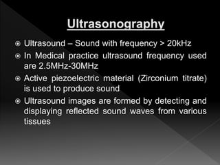
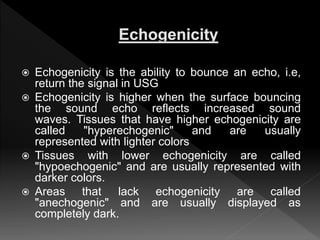
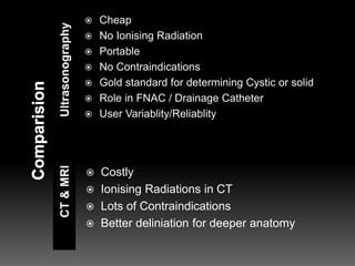
 Ultrasound – Sound with frequency > 20kHz
 In Medical practice ultrasound frequency used
are 2.5MHz-30MHz
 Active piezoelectric material (Zirconium titrate)
is used to produce sound
 Ultrasound images are formed by detecting and
displaying reflected sound waves from various
tissues
SONAR
Speed of Sound in water
1540m/sec
Distance can be Calculated
 Echogenicity is the ability to bounce an echo, i.e,
return the signal in USG
 Echogenicity is higher when the surface bouncing
the sound echo reflects increased sound
waves. Tissues that have higher echogenicity are
called "hyperechogenic" and are usually
represented with lighter colors
 Tissues with lower echogenicity are called
"hypoechogenic" and are usually represented with
darker colors.
 Areas that lack echogenicity are called
"anechogenic" and are usually displayed as
completely dark.
Transducer frequency
Typical: 3Mhz-12MHz
Resolution
High Frequency > Higher
Low Frequency > Poor
Depth of Penetration
High Frequency >
Shallow
Low Frequency > Deep
UltrasonographyCT&MRI
 Cheap
 No Ionising Radiation
 Portable
 No Contraindications
 Gold standard for determining Cystic or solid
 Role in FNAC / Drainage Catheter
 User Variablity/Reliablity
 Costly
 Ionising Radiations in CT
 Lots of Contraindications
 Better deliniation for deeper anatomy
o Doppler effect is the change in frequency or wavelength
of a wave for an observer moving relative to its source
o Sonography can be enhanced with Doppler
measurements to assess whether blood is moving towards or
away from the probe, and its relative velocity
o Lack of standards in the various laboratories for colour
coding
 Thyroid disorders
 Lymphadenopathy in Neck
 Parathyroid gland evaluation
 Salivary gland & Ducts evaluation
 Thyroglossal Cyst
 Branchial Cyst
 Ranula
 Hemangioma
 Lipoma
 Malformations
 Lymphangiomas
Cystic
Echo free Centre
Smooth Back wall
Acoustic enhancement
Solid
Many Echo reflection
Back wall indistinct
No enhancement
 Size assessment
 Evaluation of Thyroid Nodule
› Detection of focal mass
› Differentiation of Multinodular goiter/Hyperplasia
from other nodular diseases
› Extent of Thyroid Malignancy
› Follow up for residulal, reccurent or metastatic
carcinoma
› USG guided FNAC / FNA biopsy
 Deliniates Thyroid
Nodule but non
specific except for
papilary carcinoma
where some usg
features are
characteristic
 Extracapsular and
Vascular invasion
not detected
USG helps to define
thyroid enlargement
 Colour doppler
shows “Thyroid Storm”
Diagnostic of Graves
disease
 Most commonly used for evaluation of
enlarged Lymph node.
 Regional Classification of Lymph nodes in 7
levels by AJC on cancer.
 Level of Lymphnode Metastasis helps to
determine Primary
 Assessment of Lymph node number , Level
in neck and extranodal spread helps in
prognostic and therapeuatic evaluation
 USG guided FNAC to differentiate Malignancy
from reactive hyperplasia or tubercular
infiltration
 USG morphology of Malignant infiltrated lymph
node
› Round shape
› Short axis diameter 8 mm or more
› Heterogenous echotexture
› Cystic or necrotic area
› No echogenic (fatty) hilum
› Perihilar Halo
› Ill defined margins
› Invasion of surrounding tissues
› Intranodal Calcification
 Both Parotid and Submandibular are superficial
gland so can be examined best by usg
 USG deliniates pleomorphic adenoma of parotid in
95% of cases
 Stensen’s and Warton’s duct are seen on USG
when dialated
 Calculi larger than 2mm is seen as echogenic rim
with posterior acoustic shadowing
 Calculi less than 2 mm or at ostium are not seen
generally but can be indicated by duct dilation
 Parathyroid evaluation in case of adenoma and
hyperplastic gland based on echogenicity
(Echogenic) and Doppler (Vascular)
 Evaluation of Cystic lesions of Neck
› Thyroglossal cyst
› Branchial cyst
› Dysontogenetic cysts
› Laryngocoeles
› Ranula
 Evaluation of Soft tissue Neoplasms
› Lipoma
› Paraganglioma
› Lymphangioma
› Neurofibroma and schwannoma
 USG Evaluation of Maxillary Sinuses
 USG evaluation of Middle ear effusion
 Transfrontanelle evaluation
› Circle of willis
› Cephalohaematoma
› Periventricular Leukomalacia
› Hypothalamo-Pitutary axis
› Encephalocoele
› SubGaleal cyst
Usg head and neck

Usg head and neck

  • 2.
     Ultrasound –Sound with frequency > 20kHz  In Medical practice ultrasound frequency used are 2.5MHz-30MHz  Active piezoelectric material (Zirconium titrate) is used to produce sound  Ultrasound images are formed by detecting and displaying reflected sound waves from various tissues
  • 3.
    SONAR Speed of Soundin water 1540m/sec Distance can be Calculated
  • 4.
     Echogenicity isthe ability to bounce an echo, i.e, return the signal in USG  Echogenicity is higher when the surface bouncing the sound echo reflects increased sound waves. Tissues that have higher echogenicity are called "hyperechogenic" and are usually represented with lighter colors  Tissues with lower echogenicity are called "hypoechogenic" and are usually represented with darker colors.  Areas that lack echogenicity are called "anechogenic" and are usually displayed as completely dark.
  • 5.
    Transducer frequency Typical: 3Mhz-12MHz Resolution HighFrequency > Higher Low Frequency > Poor Depth of Penetration High Frequency > Shallow Low Frequency > Deep
  • 6.
    UltrasonographyCT&MRI  Cheap  NoIonising Radiation  Portable  No Contraindications  Gold standard for determining Cystic or solid  Role in FNAC / Drainage Catheter  User Variablity/Reliablity  Costly  Ionising Radiations in CT  Lots of Contraindications  Better deliniation for deeper anatomy
  • 7.
    o Doppler effectis the change in frequency or wavelength of a wave for an observer moving relative to its source o Sonography can be enhanced with Doppler measurements to assess whether blood is moving towards or away from the probe, and its relative velocity o Lack of standards in the various laboratories for colour coding
  • 8.
     Thyroid disorders Lymphadenopathy in Neck  Parathyroid gland evaluation  Salivary gland & Ducts evaluation  Thyroglossal Cyst  Branchial Cyst  Ranula  Hemangioma  Lipoma  Malformations  Lymphangiomas
  • 9.
    Cystic Echo free Centre SmoothBack wall Acoustic enhancement Solid Many Echo reflection Back wall indistinct No enhancement
  • 10.
     Size assessment Evaluation of Thyroid Nodule › Detection of focal mass › Differentiation of Multinodular goiter/Hyperplasia from other nodular diseases › Extent of Thyroid Malignancy › Follow up for residulal, reccurent or metastatic carcinoma › USG guided FNAC / FNA biopsy
  • 11.
     Deliniates Thyroid Nodulebut non specific except for papilary carcinoma where some usg features are characteristic  Extracapsular and Vascular invasion not detected
  • 12.
    USG helps todefine thyroid enlargement  Colour doppler shows “Thyroid Storm” Diagnostic of Graves disease
  • 13.
     Most commonlyused for evaluation of enlarged Lymph node.  Regional Classification of Lymph nodes in 7 levels by AJC on cancer.  Level of Lymphnode Metastasis helps to determine Primary  Assessment of Lymph node number , Level in neck and extranodal spread helps in prognostic and therapeuatic evaluation
  • 14.
     USG guidedFNAC to differentiate Malignancy from reactive hyperplasia or tubercular infiltration  USG morphology of Malignant infiltrated lymph node › Round shape › Short axis diameter 8 mm or more › Heterogenous echotexture › Cystic or necrotic area › No echogenic (fatty) hilum › Perihilar Halo › Ill defined margins › Invasion of surrounding tissues › Intranodal Calcification
  • 15.
     Both Parotidand Submandibular are superficial gland so can be examined best by usg  USG deliniates pleomorphic adenoma of parotid in 95% of cases  Stensen’s and Warton’s duct are seen on USG when dialated  Calculi larger than 2mm is seen as echogenic rim with posterior acoustic shadowing  Calculi less than 2 mm or at ostium are not seen generally but can be indicated by duct dilation
  • 16.
     Parathyroid evaluationin case of adenoma and hyperplastic gland based on echogenicity (Echogenic) and Doppler (Vascular)  Evaluation of Cystic lesions of Neck › Thyroglossal cyst › Branchial cyst › Dysontogenetic cysts › Laryngocoeles › Ranula  Evaluation of Soft tissue Neoplasms › Lipoma › Paraganglioma › Lymphangioma › Neurofibroma and schwannoma
  • 17.
     USG Evaluationof Maxillary Sinuses  USG evaluation of Middle ear effusion  Transfrontanelle evaluation › Circle of willis › Cephalohaematoma › Periventricular Leukomalacia › Hypothalamo-Pitutary axis › Encephalocoele › SubGaleal cyst

Editor's Notes

  • #8 Some : Red for artery Blue for vein Red for blood flow towards transducer blue for away Red for blood flowing away for red shift nd blue shift for blood flowing towards
  • #12 Papillary ca-hypoechoic solid nodule with punctate area of microcalcification and increased vascularity with chaotic arrangement of blood vessels Throid inferno /Thyroid storm in Graves
  • #16 Parotid-Higher echogenicity than submandibular Submandibular-Hyperechoic homogenous Pleomorphic adenoma-hypoecoic heterogenous mass with smooth lobulated outline
  • #17 Dysontogenetic cysts-epidermoid cyst,teratoma,dermoid