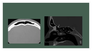
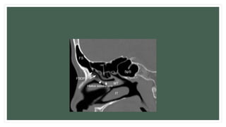
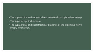
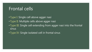
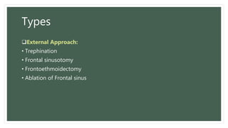
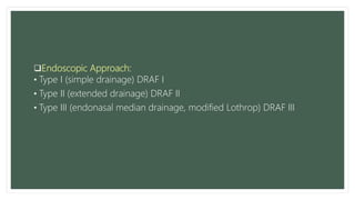
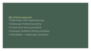
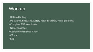
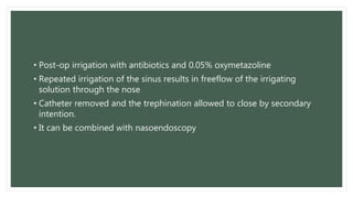
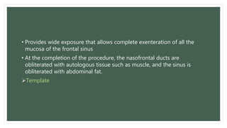
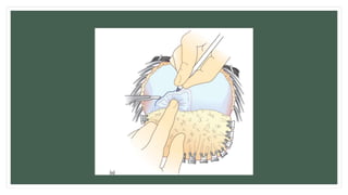
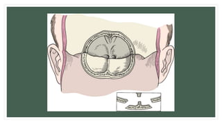
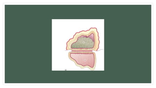
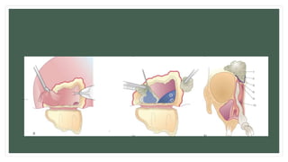
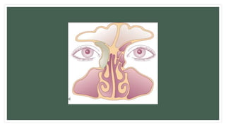
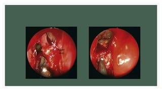
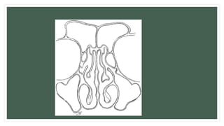
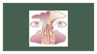
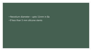
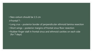
FRONTAL SINUS
SURGERIES
Dr Tabeer Arif
layout
• Introduction
• Surgical anatomy
• Types
• Indications
• Contraindications
• Workup
• Procedure
• Complications
• Conclusion
Introduction
• Frontal sinus surgery has been performed for medically unresponsive
chronic sinusitis, congenital malformations, trauma and neoplasms
• Range from opening into the sinus to complete obliteration of the
frontal sinus
Surgical Anatomy
• Mucosa lined air filled space in the frontal bone
• Bilateral and usually asymmetrical
• Frontal sinus drainage pathway into infundibulum or middle meatus
• Frontal recess is bordered by the Agger nasi (the most anterior ethmoid
cell) anteriorly and the ethmoidal bulla cells posteriorly between the
middle turbinate and lamina papyracea
• The supraorbital and supratrochlear arteries (from ophthalmic artery)
• The superior ophthalmic vein
• The supraorbital and supratrochlear branches of the trigeminal nerve
supply innervation.
Frontal cells
• Type I: Single cell above agger nasi
• Type II: Multiple cells above agger nasi
• Type III: Single cell extending from agger nasi into the frontal
sinus
• Type IV: Single isolated cell in frontal sinus
Types
External Approach:
• Trephination
• Frontal sinusotomy
• Frontoethmoidectomy
• Ablation of Frontal sinus
Endoscopic Approach:
• Type I (simple drainage) DRAF I
• Type II (extended drainage) DRAF II
• Type III (endonasal median drainage, modified Lothrop) DRAF III
Combined approach:
• Trephination with nasoendoscopy
• Endoscopic frontal sinusotomy
• Frontal sinus rescue procedure
• Intranasal modified Lothrop procedure
• Osteoplastic + endoscopic procedure
Indications
Endoscopic approach:
• Chronic frontal sinusitis unresponsive to antibiotic treatment (3 weeks
atleast)
• Polyposis
• Tumors
Open approach:
• pathology situated laterally within the sinus
• very small sinuses
• Multiple failed endoscopic procedure
• Trauma
Workup
• Detailed history
(h/o trauma, headache, watery nasal discharge, visual problems)
• Complete ENT examination
• Nasoendoscopy
• Occipitofrontal sinus X-ray
• CT scan
• MRI
OPEN APPROACHES
Trephination
• Indicated in acute purulent frontal sinusitis
• 1-cm brow incision medial to the supraorbital nerve
• Periosteum is elevated to expose the anterior wall of the frontal sinus.
• A cutting burr is used to drill into the frontal sinus
• The lateral wall of the nasal frontal recess is remove
• Irrigation with antibiotics
• Catheter placed in frontal sinus
• Closure of incision around catheter
• Post-op irrigation with antibiotics and 0.05% oxymetazoline
• Repeated irrigation of the sinus results in freeflow of the irrigating
solution through the nose
• Catheter removed and the trephination allowed to close by secondary
intention.
• It can be combined with nasoendoscopy
Frontal Sinusotomy
• Indications:
• Failure of type III drainage
• Type III drainage not possible
• Laterally located mucopyocoele
• Major destruction of posterior wall
• Major benign tumor
• Aesthetic correction
• Provides wide exposure that allows complete exenteration of all the
mucosa of the frontal sinus
• At the completion of the procedure, the nasofrontal ducts are
obliterated with autologous tissue such as muscle, and the sinus is
obliterated with abdominal fat.
Template
• Bicoronal incision
• Gull wing incision
• Temporary tarsoraphy
• The frontal bone exposed down to the supraorbital rims
• Outline the configuration of the frontal sinus using a template or a
navigation device.
• Periosteum incised
• Osteotomy into frontal sinus using a drill or a saw
• Intersinus septum fractured
• Base of the bone flap is fractured
• Diseased tissue removed
Obliteration of Frontal Sinus
• Mocosa removed
• Obliteration of the nasofrontal duct.
• Sinus obliterated with an adipose tissue graft from abdomen.
• Bone flap is fixed in its place
• Incision closed
• Compressive dressing (48-72 hrs)
• Antiobiotics
• 2 suction Drains (24-48hrs)
Complications:
• Periorbital edema
• Ecchymosis
• Headache
• Mucocoele
• Persistent frontal fullness and pain
• Difficulty in diagnosis of recurrent frontal sinus disease
FrontoEthmoidectomy
• It establishes communication between the floor of the frontal sinus and
the anterior ethmoid cells, in effect marsupializing the most anterior of
the paranasal sinuses with the middle meatus
• in patients who are not candidates for an endoscopic procedure.
• Tarsoraphy
• Incision - above medial aspect of upper eyelid curving down to the level
of medial canthus.
• Periosteum elevated
• Trephination done
• Communication established between floor of frontal sinus and anterior
ethmoid air cells which are removed until free communication with the
middle meatus is obtained.
• Removal of diseased mucosa
• Reconstruction of nasofrontal duct with nasoseptal mucosal flap
High dgree of recurrence of frontal obstruction
Supraorbital and supratrochlear nerve damage
Occular motility disturbance – double vision
Frontal Sinus Ablation
• Osteomyelitis in anterior wall of frontal sinus due to acute or chronic
pyocele
• Subperiosteal abcess of forehead(pott’s puffy tumor)
• Bicoronal incision
• The frontal sinus is ablated by complete removal of the anterior wall of
the frontal sinus, with or without the supraorbital rims
• The sinus is prepared as for a fat obliteration procedure, except that
the skin of the forehead is laid down smoothly on the healthy posterior
table of the sinus.
• Suction drain is inserted
• Pressure dressing applied
• Reconstruction
• Wound is kept dry for 5 days
• Facial sutures – 5days
• Scalp sutures – 14 days
Cranialization
Indications:
• Comminuted fracture of posterior wall
• Severe post-traumatic oedema of frontal lobe
• Foreign body of frontal lobe
• Destruction of posterior sinus wall by chronic inflamation or neoplasm
• Bone fragments removed
• Mucosa removed
• Mobilization of dura
• Duraplasty
• Obliteration of Connection with nose
• Fat graft Contraindicated if both maxillary arteries have been destroyed
ENDOSCOPIC
APPROACHES
• GA
• Local decongestant
• Local anaesthetic with vasoconstrictor
Type I
• Simple Drainage
Indications:
• Failure of conservative surgery in acute rhinosinusitis
• First time sugery
• revision after incomplete ethmoidectomy
Ethmoidectomy (including cell septa in frontal recess area)
• Inferior part of killian’s infundibulum is not disturbed
Type II
• Extended Drainage
Indications:
• Serious complication of acute
rhinosinusitis
• Medial mucopyocoele,
• Tumor surgery
• Good quality mucosa
 Resection of floor of frontal sinus between:
• Lamina papyracea and middle turbinate type IIa
• Lamina papyracea and nasal septum type IIb
anterior to ventral margin of olfactory fossa
• Wide approach to ethmoid by reducing the lacrimal bone, parts of agar
nasi and parts of frontal process of maxilla until lamina papyracea is
seen
• Frontal recess identified
• Uncapping the egg – if frontal cells are present
• Type IIa widened to type IIb with a diamond burr
• Frontal sinus opening is bordered by bone on all sides and mucosa is
left on at least one part
• Rubber finger stall (for 5 days)
• Neostium diameter – upto 11mm in IIa
• If less than 5 mm silicone stents
Type III
• Endonasal Median Drainage
Indications:
• Difficult revision surgery
• Samter’s triad
• Mucoviscidosis
• Kartagener’s syndrome
II b opening is enlarged by resecting portions of superior nasal septum in
area of frontal sinus floor
• Neo-ostium should be 1.5 cm
Frontal T :
• Long crus – posterior border of perpendicular ethmoid lamina resection
• Short wings – posterior margins of frontal sinus floor resection
• Rubber finger stall in frontal sinus and ethmoid cavities on each side
(for 7 days)
Frontal Sinus Rescue
• Indication: frontal sinus obstruction by a laterally retracted middle
turbinate.
• Resection of the scar, the anterior remnant of the mucosa covered
middle turbinate becomes visible.
• Resection of medial osseous lamella and mucosa of the middle
turbinate with the mucosa covering the skull base.
• The lateral mucosal lamella is turned medially covering the skull base.
• The frontal sinus neo-ostium is epithelized.
Rhinofrontal sinuseptomy
• Indication: several failed previous frontal sinus surgeries
• Combined intra-extranasal approach
• Through open frontoethmoidectomy frontal pathology is resected
• Total resection of frontal intersinus septum
• Partial endonasal resection of nasal septum
• B/L subtotal resection of free dependant part of middle turbinate
• B/L endoscopic ethmoidectomy
• Enlargement of isthmus area between frontal sinuses and nasal cavity
• Complete epithelization of neo-communication with free mucosal grafts
and closure of the open approach
• 91% patients were free of symptoms
• CSF leak reported in 1 patient
• In most cases type I or II is sufficient in medically unresponsive frontal
sinusitis
• Type III procedure in samter’s triad
• Post-operative sinusitis: completion of ethmoidectomy with type II a/b
– type III – osteoplastic flap frontal sinus procedure
• Frontal sinus with AP diameter more than 0.8 mm – type III
• Frontal sinus AP diameter less than 0.8 mm – obliteration
• Medial border of lesion lateral to the line through the lamina papyracea
– endonasal approach rarely possible
Congenital Malformations
• Nasal fistulas and cysts are removed completely
• Small ones endoscopically and larger via an external approach
• Neurosurgical collaboration
• Meningoencephalocoeles:
• Small, hidden in nasal cavity – micro-endoscopic approach
• Large obvious ones – external approach
Frontal Sinus Trauma
• No Wait and see policy in severely fractured frontal sinus
Riedel’s operation: radical resection of whole anterior frontal sinus wall
including all frontal sinus mucosa
• Coronal incision
Anterior and/or posterior wall trauma with Killian’s infundibulum intact –
reconstruction of anterior and posterior sinus wall
• Fragments are put together
• Metallic plate
• Biodegradeable miniplates
Severely comminuted posterior wall fractures – reconstruction of
anterior wall, resection of posterior wall, removal of all frontal sinus
mucosa : Cranialization of Frontal Sinus
• dead space obliteration
Orbital roof fractures – entrapment of superior Oblique and/or rectus
muscle
• Reduction of bone fragments, repair of peri-orbita lesion
Dural defect repair
Fibrin glue
Osteoplasty
• Frontal sinus anterior wall osteoplasty
• In frontal pneumosinus dilatans
• Horizontal full thickness bone strips are removed
Tumor Resection
• Tumor not extending lateral to the line passing through lamina
papyracea – endonasal
• Origin or fixation point in lower third if posterior wall of frontal sinus –
endonasal
• Fixation at anterior wall – external
• Intra-cranial extension
• Inverted papilloma – Drilling of mucopericondrium at the area of origin
of tumor
Malignant tumors:
• Endonasal (tumor just reaching the frontal sinus, no major bone
destruction)
• Midfacial degloving (lower sinuses involvement)
• Subcranial resection (skull base, intradural involvement)
Conclusion
• Most inflammatory frontal sinus disease requiring surgery can be
operated nasoendoscopically
• Only 5% of all frontal sinus surgeries are through an open appraoch
Thank you.

Frontal sinus surgeries

  • 1.
  • 2.
    layout • Introduction • Surgicalanatomy • Types • Indications • Contraindications • Workup • Procedure • Complications • Conclusion
  • 3.
    Introduction • Frontal sinussurgery has been performed for medically unresponsive chronic sinusitis, congenital malformations, trauma and neoplasms • Range from opening into the sinus to complete obliteration of the frontal sinus
  • 4.
    Surgical Anatomy • Mucosalined air filled space in the frontal bone • Bilateral and usually asymmetrical • Frontal sinus drainage pathway into infundibulum or middle meatus • Frontal recess is bordered by the Agger nasi (the most anterior ethmoid cell) anteriorly and the ethmoidal bulla cells posteriorly between the middle turbinate and lamina papyracea
  • 9.
    • The supraorbitaland supratrochlear arteries (from ophthalmic artery) • The superior ophthalmic vein • The supraorbital and supratrochlear branches of the trigeminal nerve supply innervation.
  • 11.
    Frontal cells • TypeI: Single cell above agger nasi • Type II: Multiple cells above agger nasi • Type III: Single cell extending from agger nasi into the frontal sinus • Type IV: Single isolated cell in frontal sinus
  • 12.
    Types External Approach: • Trephination •Frontal sinusotomy • Frontoethmoidectomy • Ablation of Frontal sinus
  • 13.
    Endoscopic Approach: • TypeI (simple drainage) DRAF I • Type II (extended drainage) DRAF II • Type III (endonasal median drainage, modified Lothrop) DRAF III
  • 14.
    Combined approach: • Trephinationwith nasoendoscopy • Endoscopic frontal sinusotomy • Frontal sinus rescue procedure • Intranasal modified Lothrop procedure • Osteoplastic + endoscopic procedure
  • 15.
    Indications Endoscopic approach: • Chronicfrontal sinusitis unresponsive to antibiotic treatment (3 weeks atleast) • Polyposis • Tumors Open approach: • pathology situated laterally within the sinus • very small sinuses • Multiple failed endoscopic procedure • Trauma
  • 16.
    Workup • Detailed history (h/otrauma, headache, watery nasal discharge, visual problems) • Complete ENT examination • Nasoendoscopy • Occipitofrontal sinus X-ray • CT scan • MRI
  • 17.
  • 18.
    Trephination • Indicated inacute purulent frontal sinusitis • 1-cm brow incision medial to the supraorbital nerve • Periosteum is elevated to expose the anterior wall of the frontal sinus. • A cutting burr is used to drill into the frontal sinus • The lateral wall of the nasal frontal recess is remove • Irrigation with antibiotics • Catheter placed in frontal sinus • Closure of incision around catheter
  • 21.
    • Post-op irrigationwith antibiotics and 0.05% oxymetazoline • Repeated irrigation of the sinus results in freeflow of the irrigating solution through the nose • Catheter removed and the trephination allowed to close by secondary intention. • It can be combined with nasoendoscopy
  • 22.
    Frontal Sinusotomy • Indications: •Failure of type III drainage • Type III drainage not possible • Laterally located mucopyocoele • Major destruction of posterior wall • Major benign tumor • Aesthetic correction
  • 23.
    • Provides wideexposure that allows complete exenteration of all the mucosa of the frontal sinus • At the completion of the procedure, the nasofrontal ducts are obliterated with autologous tissue such as muscle, and the sinus is obliterated with abdominal fat. Template
  • 25.
    • Bicoronal incision •Gull wing incision • Temporary tarsoraphy
  • 27.
    • The frontalbone exposed down to the supraorbital rims • Outline the configuration of the frontal sinus using a template or a navigation device. • Periosteum incised • Osteotomy into frontal sinus using a drill or a saw • Intersinus septum fractured • Base of the bone flap is fractured • Diseased tissue removed
  • 33.
    Obliteration of FrontalSinus • Mocosa removed • Obliteration of the nasofrontal duct. • Sinus obliterated with an adipose tissue graft from abdomen. • Bone flap is fixed in its place • Incision closed • Compressive dressing (48-72 hrs) • Antiobiotics • 2 suction Drains (24-48hrs)
  • 35.
    Complications: • Periorbital edema •Ecchymosis • Headache • Mucocoele • Persistent frontal fullness and pain • Difficulty in diagnosis of recurrent frontal sinus disease
  • 36.
    FrontoEthmoidectomy • It establishescommunication between the floor of the frontal sinus and the anterior ethmoid cells, in effect marsupializing the most anterior of the paranasal sinuses with the middle meatus • in patients who are not candidates for an endoscopic procedure.
  • 37.
    • Tarsoraphy • Incision- above medial aspect of upper eyelid curving down to the level of medial canthus. • Periosteum elevated • Trephination done • Communication established between floor of frontal sinus and anterior ethmoid air cells which are removed until free communication with the middle meatus is obtained.
  • 39.
    • Removal ofdiseased mucosa • Reconstruction of nasofrontal duct with nasoseptal mucosal flap High dgree of recurrence of frontal obstruction Supraorbital and supratrochlear nerve damage Occular motility disturbance – double vision
  • 40.
    Frontal Sinus Ablation •Osteomyelitis in anterior wall of frontal sinus due to acute or chronic pyocele • Subperiosteal abcess of forehead(pott’s puffy tumor) • Bicoronal incision • The frontal sinus is ablated by complete removal of the anterior wall of the frontal sinus, with or without the supraorbital rims • The sinus is prepared as for a fat obliteration procedure, except that the skin of the forehead is laid down smoothly on the healthy posterior table of the sinus.
  • 41.
    • Suction drainis inserted • Pressure dressing applied • Reconstruction • Wound is kept dry for 5 days • Facial sutures – 5days • Scalp sutures – 14 days
  • 42.
    Cranialization Indications: • Comminuted fractureof posterior wall • Severe post-traumatic oedema of frontal lobe • Foreign body of frontal lobe • Destruction of posterior sinus wall by chronic inflamation or neoplasm
  • 43.
    • Bone fragmentsremoved • Mucosa removed • Mobilization of dura • Duraplasty • Obliteration of Connection with nose • Fat graft Contraindicated if both maxillary arteries have been destroyed
  • 44.
  • 45.
    • GA • Localdecongestant • Local anaesthetic with vasoconstrictor
  • 46.
    Type I • SimpleDrainage Indications: • Failure of conservative surgery in acute rhinosinusitis • First time sugery • revision after incomplete ethmoidectomy Ethmoidectomy (including cell septa in frontal recess area) • Inferior part of killian’s infundibulum is not disturbed
  • 50.
    Type II • ExtendedDrainage Indications: • Serious complication of acute rhinosinusitis • Medial mucopyocoele, • Tumor surgery • Good quality mucosa  Resection of floor of frontal sinus between: • Lamina papyracea and middle turbinate type IIa • Lamina papyracea and nasal septum type IIb anterior to ventral margin of olfactory fossa
  • 53.
    • Wide approachto ethmoid by reducing the lacrimal bone, parts of agar nasi and parts of frontal process of maxilla until lamina papyracea is seen • Frontal recess identified • Uncapping the egg – if frontal cells are present • Type IIa widened to type IIb with a diamond burr • Frontal sinus opening is bordered by bone on all sides and mucosa is left on at least one part • Rubber finger stall (for 5 days)
  • 57.
    • Neostium diameter– upto 11mm in IIa • If less than 5 mm silicone stents
  • 58.
    Type III • EndonasalMedian Drainage Indications: • Difficult revision surgery • Samter’s triad • Mucoviscidosis • Kartagener’s syndrome II b opening is enlarged by resecting portions of superior nasal septum in area of frontal sinus floor
  • 59.
    • Neo-ostium shouldbe 1.5 cm Frontal T : • Long crus – posterior border of perpendicular ethmoid lamina resection • Short wings – posterior margins of frontal sinus floor resection • Rubber finger stall in frontal sinus and ethmoid cavities on each side (for 7 days)
  • 63.
    Frontal Sinus Rescue •Indication: frontal sinus obstruction by a laterally retracted middle turbinate. • Resection of the scar, the anterior remnant of the mucosa covered middle turbinate becomes visible. • Resection of medial osseous lamella and mucosa of the middle turbinate with the mucosa covering the skull base. • The lateral mucosal lamella is turned medially covering the skull base. • The frontal sinus neo-ostium is epithelized.
  • 65.
    Rhinofrontal sinuseptomy • Indication:several failed previous frontal sinus surgeries • Combined intra-extranasal approach • Through open frontoethmoidectomy frontal pathology is resected • Total resection of frontal intersinus septum • Partial endonasal resection of nasal septum • B/L subtotal resection of free dependant part of middle turbinate • B/L endoscopic ethmoidectomy
  • 66.
    • Enlargement ofisthmus area between frontal sinuses and nasal cavity • Complete epithelization of neo-communication with free mucosal grafts and closure of the open approach • 91% patients were free of symptoms • CSF leak reported in 1 patient
  • 67.
    • In mostcases type I or II is sufficient in medically unresponsive frontal sinusitis • Type III procedure in samter’s triad • Post-operative sinusitis: completion of ethmoidectomy with type II a/b – type III – osteoplastic flap frontal sinus procedure • Frontal sinus with AP diameter more than 0.8 mm – type III • Frontal sinus AP diameter less than 0.8 mm – obliteration • Medial border of lesion lateral to the line through the lamina papyracea – endonasal approach rarely possible
  • 68.
    Congenital Malformations • Nasalfistulas and cysts are removed completely • Small ones endoscopically and larger via an external approach • Neurosurgical collaboration • Meningoencephalocoeles: • Small, hidden in nasal cavity – micro-endoscopic approach • Large obvious ones – external approach
  • 69.
    Frontal Sinus Trauma •No Wait and see policy in severely fractured frontal sinus Riedel’s operation: radical resection of whole anterior frontal sinus wall including all frontal sinus mucosa • Coronal incision Anterior and/or posterior wall trauma with Killian’s infundibulum intact – reconstruction of anterior and posterior sinus wall • Fragments are put together • Metallic plate • Biodegradeable miniplates
  • 70.
    Severely comminuted posteriorwall fractures – reconstruction of anterior wall, resection of posterior wall, removal of all frontal sinus mucosa : Cranialization of Frontal Sinus • dead space obliteration Orbital roof fractures – entrapment of superior Oblique and/or rectus muscle • Reduction of bone fragments, repair of peri-orbita lesion Dural defect repair Fibrin glue
  • 71.
    Osteoplasty • Frontal sinusanterior wall osteoplasty • In frontal pneumosinus dilatans • Horizontal full thickness bone strips are removed
  • 73.
    Tumor Resection • Tumornot extending lateral to the line passing through lamina papyracea – endonasal • Origin or fixation point in lower third if posterior wall of frontal sinus – endonasal • Fixation at anterior wall – external • Intra-cranial extension • Inverted papilloma – Drilling of mucopericondrium at the area of origin of tumor
  • 74.
    Malignant tumors: • Endonasal(tumor just reaching the frontal sinus, no major bone destruction) • Midfacial degloving (lower sinuses involvement) • Subcranial resection (skull base, intradural involvement)
  • 75.
    Conclusion • Most inflammatoryfrontal sinus disease requiring surgery can be operated nasoendoscopically • Only 5% of all frontal sinus surgeries are through an open appraoch
  • 76.

Editor's Notes

  • #6 Axial section through right nasal cavity depicts the following: (A) middle turbinate (or concha); (B) ethmoid bulla; (C) drainage sites for frontal sinus (3 shaded areas); (D) uncinate process; (E) nasolacrimal duct; (F) hiatus semilunaris; (G) basal lamella; (H) septal cartilage.
  • #7 Sagittal image shows frontal sinus ostium (*) and arrow pointing to the superior compartment of the FSDP. (FS: frontal sinus, AG: agger nasi, PE: posterior ethmoid, SpS: sphenoid sinus, MT: middle turbinate, IT: inferior turbinate)
  • #8 Sagittal image with arrows demonstrating frontal sinus drainage pathway and hiatus semilunaris which drains to middle meatus. (FS: frontal sinus, SpS: sphenoid sinus, MT: middle turbinate, IT: inferior turbinate)
  • #9 Rt frontal recess bounded anteriorly and laterally by agger nasi cell (white arrow) and type 1 frontal cell (black arrow) medially by middle turbinate and posteriorly by ethmoid bulla Arrow head – nasofrontal process.. Forms the floor of the frontal sinus and demarcates the level of frontal sinus ostium
  • #11 Pulley of superior oblique
  • #12 Pneumatization of ant. Ethmoidal cells anterosuperiorly into the frontal recess
  • #19 Trephination of the frontal sinus follows the same principles involved in drainage of an abscess found in soft tissue An estimate of the depth of the frontal sinus and its relationship to the orbit and anterior cranial fossa can be obtained through review of the CT scan. The lateral wall removed to provide communication between the frontal sinus and the middle meatus.
  • #22 Restoration of function in the nasofrontal recess frequently takes 7 to 10 days. Failure to respond may be an indication for definitive surgery to correct irreversible obstruction of the nasofrontal duct.
  • #23 Anterior posterior diameter is less than 8 mm Pneumatosinus dilatans
  • #24 A template of the frontal sinus is designed from a preoperative anteroposterior radiograph taken at 6 ft of distance (Caldwell's view)
  • #25 Occipito frontal xray
  • #26  A gull-wing incision is cosmetically inferior and usually divides the supraorbital nerve, which results in troublesome paresthesias and numbness of the forehead postoperatively. The bicoronal incision affords a completely hidden scar without sensory denervation. limitation is cosmetic and only in patients with male pattern baldness.
  • #27 Dissection upto supraorbital ridge, leaving behind periosteum and preserving neurovascular bundles Scalp clamps
  • #28  With the supraorbital rims used as a landmark, the template is used Leaving the periosteum attached to the bone (osteoplastic flap)
  • #29 After the template has been used to demonstrate the extent of the frontal sinus, the pericranium is elevated and reflected inferiorly. In this way the pericranium can be used to reinforce the closure at the completion of the procedure.
  • #30 Periosteum elevated Oblique incision through the bone - replacement
  • #31 The osteoplastic flap is outlined with an oscillating saw. The osteotomy is beveled (inset) to ensure that the flap will fit securely when it is replaced at the completion of the procedure
  • #33 Bicoronal flap used to remove a mucocele of the frontal sinus
  • #34 Inner layer of bony walls must be drilled away In frontal sinus ostium region, mucosa is inverted into the nasal cavity Using temporalis fascia, bone from calvarium, conchal cartilage
  • #40 unnecessary when treating patients for fractures of the anterior wall of the frontal sinus or during removal of osteoma because the duct should not be Traumatized
  • #42 Reconstruction of the cosmetic defect should be postponed until it is ensured that the infectious process has been completely eradicated. In most circumstances, surgeons would wait 3 to 12 months before undertaking reconstruction.
  • #44 Depending on the anteroposterior diameter of sinus small – galeal periosteum, conchal cartilage Large – abdominal fat graft Cancellous bone from iliac crest and hydroxapatite Midfacial fracture
  • #47 Aspirin intolerance Asthma Frontal sinus heals due to improved drainage via ethmoid cavity
  • #53 Type II a Frontal sinus Skull base of ethmoid sinus Middle turbinate
  • #54 During surgery repeated CT scan to establish the presence of frontal cells
  • #56 Middle turbinate being dissected
  • #57 Area ant ethmoidal artery Ip lamina papyracea Mt middle turbinate Ns nsal septum Oc olfactory cleft
  • #59 Aspirin sensitivity, asthma, nasal polyps
  • #60 Resection of middle turbinate from anterior to posterior along its origin at base of skull until first olfactory fibers are seen after almost 5 mm
  • #62 Septum Middle turbinate remanant
  • #64 Lateral mucosal lamella is preserved
  • #69 Intracranial extra or intra dural extension May require duraplasty
  • #70 Sinus complications can arise as late as 48 yrs (excellent exposure & ability to harvest bone graft from temporal region) Co-polymers… L-lactide
  • #71 after any connection between the nasal cavity and the frontal sinus has been closed, e.g. with preserved dura, fascia or galea periosteum. If there is a larger gap into the nasal cavity, pinna cartilage has proved to be effective. Complete neurological examination – oculomotor n. damage Galeal periosteum, temporalis fascia Underlay between brain and dura, between dura and bone, onlay