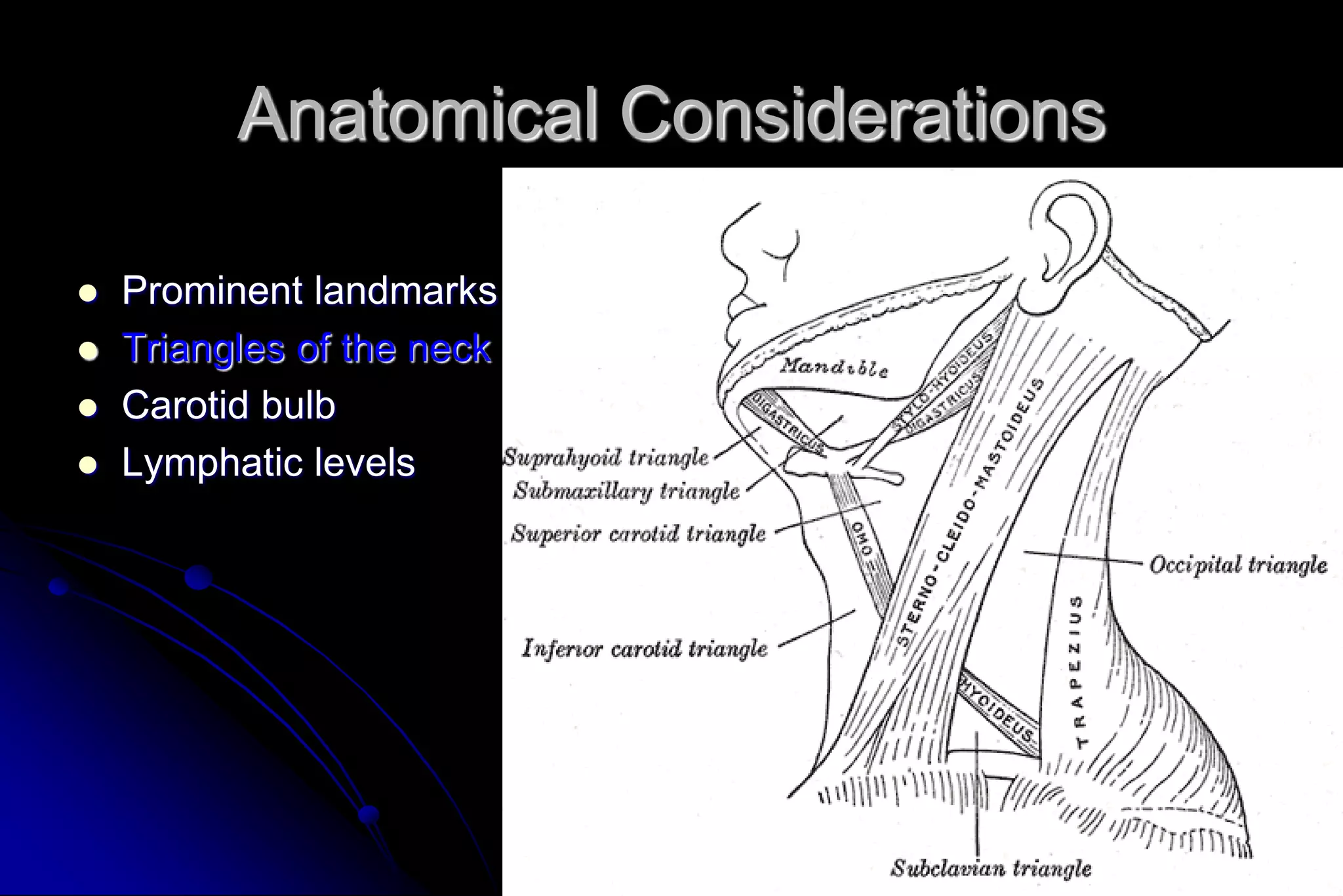
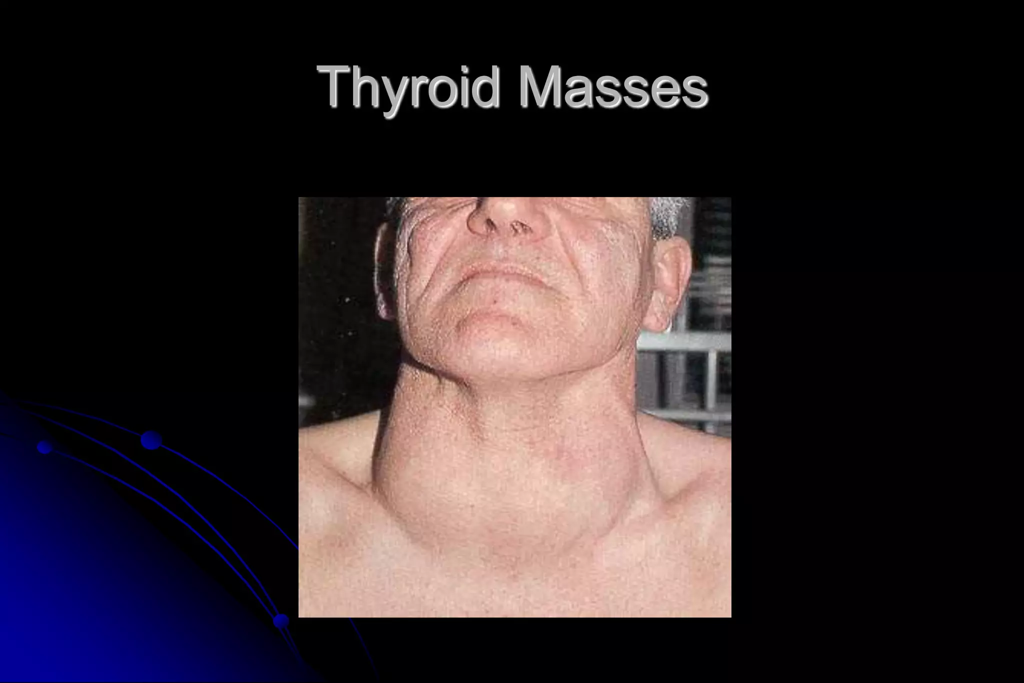
Evaluation of the Neck Mass
Akram Hajjar, MD
American Board Certified
Introduction
 Common clinical finding
 All age groups
 Very complex differential diagnosis
 Systematic approach essential
Anatomical Considerations
 Prominent landmarks
 Triangles of the neck
 Carotid bulb
 Lymphatic levels
Anatomical Considerations
 Prominent landmarks
 Triangles of the neck
 Carotid bulb
 Lymphatic levels
Anatomical Considerations
 Prominent landmarks
 Triangles of the neck
 Carotid bulb
 Lymphatic levels
Anatomical Considerations
 Prominent landmarks
 Triangles of the neck
 Carotid bulb
 Lymphatic levels
General Considerations
 Patient age
 Pediatric (0 – 15 years): 90% benign
 Young adult (16 – 40 years): similar to pediatric
 Late adult (>40 years): “rule of 80s”
 Location
 Congenital masses: consistent in location
 Metastatic masses: key to primary lesion
Metastasis Location according to Various Primary
Risk Factors for Head and Neck Cancer
 Chronic sun exposure
 Tobacco and alcohol use
 Poor dentition
 Industrial or environmental exposures
 Family history
Symptoms of Head and Neck Cancer
 Nonhealing ulcer within the oral cavity or
oropharynx
 Persistent sore throat
 Dysphagia
 Change in voice
 Recent weight loss
Diagnostic Steps
 History
 Developmental time course (age, size, acute
vs chronic)
 Associated symptoms (fever, sore throat,
cough, dysphagia, otalgia, voice)
 Personal habits (tobacco, alcohol)
 Recent travel, trauma, insect bites, pets/farm
animals
 Previous irradiation or surgery
Physical Exam
 Complete head and neck exam
 Skin, otologic examination, oropharynx, tongue
 Ulcerations, submucosal swelling, or asymmetry, particularly
in the tonsillar fossa
 Examination of the larynx and pharynx is accomplished by
indirect or flexible laryngoscopy.
 Palpation during swallowing or during a Valsalva's maneuver
may identify pathology within the larynx and thyroid gland.
 Rotation of the head in both flexion and extension aids
examination of the posterior triangle of the neck.
Empirical Antibiotics
 Inflammatory mass suspected
 Two week trial of broad spectrum antibiotics
 Follow-up 1-2 weeks for further investigation
Diagnostic Tests
 Fine needle aspiration biopsy (FNAB)
 Computed tomography (CT)
 Magnetic resonance imaging (MRI)
 Ultrasonography
 Radionucleotide scanning
Fine Needle Aspiration Biopsy
 Standard of diagnosis
 Indications
 Any neck mass that is not an obvious abscess
 Persistence after a 2 week course of antibiotics
 Progressive growth, supraclavicular, > 3 cm
 Any symptoms associated with lymphoma
 Small gauge needle
 Reduces bleeding
 Seeding of tumor – not a concern
 No contraindications (vascular ?)
Fine Needle Aspiration Biopsy
 Proper collection required
 Minimum of 4 separate passes
 Skilled cytopathologist essential
 On-site review best
Fine Needle Aspiration Biopsy
Computed Tomography
 Distinguish cystic from solid
 Extent of lesion
 Vascularity (with contrast)
 Detection of unknown primary (metastatic)
 Pathologic node (lucent, >1.5cm, loss of shape)
 Avoid contrast in thyroid lesions
Computed Tomography
Magnetic Resonance Imaging
 Similar information as CT
 Better for upper neck and skull base
 Vascular delineation with infusion
Magnetic Resonance Imaging
Ultrasonography
 Less important now with FNAB
 Solid versus cystic masses
 Congenital cysts from solid nodes/tumors
 Noninvasive (pediatric)
Ultrasonography
YROID
ASS
Radionucleotide Scanning
 Salivary and thyroid masses
 Location – glandular versus extra-glandular
 Functional information
 FNAB now preferred for for thyroid nodules
 Solitary nodules
 Multinodular goiter with new increasing nodule
 Hashimoto’s with new nodule
Radionucleotide Scanning
Nodal Mass Workup in the Adult
 Any solid asymmetric mass MUST be
considered a metastatic neoplastic lesion until
proven otherwise
 Asymptomatic cervical mass – 12% of cancer
 ~ 80% of these are SCCa
Nodal Mass Workup in the Adult
 Ipsilateral otalgia with normal otoscopy – direct
attention to tonsil, tongue base, supraglottis and
hypopharynx
 Unilateral serous otitis – direct examination of
nasopharynx
Nodal Mass Workup in the Adult
 Panendoscopy
 FNAB positive with no primary on repeat exam
 FNAB equivocal/negative in high risk patient
 Directed Biopsy
 All suspicious mucosal lesions
 Areas of concern on CT/MRI
 None observed – nasopharynx, tonsil (ipsilateral
tonsillectomy for jugulodigastric nodes), base of tongue and
piriforms
 Synchronous primaries (10 to 20%)
Nodal Mass Workup in the Adult
 Open excisional biopsy
 Only if complete workup negative
 Occurs in ~5% of patients
 Be prepared for a complete neck dissection
 Frozen section results (complete node excision)
 Inflammatory or granulomatous – culture
 Lymphoma or adenocarcinoma – close wound
Primary Tumors
 Thyroid mass
 Lymphoma
 Salivary tumors
 Lipoma
 Carotid body and glomus tumors
 Neurogenic tumors
Thyroid Masses
 Leading cause of anterior neck masses
 Children
 Most common neoplastic condition
 Male predominance
 Higher incidence of malignancy
 Adults
 Female predominance
 Mostly benign
Thyroid Masses
 Lymph node metastasis
 Initial symptom in 15% of papillary carcinomas
 40% with malignant nodules
 Histologically (microscopic) in >90%
 FNAB has replaced USG and radionucleotide scanning
 Decreases # of patients with surgery
 Increased # of malignant tumors found at surgery
 Doubled the # of cases followed up
 Unsatisfactory aspirate – repeat in 1 month
Thyroid Masses
Lymphoma
 More common in children and young adults
 Up to 80% of children with Hodgkin’s have a neck
mass
 Signs and symptoms
 Lateral neck mass only (discrete, rubbery, nontender)
 Fever
 Hepatosplenomegaly
 Diffuse adenopathy
Lymphoma
 FNAB – first line diagnostic test
 If suggestive of lymphoma – open biopsy
 Full workup – CT scans of chest, abdomen,
head and neck; bone marrow biopsy
Lymphoma
Salivary Gland Tumors
 Enlarging mass anterior/inferior to ear or at the
mandible angle is suspect
 Benign
 Asymptomatic except for mass
 Malignant
 Rapid growth, skin fixation, cranial nerve palsies
Salivary Gland Tumors
 Diagnostic tests
 Open excisional biopsy (submandibulectomy or
parotidectomy) preferred
 FNAB
 Shown to reduce surgery by 1/3 in some studies
 Delineates intra-glandular lymph node, localized sialadenitis
or benign lymphoepithelial cysts
 May facilitate surgical planning and patient counseling
 Accuracy >90% (sensitivity: ~90%; specificity: ~80%)
 CT/MRI – deep lobe tumors, intra vs. extra-parotid
 Be prepared for total parotidectomy with possible
facial nerve sacrifice
Salivary Gland Tumors
Carotid Body Tumor
 Rare in children
 Pulsatile, compressible mass
 Mobile medial/lateral not superior/inferior
 Clinical diagnosis, confirmed by angiogram or CT
 Treatment
 Irradiation or close observation in the elderly
 Surgical resection for small tumors in young patients
 Hypotensive anesthesia
 Preoperative measurement of catecholamines
Carotid Body Tumor
Lipoma
 Soft, ill-defined mass
 Usually >35 years of age
 Asymptomatic
 Clinical diagnosis – confirmed by excision
Lipoma
Neurogenic Tumors
 Arise from neural crest derivatives
 Include schwannoma, neurofibroma, and
malignant peripheral nerve sheath tumor
 Increased incidence in NF syndromes
 Schwannoma most common in head & neck
Schwannoma
 Sporadic cases mostly
 25 to 45% in neck when extracranial
 Most commonly between 20 and 50 years
 Usually mid-neck in poststyloid compartment
 Signs and symptoms
 Medial tonsillar displacement
 Hoarseness (vagus nerve)
 Horner’s syndrome (sympathetic chain)
Schwannoma
Congenital and Developmental
Mass
 Epidermal and sebaceous cysts
 Branchial cleft cysts
 Thyroglossal duct cyst
 Vascular tumors
Epidermal and Sebaceous Cysts
 Most common congenital/developmental mass
 Older age groups
 Clinical diagnosis
 Elevation and movement of overlying skin
 Skin dimple or pore
 Excisional biopsy confirms
Epidermal and Sebaceous Cysts
Branchial Cleft Cysts
 Branchial cleft anomalies
 2nd cleft most common (95%) – tract medial to
cnXII between internal and external carotids
 1st cleft less common – close association with
facial nerve possible
 3rd and 4th clefts rarely reported
 Present in older children or young adults often
following URI
Branchial Cleft Cysts
 Most common as smooth, fluctuant mass
underlying the SCM
 Skin erythema and tenderness if infected
 Treatment
 Initial control of infection
 Surgical excision, including tract
 May necessitate a total parotidectomy (1st cleft)
Branchial Cleft Cysts
Thyroglossal Duct Cyst
 Most common congenital neck mass (70%)
 50% present before age 20
 Midline (75%) or near midline (25%)
 Usually just inferior to hyoid bone (65%)
 Elevates on swallowing/protrusion of tongue
 Treatment is surgical removal (Sis trunk) after
resolution of any infection
Thyroglossal Duct Cyst
Vascular Tumors
 Lymphangiomas and hemangiomas
 Usually within 1st year of life
 Hemangiomas often resolve spontaneously,
while lymphangiomas remain unchanged
 CT/MRI may help define extent of disease
Vascular Tumors
 Treatment
 Lymphangioma – surgical excision for easily
accessible or lesions affecting vital functions;
recurrence is common
 Hemangiomas – surgical excision reserved for those
with rapid growth involving vital structures or
associated thrombocytopenia that fails medical
therapy (steroids, interferon)
Vascular Tumors (lymphangioma)
Vascular Tumors (hemangioma)
Inflammatory Disorders
 Lymphadenitis
 Granulomatous lymphadenitis
Lymphadenitis
 Very common, especially within 1st decade
 Tender node with signs of systemic infection
 Directed antibiotic therapy with follow-up
 FNAB indications (pediatric)
 Actively infectious condition with no response
 Progressively enlarging
 Solitary and asymmetric nodal mass
 Supraclavicular mass (60% malignancy)
 Persistent nodal mass without active infection
Lymphadenopathy
 Equivocal or suspicious FNAB in the pediatric
nodal mass requires open excisional biopsy to
rule out malignant or granulomatous disease
Granulomatous lymphadenitis
 Infection develops over weeks to months
 Minimal systemic complaints or findings
 Common etiologies
 TB, atypical TB, cat-scratch fever, actinomycosis,
sarcoidosis
 Firm, relatively fixed node with injection of skin
Granulomatous lymphadenitis
 Typical M. tuberculosis
 More common in adults
 Posterior triangle nodes
 Rarely seen in our population
 Usually responds to anti-TB medications
 May require excisional biopsy for further workup
Granulomatous lymphadenitis
 Atypical M. tuberculosis
 Pediatric age groups
 Anterior triangle nodes
 Brawny skin, induration and pain
 Usually responds to complete surgical excision or
curettage
Granulomatous lymphadenitis
 Cat-scratch fever (Bartonella)
 Pediatric group
 Preauricular and submandibular nodes
 Spontaneous resolution with or without antibiotics
Granulomatous lymphadenitis
Summary
 Extensive differential diagnosis
 Age of patient is important
 Accurate history and complete exam essential
 FNAB – invaluable diagnostic tool
 Possibility for malignancy in any age group
 Close follow-up and aggressive approach is best
for favorable outcomes
References
 Armstrong W, Giglio M. Is this lump in the neck anything to worry about? September
1998. Postgraduate Medicine.
 Schwetschenau E, Kelley D. The Adult Neck Mass. Sep 2002. American Family
Physician.
 Underbrink M, Bailey B. Evaluation and Management of the Patient with a Neck
Mass. www.utmb.edu/otoref/Grnds/NeckMass-200112/NeckMass2-2001

Neck mass

  • 1.
    Evaluation of theNeck Mass Akram Hajjar, MD American Board Certified
  • 2.
    Introduction  Common clinicalfinding  All age groups  Very complex differential diagnosis  Systematic approach essential
  • 4.
    Anatomical Considerations  Prominentlandmarks  Triangles of the neck  Carotid bulb  Lymphatic levels
  • 5.
    Anatomical Considerations  Prominentlandmarks  Triangles of the neck  Carotid bulb  Lymphatic levels
  • 6.
    Anatomical Considerations  Prominentlandmarks  Triangles of the neck  Carotid bulb  Lymphatic levels
  • 7.
    Anatomical Considerations  Prominentlandmarks  Triangles of the neck  Carotid bulb  Lymphatic levels
  • 8.
    General Considerations  Patientage  Pediatric (0 – 15 years): 90% benign  Young adult (16 – 40 years): similar to pediatric  Late adult (>40 years): “rule of 80s”  Location  Congenital masses: consistent in location  Metastatic masses: key to primary lesion
  • 9.
  • 10.
    Risk Factors forHead and Neck Cancer  Chronic sun exposure  Tobacco and alcohol use  Poor dentition  Industrial or environmental exposures  Family history
  • 11.
    Symptoms of Headand Neck Cancer  Nonhealing ulcer within the oral cavity or oropharynx  Persistent sore throat  Dysphagia  Change in voice  Recent weight loss
  • 12.
    Diagnostic Steps  History Developmental time course (age, size, acute vs chronic)  Associated symptoms (fever, sore throat, cough, dysphagia, otalgia, voice)  Personal habits (tobacco, alcohol)  Recent travel, trauma, insect bites, pets/farm animals  Previous irradiation or surgery
  • 13.
    Physical Exam  Completehead and neck exam  Skin, otologic examination, oropharynx, tongue  Ulcerations, submucosal swelling, or asymmetry, particularly in the tonsillar fossa  Examination of the larynx and pharynx is accomplished by indirect or flexible laryngoscopy.  Palpation during swallowing or during a Valsalva's maneuver may identify pathology within the larynx and thyroid gland.  Rotation of the head in both flexion and extension aids examination of the posterior triangle of the neck.
  • 14.
    Empirical Antibiotics  Inflammatorymass suspected  Two week trial of broad spectrum antibiotics  Follow-up 1-2 weeks for further investigation
  • 15.
    Diagnostic Tests  Fineneedle aspiration biopsy (FNAB)  Computed tomography (CT)  Magnetic resonance imaging (MRI)  Ultrasonography  Radionucleotide scanning
  • 16.
    Fine Needle AspirationBiopsy  Standard of diagnosis  Indications  Any neck mass that is not an obvious abscess  Persistence after a 2 week course of antibiotics  Progressive growth, supraclavicular, > 3 cm  Any symptoms associated with lymphoma  Small gauge needle  Reduces bleeding  Seeding of tumor – not a concern  No contraindications (vascular ?)
  • 17.
    Fine Needle AspirationBiopsy  Proper collection required  Minimum of 4 separate passes  Skilled cytopathologist essential  On-site review best
  • 18.
  • 19.
    Computed Tomography  Distinguishcystic from solid  Extent of lesion  Vascularity (with contrast)  Detection of unknown primary (metastatic)  Pathologic node (lucent, >1.5cm, loss of shape)  Avoid contrast in thyroid lesions
  • 20.
  • 21.
    Magnetic Resonance Imaging Similar information as CT  Better for upper neck and skull base  Vascular delineation with infusion
  • 22.
  • 23.
    Ultrasonography  Less importantnow with FNAB  Solid versus cystic masses  Congenital cysts from solid nodes/tumors  Noninvasive (pediatric)
  • 24.
  • 25.
    Radionucleotide Scanning  Salivaryand thyroid masses  Location – glandular versus extra-glandular  Functional information  FNAB now preferred for for thyroid nodules  Solitary nodules  Multinodular goiter with new increasing nodule  Hashimoto’s with new nodule
  • 26.
  • 27.
    Nodal Mass Workupin the Adult  Any solid asymmetric mass MUST be considered a metastatic neoplastic lesion until proven otherwise  Asymptomatic cervical mass – 12% of cancer  ~ 80% of these are SCCa
  • 28.
    Nodal Mass Workupin the Adult  Ipsilateral otalgia with normal otoscopy – direct attention to tonsil, tongue base, supraglottis and hypopharynx  Unilateral serous otitis – direct examination of nasopharynx
  • 29.
    Nodal Mass Workupin the Adult  Panendoscopy  FNAB positive with no primary on repeat exam  FNAB equivocal/negative in high risk patient  Directed Biopsy  All suspicious mucosal lesions  Areas of concern on CT/MRI  None observed – nasopharynx, tonsil (ipsilateral tonsillectomy for jugulodigastric nodes), base of tongue and piriforms  Synchronous primaries (10 to 20%)
  • 30.
    Nodal Mass Workupin the Adult  Open excisional biopsy  Only if complete workup negative  Occurs in ~5% of patients  Be prepared for a complete neck dissection  Frozen section results (complete node excision)  Inflammatory or granulomatous – culture  Lymphoma or adenocarcinoma – close wound
  • 32.
    Primary Tumors  Thyroidmass  Lymphoma  Salivary tumors  Lipoma  Carotid body and glomus tumors  Neurogenic tumors
  • 33.
    Thyroid Masses  Leadingcause of anterior neck masses  Children  Most common neoplastic condition  Male predominance  Higher incidence of malignancy  Adults  Female predominance  Mostly benign
  • 34.
    Thyroid Masses  Lymphnode metastasis  Initial symptom in 15% of papillary carcinomas  40% with malignant nodules  Histologically (microscopic) in >90%  FNAB has replaced USG and radionucleotide scanning  Decreases # of patients with surgery  Increased # of malignant tumors found at surgery  Doubled the # of cases followed up  Unsatisfactory aspirate – repeat in 1 month
  • 35.
  • 36.
    Lymphoma  More commonin children and young adults  Up to 80% of children with Hodgkin’s have a neck mass  Signs and symptoms  Lateral neck mass only (discrete, rubbery, nontender)  Fever  Hepatosplenomegaly  Diffuse adenopathy
  • 37.
    Lymphoma  FNAB –first line diagnostic test  If suggestive of lymphoma – open biopsy  Full workup – CT scans of chest, abdomen, head and neck; bone marrow biopsy
  • 38.
  • 39.
    Salivary Gland Tumors Enlarging mass anterior/inferior to ear or at the mandible angle is suspect  Benign  Asymptomatic except for mass  Malignant  Rapid growth, skin fixation, cranial nerve palsies
  • 40.
    Salivary Gland Tumors Diagnostic tests  Open excisional biopsy (submandibulectomy or parotidectomy) preferred  FNAB  Shown to reduce surgery by 1/3 in some studies  Delineates intra-glandular lymph node, localized sialadenitis or benign lymphoepithelial cysts  May facilitate surgical planning and patient counseling  Accuracy >90% (sensitivity: ~90%; specificity: ~80%)  CT/MRI – deep lobe tumors, intra vs. extra-parotid  Be prepared for total parotidectomy with possible facial nerve sacrifice
  • 41.
  • 42.
    Carotid Body Tumor Rare in children  Pulsatile, compressible mass  Mobile medial/lateral not superior/inferior  Clinical diagnosis, confirmed by angiogram or CT  Treatment  Irradiation or close observation in the elderly  Surgical resection for small tumors in young patients  Hypotensive anesthesia  Preoperative measurement of catecholamines
  • 43.
  • 44.
    Lipoma  Soft, ill-definedmass  Usually >35 years of age  Asymptomatic  Clinical diagnosis – confirmed by excision
  • 45.
  • 46.
    Neurogenic Tumors  Arisefrom neural crest derivatives  Include schwannoma, neurofibroma, and malignant peripheral nerve sheath tumor  Increased incidence in NF syndromes  Schwannoma most common in head & neck
  • 47.
    Schwannoma  Sporadic casesmostly  25 to 45% in neck when extracranial  Most commonly between 20 and 50 years  Usually mid-neck in poststyloid compartment  Signs and symptoms  Medial tonsillar displacement  Hoarseness (vagus nerve)  Horner’s syndrome (sympathetic chain)
  • 48.
  • 49.
    Congenital and Developmental Mass Epidermal and sebaceous cysts  Branchial cleft cysts  Thyroglossal duct cyst  Vascular tumors
  • 50.
    Epidermal and SebaceousCysts  Most common congenital/developmental mass  Older age groups  Clinical diagnosis  Elevation and movement of overlying skin  Skin dimple or pore  Excisional biopsy confirms
  • 51.
  • 52.
    Branchial Cleft Cysts Branchial cleft anomalies  2nd cleft most common (95%) – tract medial to cnXII between internal and external carotids  1st cleft less common – close association with facial nerve possible  3rd and 4th clefts rarely reported  Present in older children or young adults often following URI
  • 53.
    Branchial Cleft Cysts Most common as smooth, fluctuant mass underlying the SCM  Skin erythema and tenderness if infected  Treatment  Initial control of infection  Surgical excision, including tract  May necessitate a total parotidectomy (1st cleft)
  • 54.
  • 55.
    Thyroglossal Duct Cyst Most common congenital neck mass (70%)  50% present before age 20  Midline (75%) or near midline (25%)  Usually just inferior to hyoid bone (65%)  Elevates on swallowing/protrusion of tongue  Treatment is surgical removal (Sis trunk) after resolution of any infection
  • 56.
  • 57.
    Vascular Tumors  Lymphangiomasand hemangiomas  Usually within 1st year of life  Hemangiomas often resolve spontaneously, while lymphangiomas remain unchanged  CT/MRI may help define extent of disease
  • 58.
    Vascular Tumors  Treatment Lymphangioma – surgical excision for easily accessible or lesions affecting vital functions; recurrence is common  Hemangiomas – surgical excision reserved for those with rapid growth involving vital structures or associated thrombocytopenia that fails medical therapy (steroids, interferon)
  • 59.
  • 60.
  • 61.
  • 62.
    Lymphadenitis  Very common,especially within 1st decade  Tender node with signs of systemic infection  Directed antibiotic therapy with follow-up  FNAB indications (pediatric)  Actively infectious condition with no response  Progressively enlarging  Solitary and asymmetric nodal mass  Supraclavicular mass (60% malignancy)  Persistent nodal mass without active infection
  • 63.
    Lymphadenopathy  Equivocal orsuspicious FNAB in the pediatric nodal mass requires open excisional biopsy to rule out malignant or granulomatous disease
  • 64.
    Granulomatous lymphadenitis  Infectiondevelops over weeks to months  Minimal systemic complaints or findings  Common etiologies  TB, atypical TB, cat-scratch fever, actinomycosis, sarcoidosis  Firm, relatively fixed node with injection of skin
  • 65.
    Granulomatous lymphadenitis  TypicalM. tuberculosis  More common in adults  Posterior triangle nodes  Rarely seen in our population  Usually responds to anti-TB medications  May require excisional biopsy for further workup
  • 66.
    Granulomatous lymphadenitis  AtypicalM. tuberculosis  Pediatric age groups  Anterior triangle nodes  Brawny skin, induration and pain  Usually responds to complete surgical excision or curettage
  • 67.
    Granulomatous lymphadenitis  Cat-scratchfever (Bartonella)  Pediatric group  Preauricular and submandibular nodes  Spontaneous resolution with or without antibiotics
  • 68.
  • 69.
    Summary  Extensive differentialdiagnosis  Age of patient is important  Accurate history and complete exam essential  FNAB – invaluable diagnostic tool  Possibility for malignancy in any age group  Close follow-up and aggressive approach is best for favorable outcomes
  • 70.
    References  Armstrong W,Giglio M. Is this lump in the neck anything to worry about? September 1998. Postgraduate Medicine.  Schwetschenau E, Kelley D. The Adult Neck Mass. Sep 2002. American Family Physician.  Underbrink M, Bailey B. Evaluation and Management of the Patient with a Neck Mass. www.utmb.edu/otoref/Grnds/NeckMass-200112/NeckMass2-2001