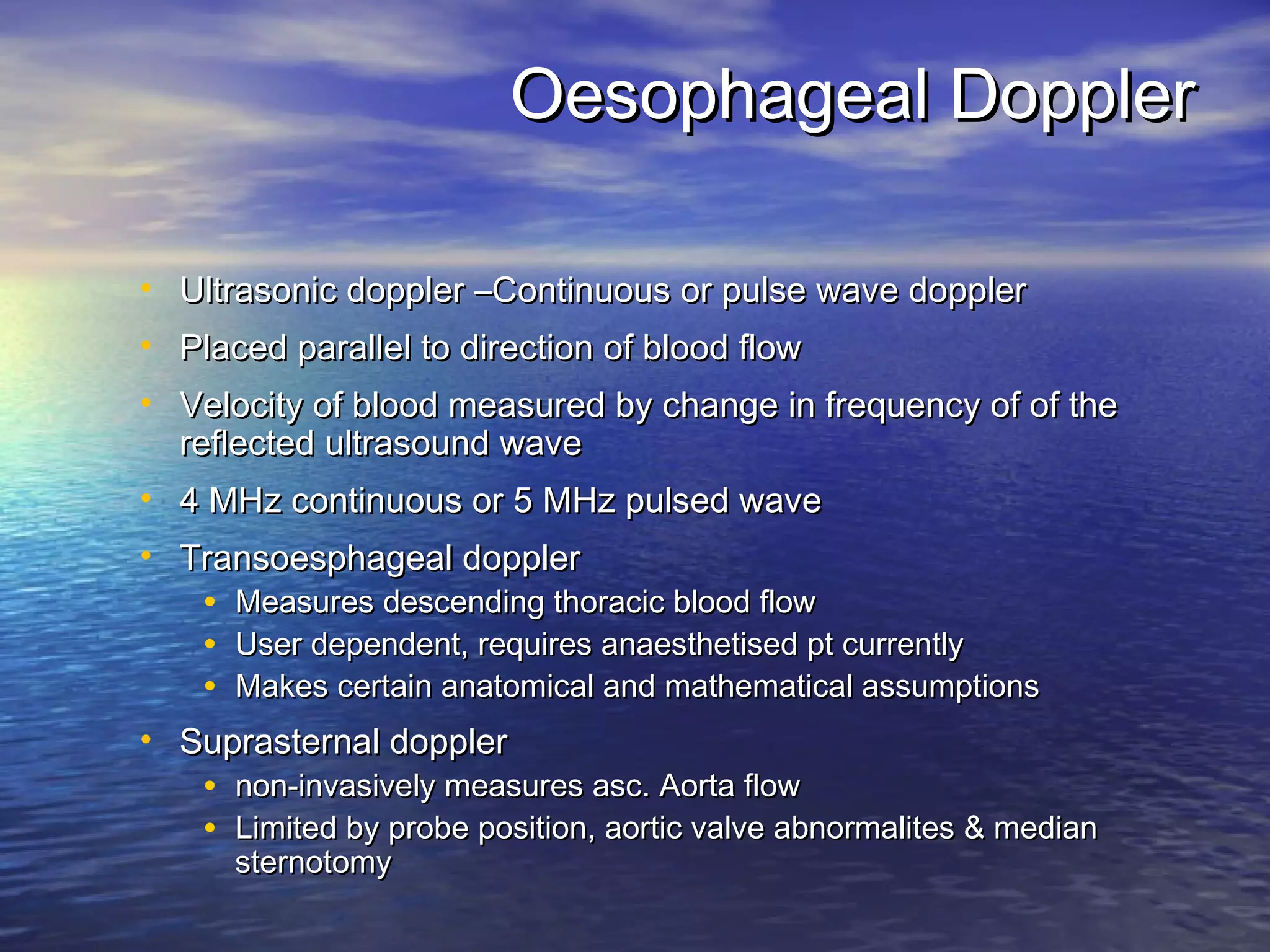
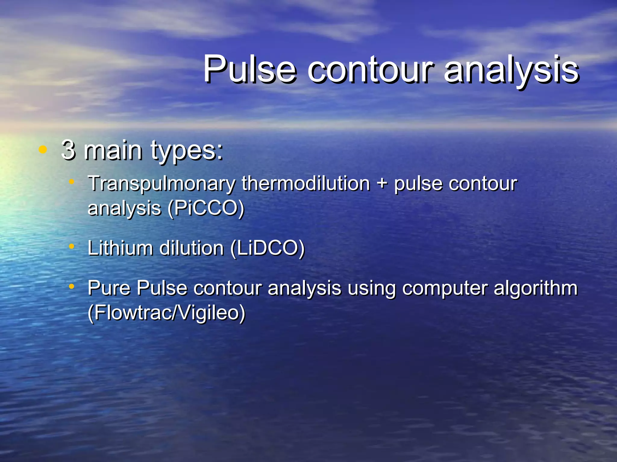
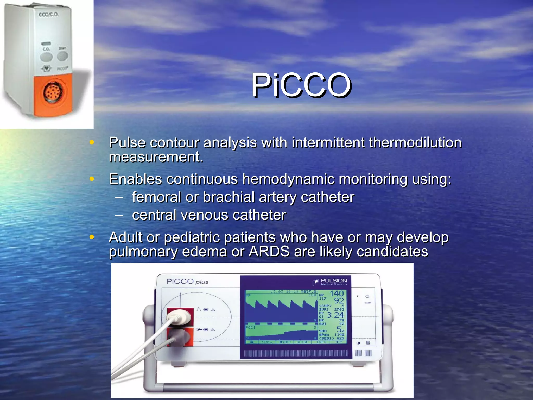
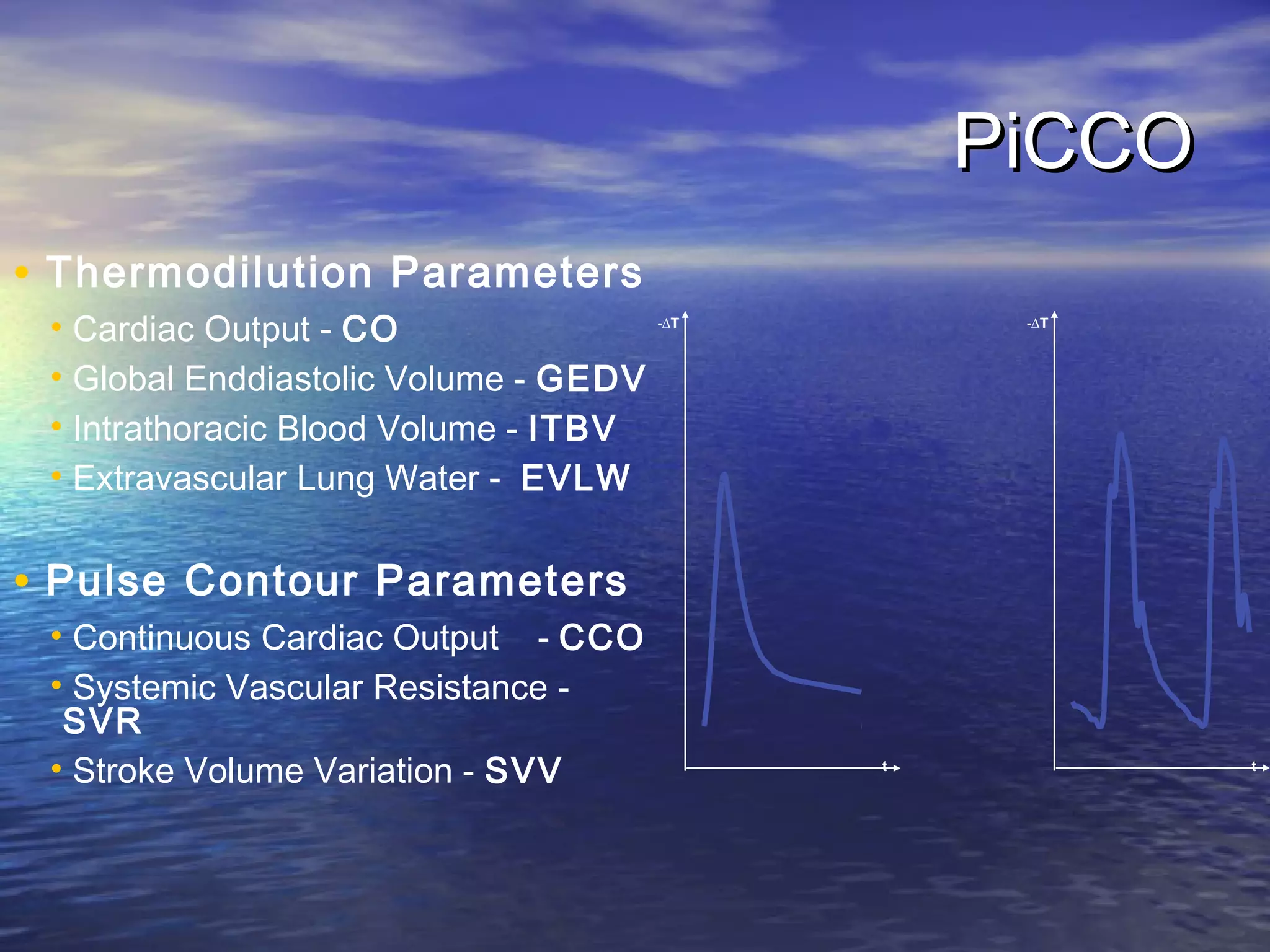
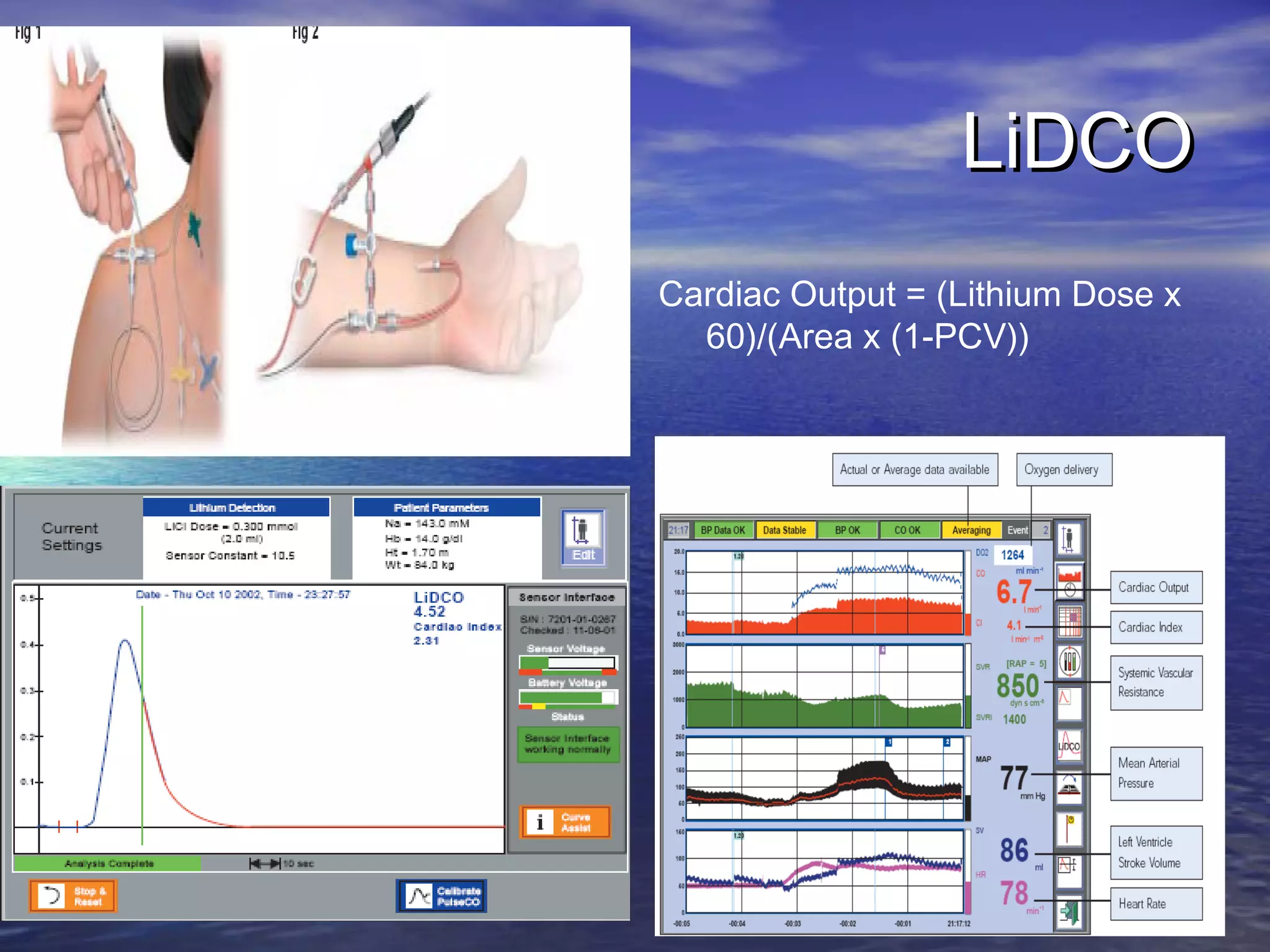
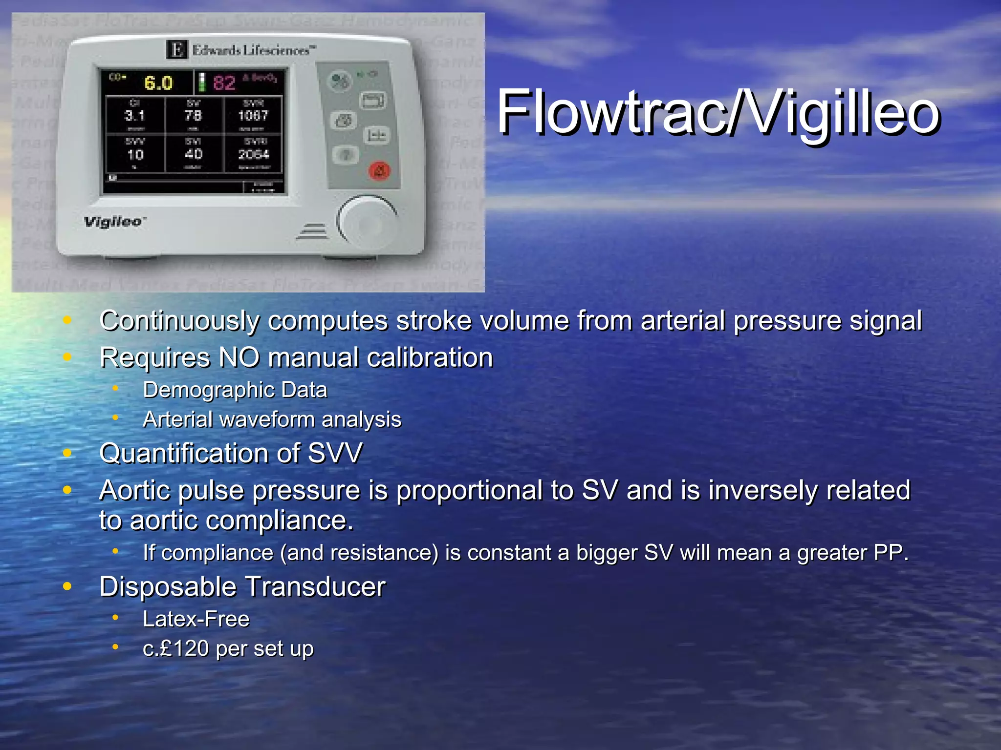
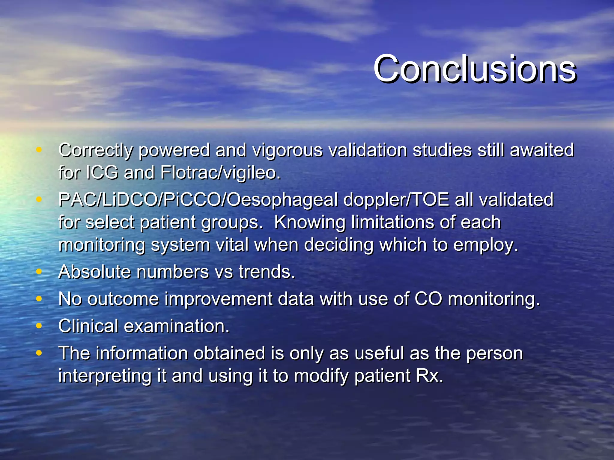
Cardiac Output Monitoring
            Dr Peter Sherren
Background
• While goal-directed therapy in severe Sepsis/septic shock has
   shown to improve outcome, CO and DO2 is only part of the goal
   directed therapy, there are many others. Rivers et al NEJM 2001;
   345.
• PACs are regarded amongst many as the GOLD standard for
   measuring CO/LVEDP and even whilst using our GOLD standard
   we have no conclusive data for improvement in patient outcome.
   Shah et al, JAMA 2005 (5051 patients) and PAC-man study, Harvey
   et al, Lancet 2005. Given this, one does question the benefit of any
   CO monitoring, no matter how close the correlation to our GOLD
   standard.
• Infact Edwards Lifesciences at the beginning of all their
   sales/presentation always have a disclaimer ‘There is NO data to
   suggest that care is improved or outcomes differ with the utilization
   of monitors that assess cardiac function’.
Techniques for monitoring CO

• Invasive
  •   PA catheter (Dye, thermodilution intermittent/continuous)
  •   Fick Principle


• Low-invasive
  • TOE
  • Oesophageal doppler
  • Arterial pulse contour analysis (PiCCO, LiDCO, Computer algorithm)


• True Non-Invasive
  •   TTE
  •   Transthoracic bio-impedence
  •   Clinical Examination!!
Pulmonary Artery Catheter
                                  Quick summary
PAC is designed to measure:
• Intra-cardiac pressures
    RAP/RVP/PAP/PAOP
•   CO/SV and indirectly many
    SVR/LVSW etc
•   True SvO2
Contra-indications:
• Tricuspid or pulmonary valve
    mechanical prosthesis
•   Right heart mass (thrombus
    and/or tumor)
•   Tricuspid or pulmonary valve
    endocarditis
•   Many relative contraindications
    reducing accuracy
PAC cont.
PA Catheter Complications


•   Infection         • Thrombocytopenia
•   Air emboli        • Catheter knotting
•   Thrombosis        • Ventricular
                        Dysrhythmias
•   PA infarction
                      • Hemothorax
•   PA rupture
                      • Pneumothorax
•   Balloon rupture
                      • Cardiac Tamponade
Fick Principle
• Fick Principle: measure volume displacement
  •   First proposed in 1870
  •   “The total uptake or release of a substance by an
      organ is the product of the blood flow through that
      organ and the arteriovenous concentration
      difference of the substance.”
  •   CO =        O2 consumption (ml/min)             art –
      mixed venous O2 conc. (ml/l)
  •   Limited by cumbersome equipment, sampling errors,
      need for invasive monitoring and steady-state
      haemodynamic and metabolic conditions
Low Invasive Monitoring
TOE
• CO can be obtained via either:
    • SV= CSA x VTI, VTI= Vmean x t
    (using PW doppler flow through either AV/PV/LVOT)
    • SV= EDV – ESV
• CO measurements using CW doppler have been validated as a
   clinical alternative to thermodilution.
• Useful tool for regional as well as overall cardiac function. RV and
   LV discrepancies. Visual tool as well as quantative.
• Recent data to suggest management benefit in general ICU setting,
  L.E. Orme et al, Br. J. Anaesth., March 2009; 102: 340 – 344 .
• Operator dependent; time consuming; no beat to beat reactivity;
   accuracy in tachycardias; training required; cost of equipment.
Oesophageal Doppler

• Ultrasonic doppler –Continuous or pulse wave doppler
• Placed parallel to direction of blood flow
• Velocity of blood measured by change in frequency of of the
  reflected ultrasound wave
• 4 MHz continuous or 5 MHz pulsed wave
• Transoesphageal doppler
    •   Measures descending thoracic blood flow
    •   User dependent, requires anaesthetised pt currently
    •   Makes certain anatomical and mathematical assumptions
• Suprasternal doppler
    •   non-invasively measures asc. Aorta flow
    •   Limited by probe position, aortic valve abnormalites & median
        sternotomy
Oesophageal Doppler principles

            • Principle of stroke volume
              calculation from aortic
              velocity (VAo)
              measurements.

            • The area under the
              maximum aortic velocity
              envelope (VTI) represents
              the stroke distance
Cardio Q
Cardio Q cont.
Oesophageal Doppler
• Disadvantages
     •   Interference by NG tube / diathermy
     •   Dislodged by movement
     •   Contraindicated in oesophageal surgery


• Advantages
     •   Min invasive
     •   Real time measurement
     •   Rapid insertion
     •   Minimal skill level
     •   Good trend monitor
Pulse contour analysis

• 3 main types:
  • Transpulmonary thermodilution + pulse contour
    analysis (PiCCO)
  • Lithium dilution (LiDCO)
  • Pure Pulse contour analysis using computer algorithm
    (Flowtrac/Vigileo)
PiCCO
• Pulse contour analysis with intermittent thermodilution
  measurement.
• Enables continuous hemodynamic monitoring using:
   – femoral or brachial artery catheter
   – central venous catheter
• Adult or pediatric patients who have or may develop
  pulmonary edema or ARDS are likely candidates
PiCCO
• Thermodilution Parameters
 • Cardiac Output - CO                 -∆T        -∆T


 • Global Enddiastolic Volume - GEDV
 • Intrathoracic Blood Volume - ITBV
 • Extravascular Lung Water - EVLW


• Pulse Contour Parameters
 • Continuous Cardiac Output - CCO
 • Systemic Vascular Resistance -
  SVR
 • Stroke Volume Variation - SVV             t           t
PiCCO
• Advantages
  •   CCO
  •   Continuous Volume responsiveness
  •   Short response time 12 secs
  •   Paediatric application >2kg

• Disadvantages
  • Needs CVP and proximal arterial line
  • Needs calibration
LiDCO
• Minimally invasive
• Safe, non-toxic rapidly redistributed and no 1st pass
  effect
• LiCl: 0.002mmol/l injected into central vein (peripheral
  administration possible as well)
• Arterial plasma conc. measured by withdrawing blood
  across lithium selective electrode at 4ml/min
• CO calculated from Li dose and area under primary
  concentration-time curve before re-circulation
LiDCO
Cardiac Output = (Lithium Dose x
  60)/(Area x (1-PCV))
Advantages
• SAFE
    • Central/peripheral venous and arterial catheters
    • Injectate is an isotonic (150 mM) solution of lithium
      chloride
    • 0.15 -0.30 mmol for an average adult
    • patient weight (>40kg) and absence of renal dysfunction or
      dialysis
•   ACCURATE
•   SIMPLE TO USE
Disadvantages

•   NMBA interfer with calibration
•   AF
•   iABP
•   People on lithium therapy
•   Renal impairment
•   Weight <40kg
•   Pregnancy 1st trimester
•   Intra cardiac shunts
Flowtrac/Vigilleo


• Continuously computes stroke volume from arterial pressure signal
• Requires NO manual calibration
    •   Demographic Data
    •   Arterial waveform analysis
• Quantification of SVV
• Aortic pulse pressure is proportional to SV and is inversely related
   to aortic compliance.
    •   If compliance (and resistance) is constant a bigger SV will mean a greater PP.
• Disposable Transducer
    •   Latex-Free
    •   c.£120 per set up
Trending Stroke Volume
                                                              Systolic
                                                              press.


                                                              PP ∝ SV

                                                              Diastolic
                                                              press.
• Arterial pressure is sampled at 100 Hz
• Changes in stroke volume will result in corresponding changes in the
  pulse pressure
• A robust “whole waveform” measure of the pulse pressure is
  achieved by taking the standard deviation of the sampled points of
  each beat
• sd(AP) ∝ Pulse Pressure ∝ Stroke Volume
• SV estimates are calculated every 20 sec
The effect of compliance on PP:
                   Age, gender and BSA factors
                  • Younger                        • Older
                                        vs.
                  • Male                vs         • Female

                  • Higher BSA
                                        .
                                        vs.        • Lower BSA


For the same
volume ➔

               • Compliance inversely affects PP
               • The algorithm compensates for the
               effects of compliance on PP based on age,
               gender and BSA
Effect of vascular tone
• The algorithm looks for characteristic changes in the arterial
  pressure waveform that reflect changes in tone (i.e., MAP,
  Skewness, Kurtosis)
• Those changes are included in the continuous calculation.


        MAP                   Skewness                  Kurtosis
Error Sources
           ICO                        CCO                       FloTrac
                                                                sensor
Comp constant                ------                       Patient data
                                                          Age
                                                          Height & weight
                                                          Gender
Clinician technique          ------                       AS/AI
Injectate Volume
                                                          Dysrhythmia
Injectate Temperature
Injection Timing
Catheter migration           Catheter migration           Sensor height
Ventilator timing            Sequential compression       Aortic balloon pump
                             device
Patient temperature shifts   Patient temperature shifts   Patient arm movement
Infusions & drips            Infusions & drips            Line bubbles
                                                          catheter whip (fem)
Valve regurgitation          Valve regurgitation          Pressure dampening
                                                          (extreme vasopressors)
Validation?
• ‘Cardiac Output Determination From the Arterial Pressure
   Wave: Clinical Testing of a Novel Algorithm That Does Not
   Require Calibration’ Journal of Cardiothorac and Vasc Anesth
   2007.


• ‘Uncalibrated pulse contour-derived stoke volume variation
   predict fluid responsiveness in mechanically ventilated patients
   undergoing liver transplantation’ BJA 2008.


• Still awaiting rigorous validation studies.
True Non-Invasive Monitoring
TTE

•   Non invasive
•   Similar method of calculation of CO to TOE
•   In addition to the limitation of TOE, obtaining
    echocardiographic windows in mechanically
    ventilated patients can be difficult.
Transthoracic bio-impedence (Impedence
                   Cardiography/ Impedance plethysmograpghy)

•   4 dual sensors with 8 lead wires
    placed on neck and chest

•   Current transmitted and seeks
    path of least resistance: blood
    filled aorta
•   Baseline impedance (resistance)
    to signal is measured
•   With each heartbeat, blood
    volume and velocity in the aorta
    change
•   Corresponding change in
    impedance is measured
•   Baseline and changes in
    impedance are used to measure
    and calculate hemodynamic
    parameters
Parameters

• Measured                     • Calculated
                                 –   Stroke Volume / Index
  –   Heart Rate
                                 –   Cardiac Output / Index
  –   Non Invasive BP            –   SVR / SVRI
  –   Velocity Index             –   LCW / LCWI
  –   Acceleration Index         –   Systolic Time Ratio
                                 –   Heather Index
  –   Thoracic Fluid Content     –   Ejection Time Ratio
  –   Pre-Ejection Time
  –   LV Ejection Time
ICG
Validation?
• Ziegler D, et al. “Comparison of cardiac output
  measurements by TEB vs. intermittent bolus
  thermodilution in mechanically ventilated
  patients” Chest. 1999; Vol. 16, No. 4 (suppl. 2):281S.

• Scott Sageman, et al. “Equivalence of
  Bioimpedance and Thermodilution in Measuring
  Cardiac Index After Cardiac Surgery” J
  Cardiothoracic & Vasc. Anesth. 16:8-14, 2002
Conclusions

• Correctly powered and vigorous validation studies still awaited
    for ICG and Flotrac/vigileo.
•   PAC/LiDCO/PiCCO/Oesophageal doppler/TOE all validated
    for select patient groups. Knowing limitations of each
    monitoring system vital when deciding which to employ.
•   Absolute numbers vs trends.
•   No outcome improvement data with use of CO monitoring.
•   Clinical examination.
•   The information obtained is only as useful as the person
    interpreting it and using it to modify patient Rx.
A fool with a tool is still a fool.

Cardiac output monitoring

  • 1.
  • 2.
    Background • While goal-directedtherapy in severe Sepsis/septic shock has shown to improve outcome, CO and DO2 is only part of the goal directed therapy, there are many others. Rivers et al NEJM 2001; 345. • PACs are regarded amongst many as the GOLD standard for measuring CO/LVEDP and even whilst using our GOLD standard we have no conclusive data for improvement in patient outcome. Shah et al, JAMA 2005 (5051 patients) and PAC-man study, Harvey et al, Lancet 2005. Given this, one does question the benefit of any CO monitoring, no matter how close the correlation to our GOLD standard. • Infact Edwards Lifesciences at the beginning of all their sales/presentation always have a disclaimer ‘There is NO data to suggest that care is improved or outcomes differ with the utilization of monitors that assess cardiac function’.
  • 3.
    Techniques for monitoringCO • Invasive • PA catheter (Dye, thermodilution intermittent/continuous) • Fick Principle • Low-invasive • TOE • Oesophageal doppler • Arterial pulse contour analysis (PiCCO, LiDCO, Computer algorithm) • True Non-Invasive • TTE • Transthoracic bio-impedence • Clinical Examination!!
  • 4.
    Pulmonary Artery Catheter Quick summary PAC is designed to measure: • Intra-cardiac pressures RAP/RVP/PAP/PAOP • CO/SV and indirectly many SVR/LVSW etc • True SvO2 Contra-indications: • Tricuspid or pulmonary valve mechanical prosthesis • Right heart mass (thrombus and/or tumor) • Tricuspid or pulmonary valve endocarditis • Many relative contraindications reducing accuracy
  • 5.
  • 6.
    PA Catheter Complications • Infection • Thrombocytopenia • Air emboli • Catheter knotting • Thrombosis • Ventricular Dysrhythmias • PA infarction • Hemothorax • PA rupture • Pneumothorax • Balloon rupture • Cardiac Tamponade
  • 7.
    Fick Principle • FickPrinciple: measure volume displacement • First proposed in 1870 • “The total uptake or release of a substance by an organ is the product of the blood flow through that organ and the arteriovenous concentration difference of the substance.” • CO = O2 consumption (ml/min) art – mixed venous O2 conc. (ml/l) • Limited by cumbersome equipment, sampling errors, need for invasive monitoring and steady-state haemodynamic and metabolic conditions
  • 8.
  • 9.
    TOE • CO canbe obtained via either: • SV= CSA x VTI, VTI= Vmean x t (using PW doppler flow through either AV/PV/LVOT) • SV= EDV – ESV • CO measurements using CW doppler have been validated as a clinical alternative to thermodilution. • Useful tool for regional as well as overall cardiac function. RV and LV discrepancies. Visual tool as well as quantative. • Recent data to suggest management benefit in general ICU setting, L.E. Orme et al, Br. J. Anaesth., March 2009; 102: 340 – 344 . • Operator dependent; time consuming; no beat to beat reactivity; accuracy in tachycardias; training required; cost of equipment.
  • 10.
    Oesophageal Doppler • Ultrasonicdoppler –Continuous or pulse wave doppler • Placed parallel to direction of blood flow • Velocity of blood measured by change in frequency of of the reflected ultrasound wave • 4 MHz continuous or 5 MHz pulsed wave • Transoesphageal doppler • Measures descending thoracic blood flow • User dependent, requires anaesthetised pt currently • Makes certain anatomical and mathematical assumptions • Suprasternal doppler • non-invasively measures asc. Aorta flow • Limited by probe position, aortic valve abnormalites & median sternotomy
  • 11.
    Oesophageal Doppler principles • Principle of stroke volume calculation from aortic velocity (VAo) measurements. • The area under the maximum aortic velocity envelope (VTI) represents the stroke distance
  • 12.
  • 13.
  • 14.
    Oesophageal Doppler • Disadvantages • Interference by NG tube / diathermy • Dislodged by movement • Contraindicated in oesophageal surgery • Advantages • Min invasive • Real time measurement • Rapid insertion • Minimal skill level • Good trend monitor
  • 15.
    Pulse contour analysis •3 main types: • Transpulmonary thermodilution + pulse contour analysis (PiCCO) • Lithium dilution (LiDCO) • Pure Pulse contour analysis using computer algorithm (Flowtrac/Vigileo)
  • 16.
    PiCCO • Pulse contouranalysis with intermittent thermodilution measurement. • Enables continuous hemodynamic monitoring using: – femoral or brachial artery catheter – central venous catheter • Adult or pediatric patients who have or may develop pulmonary edema or ARDS are likely candidates
  • 17.
    PiCCO • Thermodilution Parameters • Cardiac Output - CO -∆T -∆T • Global Enddiastolic Volume - GEDV • Intrathoracic Blood Volume - ITBV • Extravascular Lung Water - EVLW • Pulse Contour Parameters • Continuous Cardiac Output - CCO • Systemic Vascular Resistance - SVR • Stroke Volume Variation - SVV t t
  • 18.
    PiCCO • Advantages • CCO • Continuous Volume responsiveness • Short response time 12 secs • Paediatric application >2kg • Disadvantages • Needs CVP and proximal arterial line • Needs calibration
  • 19.
    LiDCO • Minimally invasive •Safe, non-toxic rapidly redistributed and no 1st pass effect • LiCl: 0.002mmol/l injected into central vein (peripheral administration possible as well) • Arterial plasma conc. measured by withdrawing blood across lithium selective electrode at 4ml/min • CO calculated from Li dose and area under primary concentration-time curve before re-circulation
  • 20.
    LiDCO Cardiac Output =(Lithium Dose x 60)/(Area x (1-PCV))
  • 21.
    Advantages • SAFE • Central/peripheral venous and arterial catheters • Injectate is an isotonic (150 mM) solution of lithium chloride • 0.15 -0.30 mmol for an average adult • patient weight (>40kg) and absence of renal dysfunction or dialysis • ACCURATE • SIMPLE TO USE
  • 22.
    Disadvantages • NMBA interfer with calibration • AF • iABP • People on lithium therapy • Renal impairment • Weight <40kg • Pregnancy 1st trimester • Intra cardiac shunts
  • 23.
    Flowtrac/Vigilleo • Continuously computesstroke volume from arterial pressure signal • Requires NO manual calibration • Demographic Data • Arterial waveform analysis • Quantification of SVV • Aortic pulse pressure is proportional to SV and is inversely related to aortic compliance. • If compliance (and resistance) is constant a bigger SV will mean a greater PP. • Disposable Transducer • Latex-Free • c.£120 per set up
  • 24.
    Trending Stroke Volume Systolic press. PP ∝ SV Diastolic press. • Arterial pressure is sampled at 100 Hz • Changes in stroke volume will result in corresponding changes in the pulse pressure • A robust “whole waveform” measure of the pulse pressure is achieved by taking the standard deviation of the sampled points of each beat • sd(AP) ∝ Pulse Pressure ∝ Stroke Volume • SV estimates are calculated every 20 sec
  • 25.
    The effect ofcompliance on PP: Age, gender and BSA factors • Younger • Older vs. • Male vs • Female • Higher BSA . vs. • Lower BSA For the same volume ➔ • Compliance inversely affects PP • The algorithm compensates for the effects of compliance on PP based on age, gender and BSA
  • 26.
    Effect of vasculartone • The algorithm looks for characteristic changes in the arterial pressure waveform that reflect changes in tone (i.e., MAP, Skewness, Kurtosis) • Those changes are included in the continuous calculation. MAP Skewness Kurtosis
  • 27.
    Error Sources ICO CCO FloTrac sensor Comp constant ------ Patient data Age Height & weight Gender Clinician technique ------ AS/AI Injectate Volume Dysrhythmia Injectate Temperature Injection Timing Catheter migration Catheter migration Sensor height Ventilator timing Sequential compression Aortic balloon pump device Patient temperature shifts Patient temperature shifts Patient arm movement Infusions & drips Infusions & drips Line bubbles catheter whip (fem) Valve regurgitation Valve regurgitation Pressure dampening (extreme vasopressors)
  • 28.
    Validation? • ‘Cardiac OutputDetermination From the Arterial Pressure Wave: Clinical Testing of a Novel Algorithm That Does Not Require Calibration’ Journal of Cardiothorac and Vasc Anesth 2007. • ‘Uncalibrated pulse contour-derived stoke volume variation predict fluid responsiveness in mechanically ventilated patients undergoing liver transplantation’ BJA 2008. • Still awaiting rigorous validation studies.
  • 29.
  • 30.
    TTE • Non invasive • Similar method of calculation of CO to TOE • In addition to the limitation of TOE, obtaining echocardiographic windows in mechanically ventilated patients can be difficult.
  • 31.
    Transthoracic bio-impedence (Impedence Cardiography/ Impedance plethysmograpghy) • 4 dual sensors with 8 lead wires placed on neck and chest • Current transmitted and seeks path of least resistance: blood filled aorta • Baseline impedance (resistance) to signal is measured • With each heartbeat, blood volume and velocity in the aorta change • Corresponding change in impedance is measured • Baseline and changes in impedance are used to measure and calculate hemodynamic parameters
  • 32.
    Parameters • Measured • Calculated – Stroke Volume / Index – Heart Rate – Cardiac Output / Index – Non Invasive BP – SVR / SVRI – Velocity Index – LCW / LCWI – Acceleration Index – Systolic Time Ratio – Heather Index – Thoracic Fluid Content – Ejection Time Ratio – Pre-Ejection Time – LV Ejection Time
  • 33.
  • 34.
    Validation? • Ziegler D,et al. “Comparison of cardiac output measurements by TEB vs. intermittent bolus thermodilution in mechanically ventilated patients” Chest. 1999; Vol. 16, No. 4 (suppl. 2):281S. • Scott Sageman, et al. “Equivalence of Bioimpedance and Thermodilution in Measuring Cardiac Index After Cardiac Surgery” J Cardiothoracic & Vasc. Anesth. 16:8-14, 2002
  • 35.
    Conclusions • Correctly poweredand vigorous validation studies still awaited for ICG and Flotrac/vigileo. • PAC/LiDCO/PiCCO/Oesophageal doppler/TOE all validated for select patient groups. Knowing limitations of each monitoring system vital when deciding which to employ. • Absolute numbers vs trends. • No outcome improvement data with use of CO monitoring. • Clinical examination. • The information obtained is only as useful as the person interpreting it and using it to modify patient Rx.
  • 36.
    A fool witha tool is still a fool.

Editor's Notes

  • #5 High PEEP,Tamponade, Pneumothorax, reduced myocardial compliance, Mitral stenosis, Pulmonary occlusive disease, severe MR
  • #25 Arterial pressure is sampled at 100 times per second (100 Hz). Each sample is a pressure data point measured in mm Hg. This data is analyzed and updated every 20 seconds, utilizing the 2000 data points (20 sec x 100 Hz) collected. The standard deviation (sd(AP)) of these data points is proportional to the pulse pressure, which is proportional to the stroke volume. This method is robust in its assessment of pulse pressure because it looks at the whole waveform. sd(AP) = a measure of variance The Stroke Volume is averaged and displayed every 20 seconds. The user has the capability to choose between a 20 second calculation or 5 minute moving average option.
  • #26 There is a direct relationship (i.e., the shape of the trend shown above) between arterial pressure and large vessel compliance as it relates to a human’s age or gender. That is, a male will typically have a more compliant aorta than a female of the same age, and a younger person will have a more compliant aorta than an elderly person. This relationship was quantified and mathematically modeled by Langewouters. Through development of the algorithm, it has also been found that there is a relationship between aortic compliance and BSA. For example, a larger person (higher BSA) will typically have more compliant vessels than a smaller person (lower BSA). As part of the assessment for vascular tone, estimates of aortic compliance based on the above principles are important but not all encompassing. Therefore, in addition to Langewouter’s mathematical model, further waveform analysis is conducted by the algorithm to take into account patient specific, real time effects of vascular tone on the waveform.
  • #27 The aortic compliance function (i.e., age, gender, BSA function) within X helps to provide a “ballpark estimate” of the patient’s likely vascular tone. Further analysis of the waveform shape is also a significant factor to providing vascular tone estimates essential to the SV calculation. This method allows for reliable calculation of key flow parameters without manual calibration. X (pronounced “khi”) = a symbol for dynamic polynomial functions, a function that continuously adjusts to multiple changing variables Depending upon the state of the patient’s vasculature, the arterial waveform will take a shape that can be characterized mathematically. The statistical tools used, in addition to the aforementioned standard deviation, are the mean, skewness and kurtosis. Mean , clinically known as MAP, can provide an indication of the increase or decrease in resistance. Skewness , or symmetry of the data, is also often associated with the stiffness of the patient’s vasculature. Kurtosis , a measure of how peaked or flat the data distribution is, provides an indication of the nature of vasculature as well. For example, larger vessels will typically have a “flat” distribution as compared to peripheral vasculature. Skewness : The angle or slope exhibited on the rise of the waveform. Kurtosis : How flat and wide the waveform is If the shape changes, the mathematical calculation will change, providing a patient-specific, real time assessment of shifts in vascular tone. The greater the magnitude increase in tone, as calculated as a decrease in X, the lesser the weight pulse pressure estimates (i.e., sd(AP)) will have in the SV calculation. Furthermore, the effect of differences in arterial sites on the arterial waveform are neutralized within the X function. Therefore, FloTrac can be used with any existing peripheral arterial catheter. This is possible through the analysis of arterial waveform characteristics that are specific to different points within the vascular tree. Two key variables in this portion of the function are the standard deviation (pulsatility) and the kurtosis (peaked/flat nature) of arterial pressure.
  • #28 Additional sources of error are noted and should be taken into consideration when comparing these two very different technologies.