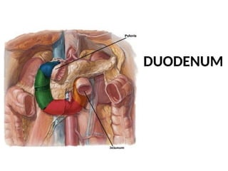
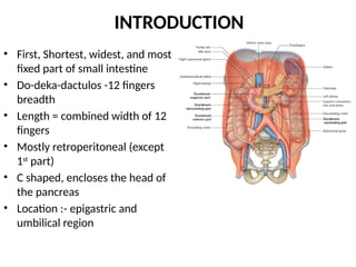
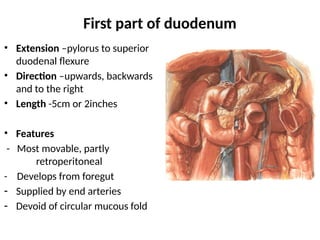
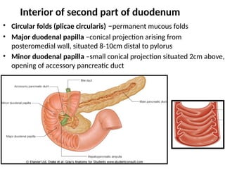
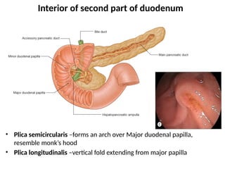
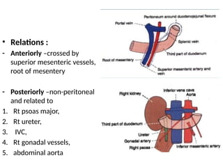
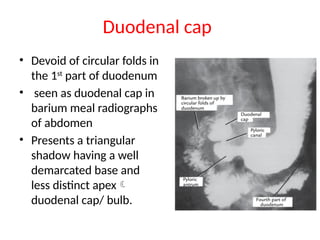
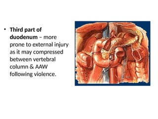
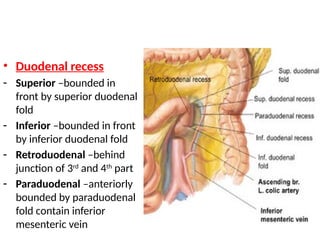
DUODENUM
INTRODUCTION
• First, Shortest, widest, and most
fixed part of small intestine
• Do-deka-dactulos -12 fingers
breadth
• Length = combined width of 12
fingers
• Mostly retroperitoneal (except
1st
part)
• C shaped, encloses the head of
the pancreas
• Location :- epigastric and
umbilical region
Subdivided into four parts
• First or upper part –2inches/ 5cm
• Second or vertical part –3inches/ 7.5cm
• Third or horizontal part –4inches/10cm
• Fourth or ascending part – 1inch/ 2.5cm
First part of duodenum
• Extension –pylorus to superior
duodenal flexure
• Direction –upwards, backwards
and to the right
• Length -5cm or 2inches
• Features
- Most movable, partly
retroperitoneal
- Develops from foregut
- Supplied by end arteries
- Devoid of circular mucous fold
• Relations :
- Anteriorly – covered with
peritoneum of the greater sac,
and is related to
i. Quadrate lobe of liver
ii. Neck and body of gall
bladder
- Posteriorly –non-peritoneal
related to
i. Portal vein,
ii. Bile duct,
iii. Gastroduodenal artery,
iv. IVC
Relations
• Superiorly –forms floor of
epiploic foramen and
related to hepatic artery
• Inferiorly –head and neck of
pancreas
Second part of duodenum
• Extension –from superior
duodenal flexure to
inferior duodenal flexure
• Upper ½ develops from
foregut & lower ½ from
midgut.
• Direction –downward in
front of hilum of kidney
• Length –3inches or 7.5cm
• Relations :
- Anteriorly –
i. Rt lobe of liver
ii. Fundus and body of gall
bladder
iii. Transverse colon
- Posteriorly –
i. Rt kidney, Rt renal
vessels, pelvis of Rt
ureter
ii. Rt psoas major muscle
iii. Rt edge of IVC
- Laterally –
Rt colic flexure
- Medially-
i. Head of pancreas
ii. Anastomosis of Superior and
inferior pancreatico-
duodenal vessels
iii. Bile duct and pancreatic duct
–unite to form ampulla of
Vater
Interior of second part of duodenum
• Circular folds (plicae circularis) –permanent mucous folds
• Major duodenal papilla –conical projection arising from
posteromedial wall, situated 8-10cm distal to pylorus
• Minor duodenal papilla –small conical projection situated 2cm above,
opening of accessory pancreatic duct
Interior of second part of duodenum
• Plica semicircularis –forms an arch over Major duodenal papilla,
resemble monk’s hood
• Plica longitudinalis –vertical fold extending from major papilla
Third part of duodenum
• Extension –from inferior
duodenal flexure to front of
aorta
• Length – 4 inches or 10 cm
• Relations :
- Anteriorly –crossed by
superior mesenteric vessels,
root of mesentery
- Posteriorly –non-peritoneal
and related to
1. Rt psoas major,
2. Rt ureter,
3. IVC,
4. Rt gonadal vessels,
5. abdominal aorta
Relations
- Superiorly –head of
pancreas, inferior
pancreaticoduodenal
vessels
- Inferiorly –coils of jejunum
Fourth part of duodenum
• Extension –from front of
aorta to DJ flexure
• Length –1 inch or 2.5cm
• Flexure is kept in position by
suspensory muscle of
duodenum
• Relations :
- Anteriorly –covered with
peritoneum, related to
transverse colon and
mesocolon
- Posteriorly –Lt crus of
diaphragm, Lt psoas major, Lt
sympathetic trunk, Lt gonadal
vessels, Lt suprarenal vein,
inferior mesenteric vein
- Rt side –uncinate process
- Lt side –Lt kidney, ureter
- Above –body of pancreas
Suspensory muscle of duodenum
OR
Suspensory ligament of treitz
• Fibro-muscular band arises
from Rt crus of diaphragm
• Passes downward and
forward by side of coeliac
artery
• The band is attached to
posterior surface of DJ flexure
• Upper 1/3 –striated muscle
middle 1/3 –elastic fibres
lower 1/3 –nonstriated
muscle
• Function –flexes Dj flexure
and prevent it from being
dragged down
- Landmark in radiological
diagnosis of incomplete
rotation or mal rotation of
small intestine
Structure of duodenum
• Serosa –derived from
peritoneum, incomplete
• Muscularis externa –outer
longitudinal, inner circular
separated by myenteric
plexus of nerves
• Submucosa –loose areolar
tissue contain plexus of blood
vessels, lymphatics, meissner’s
plexus, Brunner’s gland
• Mucosa –surface epithelium,
lamina propria, muscularis
mucosa
ENDOSCOPIC APPEARANCE
A) 1ST
part
B) 2nd
part
C) Major duodenal
papilla
D) 3rd
part
Blood supply of duodenum
• Superior pancreaticoduodenal artery
• Inferior pancreaticoduodenal artery
• Supraduodenal artery of Wilkie br of Gastroduodenal art
• Retroduodenal branch of gastroduodenal artery
• Leash of branches of hepatic artery
• Branches from Rt gastroepiploic artery
• Artery from first jejunal branch of SMA
Venous drainage
• Veins corresponding to
arteries drain into SMV
and portal vein
Lymphatic drainage
• Pancreaticoduodenal lymph
nodes
• coeliac and superior
mesenteric groups of pre-
aortic lymph nodes
Nerve supply
• Sympathetic – from T6-T9
• Parasympathetic –from
both vagi through celiac
and superior mesenteric
plexus
Applied
• First part of duodenum –prone
to peptic ulcer, supplied by
series of end arteries
1. Mucosal folds are absent
2. Acidic contents of stomach
enters
- Causes –trauma, alcohol,
smoking, ulcerogenic drugs
(NSAID)
- Sometimes gastroduodenal
artery passing behind first part
and get erroded by peptic ulcer,
producing severe haemorrhage
Duodenal cap
• Devoid of circular folds in
the 1st
part of duodenum
• seen as duodenal cap in
barium meal radiographs
of abdomen
• Presents a triangular
shadow having a well
demarcated base and
less distinct apex
duodenal cap/ bulb.
• Second part –more
protected, opening of bile
duct and pancreatic duct
• Small bile stones impacted on
summit of major duodenal
papilla –obstructive jaundice
• Annular type of head of
pancreas –encircles the
duodenum – malignant
growth of pancreatic head –
duodenal obstruction
• Third part of
duodenum – more
prone to external injury
as it may compressed
between vertebral
column & AAW
following violence.
• Duodenal recess
- Superior –bounded in
front by superior duodenal
fold
- Inferior –bounded in front
by inferior duodenal fold
- Retroduodenal –behind
junction of 3rd
and 4th
part
- Paraduodenal –anteriorly
bounded by paraduodenal
fold contain inferior
mesenteric vein
• A loop of small gut may herniate into any of
the recess
• If the gut fails to return, the internal hernia
thus formed produces intestinal obstruction
Duodenum.pptx...........................

Duodenum.pptx...........................

  • 1.
  • 2.
    INTRODUCTION • First, Shortest,widest, and most fixed part of small intestine • Do-deka-dactulos -12 fingers breadth • Length = combined width of 12 fingers • Mostly retroperitoneal (except 1st part) • C shaped, encloses the head of the pancreas • Location :- epigastric and umbilical region
  • 3.
    Subdivided into fourparts • First or upper part –2inches/ 5cm • Second or vertical part –3inches/ 7.5cm • Third or horizontal part –4inches/10cm • Fourth or ascending part – 1inch/ 2.5cm
  • 4.
    First part ofduodenum • Extension –pylorus to superior duodenal flexure • Direction –upwards, backwards and to the right • Length -5cm or 2inches • Features - Most movable, partly retroperitoneal - Develops from foregut - Supplied by end arteries - Devoid of circular mucous fold
  • 5.
    • Relations : -Anteriorly – covered with peritoneum of the greater sac, and is related to i. Quadrate lobe of liver ii. Neck and body of gall bladder - Posteriorly –non-peritoneal related to i. Portal vein, ii. Bile duct, iii. Gastroduodenal artery, iv. IVC
  • 6.
    Relations • Superiorly –formsfloor of epiploic foramen and related to hepatic artery • Inferiorly –head and neck of pancreas
  • 7.
    Second part ofduodenum • Extension –from superior duodenal flexure to inferior duodenal flexure • Upper ½ develops from foregut & lower ½ from midgut. • Direction –downward in front of hilum of kidney • Length –3inches or 7.5cm
  • 8.
    • Relations : -Anteriorly – i. Rt lobe of liver ii. Fundus and body of gall bladder iii. Transverse colon
  • 9.
    - Posteriorly – i.Rt kidney, Rt renal vessels, pelvis of Rt ureter ii. Rt psoas major muscle iii. Rt edge of IVC
  • 10.
    - Laterally – Rtcolic flexure - Medially- i. Head of pancreas ii. Anastomosis of Superior and inferior pancreatico- duodenal vessels iii. Bile duct and pancreatic duct –unite to form ampulla of Vater
  • 12.
    Interior of secondpart of duodenum • Circular folds (plicae circularis) –permanent mucous folds • Major duodenal papilla –conical projection arising from posteromedial wall, situated 8-10cm distal to pylorus • Minor duodenal papilla –small conical projection situated 2cm above, opening of accessory pancreatic duct
  • 13.
    Interior of secondpart of duodenum • Plica semicircularis –forms an arch over Major duodenal papilla, resemble monk’s hood • Plica longitudinalis –vertical fold extending from major papilla
  • 14.
    Third part ofduodenum • Extension –from inferior duodenal flexure to front of aorta • Length – 4 inches or 10 cm
  • 15.
    • Relations : -Anteriorly –crossed by superior mesenteric vessels, root of mesentery - Posteriorly –non-peritoneal and related to 1. Rt psoas major, 2. Rt ureter, 3. IVC, 4. Rt gonadal vessels, 5. abdominal aorta
  • 16.
    Relations - Superiorly –headof pancreas, inferior pancreaticoduodenal vessels - Inferiorly –coils of jejunum
  • 17.
    Fourth part ofduodenum • Extension –from front of aorta to DJ flexure • Length –1 inch or 2.5cm • Flexure is kept in position by suspensory muscle of duodenum
  • 18.
    • Relations : -Anteriorly –covered with peritoneum, related to transverse colon and mesocolon - Posteriorly –Lt crus of diaphragm, Lt psoas major, Lt sympathetic trunk, Lt gonadal vessels, Lt suprarenal vein, inferior mesenteric vein - Rt side –uncinate process - Lt side –Lt kidney, ureter - Above –body of pancreas
  • 19.
    Suspensory muscle ofduodenum OR Suspensory ligament of treitz • Fibro-muscular band arises from Rt crus of diaphragm • Passes downward and forward by side of coeliac artery • The band is attached to posterior surface of DJ flexure • Upper 1/3 –striated muscle middle 1/3 –elastic fibres lower 1/3 –nonstriated muscle
  • 20.
    • Function –flexesDj flexure and prevent it from being dragged down - Landmark in radiological diagnosis of incomplete rotation or mal rotation of small intestine
  • 21.
    Structure of duodenum •Serosa –derived from peritoneum, incomplete • Muscularis externa –outer longitudinal, inner circular separated by myenteric plexus of nerves • Submucosa –loose areolar tissue contain plexus of blood vessels, lymphatics, meissner’s plexus, Brunner’s gland • Mucosa –surface epithelium, lamina propria, muscularis mucosa
  • 22.
    ENDOSCOPIC APPEARANCE A) 1ST part B)2nd part C) Major duodenal papilla D) 3rd part
  • 23.
    Blood supply ofduodenum • Superior pancreaticoduodenal artery • Inferior pancreaticoduodenal artery • Supraduodenal artery of Wilkie br of Gastroduodenal art • Retroduodenal branch of gastroduodenal artery • Leash of branches of hepatic artery • Branches from Rt gastroepiploic artery • Artery from first jejunal branch of SMA
  • 24.
    Venous drainage • Veinscorresponding to arteries drain into SMV and portal vein
  • 25.
    Lymphatic drainage • Pancreaticoduodenallymph nodes • coeliac and superior mesenteric groups of pre- aortic lymph nodes
  • 26.
    Nerve supply • Sympathetic– from T6-T9 • Parasympathetic –from both vagi through celiac and superior mesenteric plexus
  • 27.
    Applied • First partof duodenum –prone to peptic ulcer, supplied by series of end arteries 1. Mucosal folds are absent 2. Acidic contents of stomach enters - Causes –trauma, alcohol, smoking, ulcerogenic drugs (NSAID) - Sometimes gastroduodenal artery passing behind first part and get erroded by peptic ulcer, producing severe haemorrhage
  • 28.
    Duodenal cap • Devoidof circular folds in the 1st part of duodenum • seen as duodenal cap in barium meal radiographs of abdomen • Presents a triangular shadow having a well demarcated base and less distinct apex duodenal cap/ bulb.
  • 29.
    • Second part–more protected, opening of bile duct and pancreatic duct • Small bile stones impacted on summit of major duodenal papilla –obstructive jaundice • Annular type of head of pancreas –encircles the duodenum – malignant growth of pancreatic head – duodenal obstruction
  • 30.
    • Third partof duodenum – more prone to external injury as it may compressed between vertebral column & AAW following violence.
  • 31.
    • Duodenal recess -Superior –bounded in front by superior duodenal fold - Inferior –bounded in front by inferior duodenal fold - Retroduodenal –behind junction of 3rd and 4th part - Paraduodenal –anteriorly bounded by paraduodenal fold contain inferior mesenteric vein
  • 32.
    • A loopof small gut may herniate into any of the recess • If the gut fails to return, the internal hernia thus formed produces intestinal obstruction