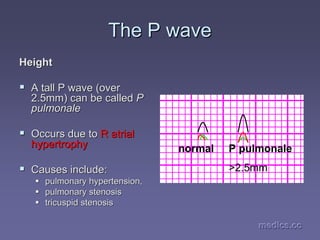
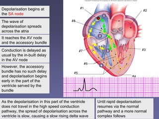
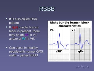
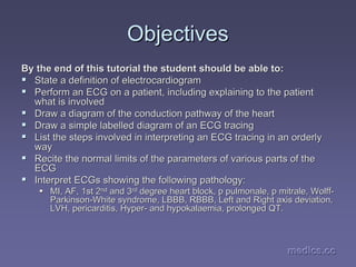
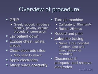
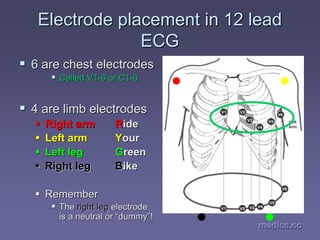
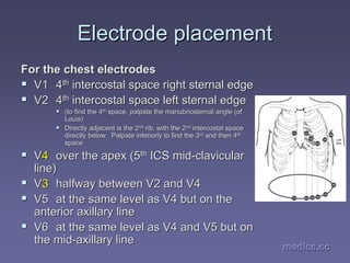
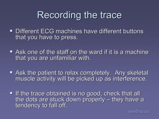
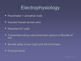
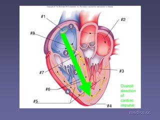
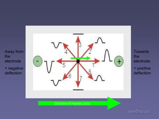
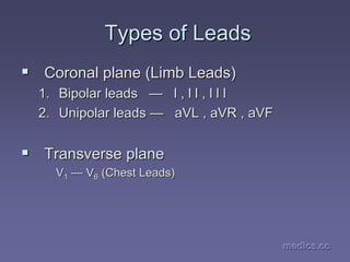
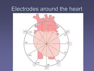
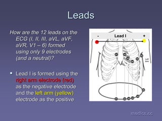
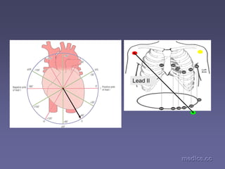
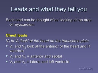
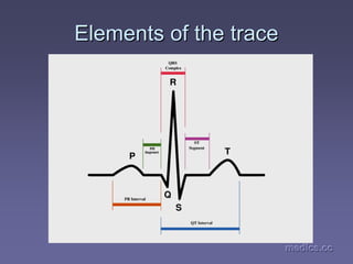
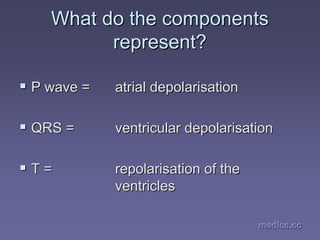
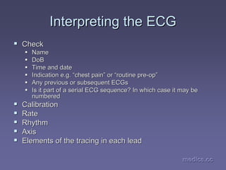
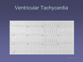
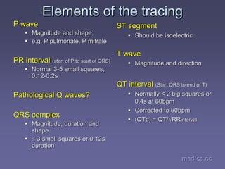
This document provides an overview of performing and interpreting electrocardiograms (ECGs). It outlines the objectives of understanding ECGs, including defining an ECG, performing one, and interpreting various cardiac pathologies. The document explains that an ECG is a tracing of the heart's electrical activity and describes the process for recording one, including electrode placement and the cardiac conduction system. It also provides a high-level overview of the typical waves, segments and intervals seen on an ECG tracing and how the different leads view the heart.
crème de la crème basics to understand electrocardiographic analysis in an easy & simple way with some specifications to its use in Emergency medicine/clinical toxicology practice.
ECG Rhythm Interpretation
ST Elevation and non-ST Elevation MIs
ECG Changes
ECG Changes & the Evolving MI
Left Ventricular Hypertrophy
Normal Impulse Conduction
Bundle Branch Blocks
ECG Lecture: Sinus arrest, sinoatrial exit block, AV block and escape rhythmsMichael-Joseph Agbayani
Simple ECG lecture about sinus arrest, sinoatrial exit block, AV block and escape rhythms. Slideshow was made with an audience of medical professionals in mind.
A 45 years old lady presented with generalized weakness and palpitations. She is a diagnosed case of chronic renal failure with Diabetes mellitus and Hypertension. Her serum K+ level is 6.8 meq/L. She had the following ECG.
Case; A 54 years old gentleman complained of chest discomfort on exertion for the last 5 months. He is smoker for 10 years, diabetic for 5 years and hypertensive for 3 years. He had the following ECG.
Case: A 25 years old gentleman presented with chest pain and fever .He was normotensive, non-smoker and non-diabetic. His pulse 128b/min and BP-130/80 mm Hg. Troponin I was normal.
Case: A 58 years old gentleman complained of severe central chest pain with excessive sweating 5 days back. He is smoker for 7 years, diabetic for 5 years and hypertensive for 4 years. His BP-90/70 mm Hg. He had the following ECG.
-Anatomical description of duodenum.
-Physiological functions of duodenum.
-Histology of duodenum.
-Duodenum blood supply and its innervation.
-Some disease and disorders that affect duodenum and its function.
-Anatomical description of kidney.
-Physiological functions of kidney.
-Kidney blood supply and its innervation.
-Some disease and disorders that affect kidneys and its function.
anatomical description of thyroid gland.
physiological functions of thyroid gland.
blood supply and its innervation.
some disease and disorders that affect thyroid glad and its function.
Factory Supply Best Quality Pmk Oil CAS 28578–16–7 PMK Powder in Stockrebeccabio
Factory Supply Best Quality Pmk Oil CAS 28578–16–7 PMK Powder in Stock
Telegram: bmksupplier
signal: +85264872720
threema: TUD4A6YC
You can contact me on Telegram or Threema
Communicate promptly and reply
Free of customs clearance, Double Clearance 100% pass delivery to USA, Canada, Spain, Germany, Netherland, Poland, Italy, Sweden, UK, Czech Republic, Australia, Mexico, Russia, Ukraine, Kazakhstan.Door to door service
Hot Selling Organic intermediates
Tom Selleck Health: A Comprehensive Look at the Iconic Actor’s Wellness Journeygreendigital
Tom Selleck, an enduring figure in Hollywood. has captivated audiences for decades with his rugged charm, iconic moustache. and memorable roles in television and film. From his breakout role as Thomas Magnum in Magnum P.I. to his current portrayal of Frank Reagan in Blue Bloods. Selleck's career has spanned over 50 years. But beyond his professional achievements. fans have often been curious about Tom Selleck Health. especially as he has aged in the public eye.
Follow us on: Pinterest
Introduction
Many have been interested in Tom Selleck health. not only because of his enduring presence on screen but also because of the challenges. and lifestyle choices he has faced and made over the years. This article delves into the various aspects of Tom Selleck health. exploring his fitness regimen, diet, mental health. and the challenges he has encountered as he ages. We'll look at how he maintains his well-being. the health issues he has faced, and his approach to ageing .
Early Life and Career
Childhood and Athletic Beginnings
Tom Selleck was born on January 29, 1945, in Detroit, Michigan, and grew up in Sherman Oaks, California. From an early age, he was involved in sports, particularly basketball. which played a significant role in his physical development. His athletic pursuits continued into college. where he attended the University of Southern California (USC) on a basketball scholarship. This early involvement in sports laid a strong foundation for his physical health and disciplined lifestyle.
Transition to Acting
Selleck's transition from an athlete to an actor came with its physical demands. His first significant role in "Magnum P.I." required him to perform various stunts and maintain a fit appearance. This role, which he played from 1980 to 1988. necessitated a rigorous fitness routine to meet the show's demands. setting the stage for his long-term commitment to health and wellness.
Fitness Regimen
Workout Routine
Tom Selleck health and fitness regimen has evolved. adapting to his changing roles and age. During his "Magnum, P.I." days. Selleck's workouts were intense and focused on building and maintaining muscle mass. His routine included weightlifting, cardiovascular exercises. and specific training for the stunts he performed on the show.
Selleck adjusted his fitness routine as he aged to suit his body's needs. Today, his workouts focus on maintaining flexibility, strength, and cardiovascular health. He incorporates low-impact exercises such as swimming, walking, and light weightlifting. This balanced approach helps him stay fit without putting undue strain on his joints and muscles.
Importance of Flexibility and Mobility
In recent years, Selleck has emphasized the importance of flexibility and mobility in his fitness regimen. Understanding the natural decline in muscle mass and joint flexibility with age. he includes stretching and yoga in his routine. These practices help prevent injuries, improve posture, and maintain mobilit
Ozempic: Preoperative Management of Patients on GLP-1 Receptor Agonists Saeid Safari
Preoperative Management of Patients on GLP-1 Receptor Agonists like Ozempic and Semiglutide
ASA GUIDELINE
NYSORA Guideline
2 Case Reports of Gastric Ultrasound
These lecture slides, by Dr Sidra Arshad, offer a quick overview of physiological basis of a normal electrocardiogram.
Learning objectives:
1. Define an electrocardiogram (ECG) and electrocardiography
2. Describe how dipoles generated by the heart produce the waveforms of the ECG
3. Describe the components of a normal electrocardiogram of a typical bipolar leads (limb II)
4. Differentiate between intervals and segments
5. Enlist some common indications for obtaining an ECG
Study Resources:
1. Chapter 11, Guyton and Hall Textbook of Medical Physiology, 14th edition
2. Chapter 9, Human Physiology - From Cells to Systems, Lauralee Sherwood, 9th edition
3. Chapter 29, Ganong’s Review of Medical Physiology, 26th edition
4. Electrocardiogram, StatPearls - https://www.ncbi.nlm.nih.gov/books/NBK549803/
5. ECG in Medical Practice by ABM Abdullah, 4th edition
6. ECG Basics, http://www.nataliescasebook.com/tag/e-c-g-basics
Flu Vaccine Alert in Bangalore Karnatakaaddon Scans
As flu season approaches, health officials in Bangalore, Karnataka, are urging residents to get their flu vaccinations. The seasonal flu, while common, can lead to severe health complications, particularly for vulnerable populations such as young children, the elderly, and those with underlying health conditions.
Dr. Vidisha Kumari, a leading epidemiologist in Bangalore, emphasizes the importance of getting vaccinated. "The flu vaccine is our best defense against the influenza virus. It not only protects individuals but also helps prevent the spread of the virus in our communities," he says.
This year, the flu season is expected to coincide with a potential increase in other respiratory illnesses. The Karnataka Health Department has launched an awareness campaign highlighting the significance of flu vaccinations. They have set up multiple vaccination centers across Bangalore, making it convenient for residents to receive their shots.
To encourage widespread vaccination, the government is also collaborating with local schools, workplaces, and community centers to facilitate vaccination drives. Special attention is being given to ensuring that the vaccine is accessible to all, including marginalized communities who may have limited access to healthcare.
Residents are reminded that the flu vaccine is safe and effective. Common side effects are mild and may include soreness at the injection site, mild fever, or muscle aches. These side effects are generally short-lived and far less severe than the flu itself.
Healthcare providers are also stressing the importance of continuing COVID-19 precautions. Wearing masks, practicing good hand hygiene, and maintaining social distancing are still crucial, especially in crowded places.
Protect yourself and your loved ones by getting vaccinated. Together, we can help keep Bangalore healthy and safe this flu season. For more information on vaccination centers and schedules, residents can visit the Karnataka Health Department’s official website or follow their social media pages.
Stay informed, stay safe, and get your flu shot today!
Pulmonary Thromboembolism - etilogy, types, medical- Surgical and nursing man...VarunMahajani
Disruption of blood supply to lung alveoli due to blockage of one or more pulmonary blood vessels is called as Pulmonary thromboembolism. In this presentation we will discuss its causes, types and its management in depth.
These simplified slides by Dr. Sidra Arshad present an overview of the non-respiratory functions of the respiratory tract.
Learning objectives:
1. Enlist the non-respiratory functions of the respiratory tract
2. Briefly explain how these functions are carried out
3. Discuss the significance of dead space
4. Differentiate between minute ventilation and alveolar ventilation
5. Describe the cough and sneeze reflexes
Study Resources:
1. Chapter 39, Guyton and Hall Textbook of Medical Physiology, 14th edition
2. Chapter 34, Ganong’s Review of Medical Physiology, 26th edition
3. Chapter 17, Human Physiology by Lauralee Sherwood, 9th edition
4. Non-respiratory functions of the lungs https://academic.oup.com/bjaed/article/13/3/98/278874
Couples presenting to the infertility clinic- Do they really have infertility...Sujoy Dasgupta
Dr Sujoy Dasgupta presented the study on "Couples presenting to the infertility clinic- Do they really have infertility? – The unexplored stories of non-consummation" in the 13th Congress of the Asia Pacific Initiative on Reproduction (ASPIRE 2024) at Manila on 24 May, 2024.
micro teaching on communication m.sc nursing.pdfAnurag Sharma
Microteaching is a unique model of practice teaching. It is a viable instrument for the. desired change in the teaching behavior or the behavior potential which, in specified types of real. classroom situations, tends to facilitate the achievement of specified types of objectives.
Ethanol (CH3CH2OH), or beverage alcohol, is a two-carbon alcohol
that is rapidly distributed in the body and brain. Ethanol alters many
neurochemical systems and has rewarding and addictive properties. It
is the oldest recreational drug and likely contributes to more morbidity,
mortality, and public health costs than all illicit drugs combined. The
5th edition of the Diagnostic and Statistical Manual of Mental Disorders
(DSM-5) integrates alcohol abuse and alcohol dependence into a single
disorder called alcohol use disorder (AUD), with mild, moderate,
and severe subclassifications (American Psychiatric Association, 2013).
In the DSM-5, all types of substance abuse and dependence have been
combined into a single substance use disorder (SUD) on a continuum
from mild to severe. A diagnosis of AUD requires that at least two of
the 11 DSM-5 behaviors be present within a 12-month period (mild
AUD: 2–3 criteria; moderate AUD: 4–5 criteria; severe AUD: 6–11 criteria).
The four main behavioral effects of AUD are impaired control over
drinking, negative social consequences, risky use, and altered physiological
effects (tolerance, withdrawal). This chapter presents an overview
of the prevalence and harmful consequences of AUD in the U.S.,
the systemic nature of the disease, neurocircuitry and stages of AUD,
comorbidities, fetal alcohol spectrum disorders, genetic risk factors, and
pharmacotherapies for AUD.
Lung Cancer: Artificial Intelligence, Synergetics, Complex System Analysis, S...Oleg Kshivets
RESULTS: Overall life span (LS) was 2252.1±1742.5 days and cumulative 5-year survival (5YS) reached 73.2%, 10 years – 64.8%, 20 years – 42.5%. 513 LCP lived more than 5 years (LS=3124.6±1525.6 days), 148 LCP – more than 10 years (LS=5054.4±1504.1 days).199 LCP died because of LC (LS=562.7±374.5 days). 5YS of LCP after bi/lobectomies was significantly superior in comparison with LCP after pneumonectomies (78.1% vs.63.7%, P=0.00001 by log-rank test). AT significantly improved 5YS (66.3% vs. 34.8%) (P=0.00000 by log-rank test) only for LCP with N1-2. Cox modeling displayed that 5YS of LCP significantly depended on: phase transition (PT) early-invasive LC in terms of synergetics, PT N0—N12, cell ratio factors (ratio between cancer cells- CC and blood cells subpopulations), G1-3, histology, glucose, AT, blood cell circuit, prothrombin index, heparin tolerance, recalcification time (P=0.000-0.038). Neural networks, genetic algorithm selection and bootstrap simulation revealed relationships between 5YS and PT early-invasive LC (rank=1), PT N0—N12 (rank=2), thrombocytes/CC (3), erythrocytes/CC (4), eosinophils/CC (5), healthy cells/CC (6), lymphocytes/CC (7), segmented neutrophils/CC (8), stick neutrophils/CC (9), monocytes/CC (10); leucocytes/CC (11). Correct prediction of 5YS was 100% by neural networks computing (area under ROC curve=1.0; error=0.0).
CONCLUSIONS: 5YS of LCP after radical procedures significantly depended on: 1) PT early-invasive cancer; 2) PT N0--N12; 3) cell ratio factors; 4) blood cell circuit; 5) biochemical factors; 6) hemostasis system; 7) AT; 8) LC characteristics; 9) LC cell dynamics; 10) surgery type: lobectomy/pneumonectomy; 11) anthropometric data. Optimal diagnosis and treatment strategies for LC are: 1) screening and early detection of LC; 2) availability of experienced thoracic surgeons because of complexity of radical procedures; 3) aggressive en block surgery and adequate lymph node dissection for completeness; 4) precise prediction; 5) adjuvant chemoimmunoradiotherapy for LCP with unfavorable prognosis.
3. medics.cc
medics.cc
medics.cc
medics.cc
medics.cc
medics.cc
Objectives
Objectives
By the end of this tutorial the student should be able to:
ƒ
ƒ State a definition of electrocardiogram
State a definition of electrocardiogram
ƒ
ƒ Perform an ECG on a patient, including explaining to the patient
Perform an ECG on a patient, including explaining to the patient
what is involved
what is involved
ƒ
ƒ Draw a diagram of the conduction pathway of the heart
Draw a diagram of the conduction pathway of the heart
ƒ
ƒ Draw a simple labelled diagram of an ECG tracing
Draw a simple labelled diagram of an ECG tracing
ƒ
ƒ List the steps involved in interpreting an ECG tracing in an ord
List the steps involved in interpreting an ECG tracing in an orderly
erly
way
way
ƒ
ƒ Recite the normal limits of the parameters of various parts of t
Recite the normal limits of the parameters of various parts of the
he
ECG
ECG
ƒ
ƒ Interpret
Interpret ECGs
ECGs showing the following pathology:
showing the following pathology:
ƒ
ƒ MI, AF, 1st 2
MI, AF, 1st 2nd
nd and 3
and 3rd
rd degree heart block, p
degree heart block, p pulmonale
pulmonale, p
, p mitrale
mitrale, Wolff
, Wolff-
-
Parkinson
Parkinson-
-White syndrome, LBBB, RBBB, Left and Right axis deviation,
White syndrome, LBBB, RBBB, Left and Right axis deviation,
LVH,
LVH, pericarditis
pericarditis, Hyper
, Hyper-
- and
and hypokalaemia
hypokalaemia, prolonged QT.
, prolonged QT.
6. medics.cc
medics.cc
medics.cc
medics.cc
medics.cc
medics.cc
Overview of procedure
ƒ
ƒ GRIP
GRIP
ƒ
ƒ Greet, rapport, introduce,
Greet, rapport, introduce,
identify, privacy, explain
procedure, permission
ƒ
ƒ Lay patient down
Lay patient down
ƒ
ƒ Expose chest, wrists,
Expose chest, wrists,
ankles
ankles
ƒ
ƒ Clean electrode sites
Clean electrode sites
ƒ
ƒ May need to shave
May need to shave
ƒ
ƒ Apply electrodes
Apply electrodes
ƒ
ƒ Attach wires
Attach wires correctly
correctly
ƒ
ƒ Turn on machine
Turn on machine
ƒ
ƒ Calibrate to 10mm/mV
Calibrate to 10mm/mV
ƒ
ƒ Rate at 25mm/s
Rate at 25mm/s
ƒ
ƒ Record and print
Record and print
ƒ
ƒ Label
Label the tracing
the tracing
ƒ
ƒ Name,
Name, DoB
DoB, hospital
, hospital
number, date and
number, date and
time, reason for
time, reason for
recording
recording
ƒ
ƒ Disconnect if
Disconnect if
adequate and remove
adequate and remove
electrodes
electrodes
7. medics.cc
medics.cc
medics.cc
medics.cc
medics.cc
medics.cc
Electrode placement
Electrode placement
ƒ
ƒ 10 electrodes in total are placed on the
10 electrodes in total are placed on the
patient
patient
ƒ
ƒ Firstly self
Firstly self-
-adhesive
adhesive ‘
‘dots
dots’
’ are attached to
are attached to
the patient. These have single electrical
contacts on them
contacts on them
ƒ
ƒ The 10 leads on the ECG machine are
The 10 leads on the ECG machine are
then clipped onto the contacts of the
then clipped onto the contacts of the ‘
‘dots
dots’
’
8. medics.cc
medics.cc
medics.cc
medics.cc
medics.cc
medics.cc
Electrode placement in 12 lead
ECG
ECG
ƒ
ƒ 6 are chest electrodes
6 are chest electrodes
ƒ
ƒ Called V1
Called V1-
-6 or C1
6 or C1-
-6
6
ƒ
ƒ 4 are limb electrodes
4 are limb electrodes
ƒ
ƒ Right arm
Right arm R
Ride
ide
ƒ
ƒ Left arm
Left arm Y
Your
our
ƒ
ƒ Left leg
Left leg G
Green
reen
ƒ
ƒ Right leg
Right leg B
Bike
ike
ƒ
ƒ Remember
Remember
ƒ
ƒ The
The right leg
right leg electrode
electrode
is a neutral or
is a neutral or “
“dummy
dummy”
”!
!
9. medics.cc
medics.cc
medics.cc
medics.cc
medics.cc
medics.cc
Electrode placement
Electrode placement
For the chest electrodes
ƒ
ƒ V1
V1 4
4th
th intercostal
intercostal space right
space right sternal
sternal edge
edge
ƒ
ƒ V2
V2 4
4th
th intercostal
intercostal space left
space left sternal
sternal edge
edge
ƒ
ƒ (to find the 4
(to find the 4th
th
space, palpate the
space, palpate the manubriosternal
manubriosternal angle (of
angle (of
Louis)
Louis)
ƒ
ƒ Directly adjacent is the 2
Directly adjacent is the 2nd
nd
rib, with the 2
rib, with the 2nd
nd
intercostal
intercostal space
space
directly below. Palpate inferiorly to find the 3
directly below. Palpate inferiorly to find the 3rd
rd
and then 4
and then 4th
th
space
space
ƒ
ƒ V
V4
4 over the apex (5
over the apex (5th
th ICS mid
ICS mid-
-clavicular
clavicular
line)
line)
ƒ
ƒ V
V3
3 halfway between V2 and V4
halfway between V2 and V4
ƒ
ƒ V5
V5 at the same level as V4 but on the
at the same level as V4 but on the
anterior axillary line
ƒ
ƒ V6
V6 at the same level as V4 and V5 but on
at the same level as V4 and V5 but on
the mid
the mid-
-axillary
axillary line
line
10. medics.cc
medics.cc
medics.cc
medics.cc
medics.cc
medics.cc
Recording the trace
Recording the trace
ƒ
ƒ Different ECG machines have different buttons
Different ECG machines have different buttons
that you have to press.
ƒ
ƒ Ask one of the staff on the ward if it is a machine
Ask one of the staff on the ward if it is a machine
that you are unfamiliar with.
ƒ
ƒ Ask the patient to relax completely. Any skeletal
Ask the patient to relax completely. Any skeletal
muscle activity will be picked up as interference.
ƒ
ƒ If the trace obtained is no good, check that all
If the trace obtained is no good, check that all
the dots are stuck down properly
the dots are stuck down properly –
– they have a
they have a
tendency to fall off.
12. medics.cc
medics.cc
medics.cc
medics.cc
medics.cc
medics.cc
Electrophysiology
Electrophysiology
ƒ
ƒ Pacemaker =
Pacemaker = sinoatrial
sinoatrial node
node
ƒ
ƒ Impulse travels across atria
Impulse travels across atria
ƒ
ƒ Reaches AV node
Reaches AV node
ƒ
ƒ Transmitted along
Transmitted along interventricular
interventricular septum in Bundle of
septum in Bundle of
His
His
ƒ
ƒ Bundle splits in two (right and left branches)
Bundle splits in two (right and left branches)
ƒ
ƒ Purkinje fibres
Purkinje fibres
14. medics.cc
medics.cc
medics.cc
medics.cc
medics.cc
medics.cc
How does the ECG work?
ƒ
ƒ Electrical impulse (wave of depolarisation) picked up by
Electrical impulse (wave of depolarisation) picked up by
placing electrodes on patient
ƒ
ƒ The voltage change is sensed by measuring the current
The voltage change is sensed by measuring the current
change across 2 electrodes
change across 2 electrodes –
– a positive electrode and a
a positive electrode and a
negative electrode
negative electrode
ƒ
ƒ If the electrical impulse travels
If the electrical impulse travels towards
towards the positive
the positive
electrode this results in a
electrode this results in a positive
positive deflection
deflection
ƒ
ƒ If the impulse travels
If the impulse travels away
away from the positive electrode
from the positive electrode
this results in a
this results in a negative
negative deflection
deflection
18. medics.cc
medics.cc
medics.cc
medics.cc
medics.cc
medics.cc
Leads
Leads
How are the 12 leads on the
ECG (I, II, III,
ECG (I, II, III, aVL
aVL,
, aVF
aVF,
,
aVR
aVR, V1
, V1 –
– 6) formed
6) formed
using only 9 electrodes
(and a neutral)?
(and a neutral)?
ƒ
ƒ Lead I is formed using the
Lead I is formed using the
right arm electrode (red)
as the negative electrode
and the
and the left arm (yellow)
left arm (yellow)
electrode as the positive
- Lead I +
22. medics.cc
medics.cc
medics.cc
medics.cc
medics.cc
medics.cc
Leads
Leads
ƒ
ƒ Lead III is formed using the
Lead III is formed using the left arm
left arm
electrode
electrode as the negative electrode and
as the negative electrode and
the
the left leg electrode
left leg electrode as the positive
as the positive
ƒ
ƒ aVL
aVL,
, aVF
aVF, and
, and aVR
aVR are
are composite leads
composite leads,
,
computed using the information from the
other leads
other leads
23. medics.cc
medics.cc
medics.cc
medics.cc
medics.cc
medics.cc
Leads and what they tell you
Limb leads
Limb leads
Limb leads look at the heart in the coronal
plane
plane
ƒ
ƒ aVL
aVL, I and II = lateral
, I and II = lateral
ƒ
ƒ II, III and
II, III and aVF
aVF = inferior
= inferior
ƒ
ƒ aVR
aVR = right side of the heart
= right side of the heart
24. Leads look at the heart from
Leads look at the heart from
different directions
axis
25. medics.cc
medics.cc
medics.cc
medics.cc
medics.cc
medics.cc
Leads and what they tell you
Each lead can be thought of as
Each lead can be thought of as ‘
‘looking at
looking at’
’ an area
an area
of myocardium
of myocardium
Chest leads
Chest leads
V
V1
1 to V
to V6
6 ‘
‘look
look’
’ at the heart on the transverse plain
at the heart on the transverse plain
ƒ
ƒ V
V1
1 and V
and V2
2 look at the anterior of the heart and R
look at the anterior of the heart and R
ventricle
ventricle
ƒ
ƒ V
V3
3 and V
and V4
4 = anterior and
= anterior and septal
septal
ƒ
ƒ V
V5
5 and V
and V6
6 = lateral and left ventricle
= lateral and left ventricle
27. medics.cc
medics.cc
medics.cc
medics.cc
medics.cc
medics.cc
What do the components
represent?
represent?
ƒ
ƒ P wave =
P wave = atrial
atrial depolarisation
depolarisation
ƒ
ƒ QRS =
QRS = ventricular depolarisation
ventricular depolarisation
ƒ
ƒ T =
T = repolarisation of the
repolarisation of the
ventricles
ventricles
29. medics.cc
medics.cc
medics.cc
medics.cc
medics.cc
medics.cc
Interpreting the ECG
ƒ
ƒ Check
Check
ƒ
ƒ Name
Name
ƒ
ƒ DoB
DoB
ƒ
ƒ Time and date
Time and date
ƒ
ƒ Indication e.g.
Indication e.g. “
“chest pain
chest pain”
” or
or “
“routine pre
routine pre-
-op
op”
”
ƒ
ƒ Any previous or subsequent
Any previous or subsequent ECGs
ECGs
ƒ
ƒ Is it part of a serial ECG sequence? In which case it may be
Is it part of a serial ECG sequence? In which case it may be
numbered
numbered
ƒ
ƒ Calibration
Calibration
ƒ
ƒ Rate
Rate
ƒ
ƒ Rhythm
Rhythm
ƒ
ƒ Axis
Axis
ƒ
ƒ Elements of the tracing in each lead
Elements of the tracing in each lead
30. medics.cc
medics.cc
medics.cc
medics.cc
medics.cc
medics.cc
Calibration
Calibration
Check that your ECG is calibrated correctly
Height
Height
ƒ
ƒ 10mm = 1mV
10mm = 1mV
ƒ
ƒ Look for a reference pulse which should be the
Look for a reference pulse which should be the
rectangular looking wave somewhere near the
left of the paper. It should be 10mm (10 small
squares) tall
squares) tall
Paper speed
Paper speed
ƒ
ƒ 25mm/s
25mm/s
ƒ
ƒ 25 mm (25 small squares / 5 large squares)
25 mm (25 small squares / 5 large squares)
equals one second
equals one second
31. medics.cc
medics.cc
medics.cc
medics.cc
medics.cc
medics.cc
Rate
Rate
ƒ
ƒ If the heart rate is
If the heart rate is regular
regular
ƒ
ƒ Count the number of large squares between
Count the number of large squares between
R waves
R waves
ƒ
ƒ i.e. the RR interval in large squares
i.e. the RR interval in large squares
ƒ
ƒ Rate =
Rate = 300
300
RR
RR
e.g. RR =
e.g. RR = 4
4 large squares
large squares
300/
300/4
4 = 75 beats per minute
= 75 beats per minute
32. medics.cc
medics.cc
medics.cc
medics.cc
medics.cc
medics.cc
Rate
Rate
If the rhythm is
If the rhythm is irregular
irregular (see next slide on rhythm
(see next slide on rhythm
to check whether your rhythm is regular or not) it
may be better to estimate the rate using the
rhythm strip at the bottom of the ECG (usually
lead II)
lead II)
The rhythm strip is usually 25cm long (250mm i.e.
10 seconds)
10 seconds)
If you count the number of R waves on that strip
and multiple by 6 you will get the rate
33. medics.cc
medics.cc
medics.cc
medics.cc
medics.cc
medics.cc
Rhythm
Rhythm
Is the rhythm regular?
ƒ
ƒ The easiest way to tell is to take a sheet of paper and line up
The easiest way to tell is to take a sheet of paper and line up one
one
edge with the tips of the R waves on the rhythm strip.
ƒ
ƒ Mark off on the paper the positions of 3 or 4 R wave tips
Mark off on the paper the positions of 3 or 4 R wave tips
ƒ
ƒ Move the paper along the rhythm strip so that your first mark li
Move the paper along the rhythm strip so that your first mark lines
nes
up with another R wave tip
ƒ
ƒ See if the subsequent R wave tips line up with the subsequent
See if the subsequent R wave tips line up with the subsequent
marks on your paper
marks on your paper
ƒ
ƒ If they do line up, the rhythm is regular. If not, the rhythm i
If they do line up, the rhythm is regular. If not, the rhythm is irregular
s irregular
34. medics.cc
medics.cc
medics.cc
medics.cc
medics.cc
medics.cc
Rhythm
Rhythm
Sinus Rhythm
Sinus Rhythm
ƒ
ƒ Definition
Definition Cardiac impulse originates from the
Cardiac impulse originates from the
sinus node. Every QRS must be
preceded by a P wave.
ƒ
ƒ (This does not mean that every P wave must be
(This does not mean that every P wave must be
followed by a QRS
followed by a QRS –
– such as in 2
such as in 2nd
nd degree heart
degree heart
block where some P waves are not followed by a
QRS, however every QRS is preceded by a P wave
and the rhythm originates in the sinus node, hence it
is a sinus rhythm. It could be said that it is not a
normal
normal sinus rhythm)
sinus rhythm)
35. medics.cc
medics.cc
medics.cc
medics.cc
medics.cc
medics.cc
Rhythm
Rhythm
Sinus arrhythmia
Sinus arrhythmia
ƒ
ƒ There is a change in heart rate depending on the phase of
There is a change in heart rate depending on the phase of
respiration
respiration
ƒ
ƒ Q. If a person with sinus arrhythmia inspires, what happens to t
Q. If a person with sinus arrhythmia inspires, what happens to their
heir
heart rate?
heart rate?
ƒ
ƒ A. The heart rate speeds up. This is because on inspiration th
A. The heart rate speeds up. This is because on inspiration there is
ere is
a
a decrease
decrease in
in intrathoracic
intrathoracic pressure, this leads to an increased
pressure, this leads to an increased
venous return to the right atrium. Increased stretching of the
venous return to the right atrium. Increased stretching of the right
right
atrium sets off a brainstem reflex (Bainbridge
atrium sets off a brainstem reflex (Bainbridge’
’s reflex) that leads to
s reflex) that leads to
sympathetic activation of the heart, hence it speeds up)
ƒ
ƒ This physiological phenomenon is more apparent in children and
This physiological phenomenon is more apparent in children and
young adults
young adults
37. medics.cc
medics.cc
medics.cc
medics.cc
medics.cc
medics.cc
Axis
Axis
ƒ
ƒ The axis can be though of as the overall
The axis can be though of as the overall
direction of the cardiac impulse or wave of
depolarisation of the heart
ƒ
ƒ An abnormal axis (axis deviation) can give
An abnormal axis (axis deviation) can give
a clue to possible pathology
38. Axis
Axis
A normal axis
can lie
anywhere
between -30
and +90
degrees
or +120
degrees
according to
some
An axis falling
outside the normal
range can be left
axis deviation
right axis
deviation
or extreme
axis
deviation
39. medics.cc
medics.cc
medics.cc
medics.cc
medics.cc
medics.cc
Axis deviation
Axis deviation -
- Causes
Causes
ƒ
ƒ Wolff
Wolff-
-Parkinson
Parkinson-
-White
White
syndrome can cause both Left
and Right axis deviation
A useful mnemonic:
A useful mnemonic:
ƒ
ƒ “
“RAD RALPH
RAD RALPH the
the LAD
LAD from
from
VILLA
VILLA”
”
ƒ
ƒ R
Right
ight A
Axis
xis D
Deviation
eviation
ƒ
ƒ R
Right ventricular hypertrophy
ight ventricular hypertrophy
ƒ
ƒ A
Anterolateral
nterolateral MI
MI
ƒ
ƒ L
Left
eft P
Posterior
osterior H
Hemiblock
emiblock
ƒ
ƒ L
Left
eft A
Axis
xis D
Deviation
eviation
ƒ
ƒ V
Ventricular tachycardia
entricular tachycardia
ƒ
ƒ I
Inferior MI
nferior MI
ƒ
ƒ L
Left ventricular hypertrophy
eft ventricular hypertrophy
ƒ
ƒ L
Left
eft A
Anterior
nterior hemiblock
hemiblock
40. medics.cc
medics.cc
medics.cc
medics.cc
medics.cc
medics.cc
The P wave
The P wave
The P wave represents
The P wave represents atrial
atrial
depolarisation
depolarisation
It can be thought of as being
made up of two separate
waves due to
waves due to right
right atrial
atrial
depolarisation and
depolarisation and left
left atrial
atrial
depolarisation.
depolarisation.
Which occurs first?
Which occurs first?
Right
Right atrial
atrial depolarisation
depolarisation
right atrial depolarisation
Sum of
right and
left waves
left atrial depolarisation
41. medics.cc
medics.cc
medics.cc
medics.cc
medics.cc
medics.cc
The P wave
The P wave
Dimensions
Dimensions
ƒ
ƒ No hard and fast rules
No hard and fast rules
Height
Height
ƒ
ƒ a P wave over 2.5mm should arouse suspicion
a P wave over 2.5mm should arouse suspicion
Length
Length
ƒ
ƒ a P wave longer than 0.08s (2 small squares) should
a P wave longer than 0.08s (2 small squares) should
arouse suspicion
arouse suspicion
42. medics.cc
medics.cc
medics.cc
medics.cc
medics.cc
medics.cc
The P wave
The P wave
Height
Height
ƒ
ƒ A tall P wave (over
A tall P wave (over
2.5mm) can be called
2.5mm) can be called P
P
pulmonale
pulmonale
ƒ
ƒ Occurs due to
Occurs due to R
R atrial
atrial
hypertrophy
hypertrophy
ƒ
ƒ Causes include:
Causes include:
ƒ
ƒ pulmonary hypertension,
pulmonary hypertension,
ƒ
ƒ pulmonary
pulmonary stenosis
stenosis
ƒ
ƒ tricuspid
tricuspid stenosis
stenosis
normal P pulmonale
>2.5mm
43. medics.cc
medics.cc
medics.cc
medics.cc
medics.cc
medics.cc
The P wave
The P wave
Length
Length
ƒ
ƒ A P wave with a length
A P wave with a length
>0.08 seconds (2 small
squares) and a bifid
shape is called
shape is called P
P mitrale
mitrale
ƒ
ƒ It is caused by left
It is caused by left atrial
atrial
hypertrophy and delayed
left
left atrial
atrial depolarisation
depolarisation
ƒ
ƒ Causes include:
Causes include:
ƒ
ƒ Mitral valve disease
Mitral valve disease
ƒ
ƒ LVH
LVH
normal P mitrale
44. medics.cc
medics.cc
medics.cc
medics.cc
medics.cc
medics.cc
The PR interval
The PR interval
ƒ
ƒ The PR interval is measured between the
The PR interval is measured between the
start of the P wave to the start of the QRS
complex
complex
ƒ
ƒ (therefore if there is a Q wave before the R
(therefore if there is a Q wave before the R
wave the PR interval is measured from the
start of the P wave to the start of the
start of the P wave to the start of the Q
Q
wave, not the start of the R wave)
45. medics.cc
medics.cc
medics.cc
medics.cc
medics.cc
medics.cc
The PR interval
The PR interval
ƒ
ƒ The PR interval corresponds to the time
The PR interval corresponds to the time
period between depolarisation of the atria
and ventricular depolarisation.
ƒ
ƒ A normal PR interval is between 0.12 and
A normal PR interval is between 0.12 and
0.2 seconds ( 3
0.2 seconds ( 3-
-5 small squares)
5 small squares)
46. medics.cc
medics.cc
medics.cc
medics.cc
medics.cc
medics.cc
The PR interval
The PR interval
ƒ
ƒ If the PR interval is short (less than 3 small
If the PR interval is short (less than 3 small
squares) it may signify that there is an accessory
electrical pathway between the atria and the
ventricles, hence the ventricles depolarise early
giving a short PR interval.
ƒ
ƒ One example of this is Wolff
One example of this is Wolff-
-Parkinson
Parkinson-
-White
White
syndrome where the accessory pathway is
called the bundle of Kent. See next slide for an
animation to explain this
47. medics.cc
medics.cc
medics.cc
medics.cc
medics.cc
medics.cc
Depolarisation begins at
the SA node
The wave of
depolarisation spreads
across the atria
It reaches the AV node
and the accessory bundle
Conduction is delayed as
usual by the in-built delay
in the AV node
However, the accessory
bundle has no such delay
and depolarisation begins
early in the part of the
ventricle served by the
bundle
As the depolarisation in this part of the ventricle
does not travel in the high speed conduction
pathway, the spread of depolarisation across the
ventricle is slow, causing a slow rising delta wave
Until rapid depolarisation
resumes via the normal
pathway and a more normal
complex follows
48. medics.cc
medics.cc
medics.cc
medics.cc
medics.cc
medics.cc
The PR interval
The PR interval
ƒ
ƒ If the PR interval is long (>5 small squares
If the PR interval is long (>5 small squares
or 0.2s):
or 0.2s):
ƒ
ƒ If there is a constant long PR interval 1
If there is a constant long PR interval 1st
st
degree heart block is present
ƒ
ƒ First degree heart block is a longer than
First degree heart block is a longer than
normal delay in conduction at the AV node
49. medics.cc
medics.cc
medics.cc
medics.cc
medics.cc
medics.cc
The PR interval
The PR interval
ƒ
ƒ If the PR interval looks as though it is
If the PR interval looks as though it is widening
widening
every beat and then a QRS complex is missing,
there is
there is 2
2nd
nd degree heart block,
degree heart block, Mobitz
Mobitz type I
type I.
.
The lengthening of the PR interval in
subsequent beats is known as the
subsequent beats is known as the Wenckebach
Wenckebach
phenomenon
phenomenon
ƒ
ƒ (remember (
(remember (w
w)one,
)one, W
Wenckebach
enckebach,
, w
widens)
idens)
ƒ
ƒ If the PR interval is
If the PR interval is constant
constant but then there is a
but then there is a
missed QRS complex then there is
missed QRS complex then there is 2
2nd
nd degree
degree
heart block,
heart block, Mobitz
Mobitz type II
type II
50. medics.cc
medics.cc
medics.cc
medics.cc
medics.cc
medics.cc
The PR interval
The PR interval
ƒ
ƒ If there is
If there is no discernable relationship
no discernable relationship
between the P waves and the QRS
complexes, then
complexes, then 3
3rd
rd
degree heart
degree heart block is
block is
present
present
51. medics.cc
medics.cc
medics.cc
medics.cc
medics.cc
medics.cc
Heart block (AV node block)
Summary
Summary
ƒ
ƒ 1
1st
st degree
degree
ƒ
ƒ constant PR, >0.2 seconds
constant PR, >0.2 seconds
ƒ
ƒ 2
2nd
nd degree type 1 (
degree type 1 (Wenckebach
Wenckebach)
)
ƒ
ƒ PR widens over subsequent beats then a QRS is dropped
PR widens over subsequent beats then a QRS is dropped
ƒ
ƒ 2
2nd
nd degree type 2
degree type 2
ƒ
ƒ PR is constant then a QRS is dropped
PR is constant then a QRS is dropped
ƒ
ƒ 3
3rd
rd degree
degree
ƒ
ƒ No discernable relationship between p waves and QRS
No discernable relationship between p waves and QRS
complexes
complexes
52. medics.cc
medics.cc
medics.cc
medics.cc
medics.cc
medics.cc
The Q wave
The Q wave
Are there any pathological Q
waves?
waves?
ƒ
ƒ A Q wave can be pathological
A Q wave can be pathological
if it is:
if it is:
ƒ
ƒ Deeper than 2 small squares
Deeper than 2 small squares
(0.2mV)
(0.2mV)
and/or
and/or
ƒ
ƒ Wider than 1 small square
Wider than 1 small square
(0.04s)
(0.04s)
and/or
and/or
ƒ
ƒ In a lead other than III or one
In a lead other than III or one
of the leads that look at the
heart from the left (I, II,
heart from the left (I, II, aVL
aVL,
,
V5 and V6) where small Qs
(i.e. not meeting the criteria
above) can be normal
Normal if in
I,II,III,aVL,V5-6
Pathological
anywhere
54. medics.cc
medics.cc
medics.cc
medics.cc
medics.cc
medics.cc
The QRS height
The QRS height
ƒ
ƒ If the complexes in the chest leads look
If the complexes in the chest leads look
very tall, consider left ventricular
hypertrophy (LVH)
hypertrophy (LVH)
ƒ
ƒ If the depth of the S wave in V
If the depth of the S wave in V1
1 added to
added to
the height of the R wave in V
the height of the R wave in V6
6 comes to
comes to
more than 35mm, LVH is present
55. medics.cc
medics.cc
medics.cc
medics.cc
medics.cc
medics.cc
QRS width
QRS width
ƒ
ƒ The width of the QRS complex should be less
The width of the QRS complex should be less
than 0.12 seconds (3 small squares)
ƒ
ƒ Some texts say less than 0.10 seconds (2.5
Some texts say less than 0.10 seconds (2.5
small squares)
small squares)
ƒ
ƒ If the QRS is wider than this, it suggests a
If the QRS is wider than this, it suggests a
ventricular conduction problem
ventricular conduction problem –
– usually
usually right or
right or
left bundle branch block (RBBB or LBBB)
56. medics.cc
medics.cc
medics.cc
medics.cc
medics.cc
medics.cc
LBBB
LBBB
ƒ
ƒ If
If left
left bundle branch block
bundle branch block
is present, the QRS
is present, the QRS
complex may look like a
‘
‘W
W’
’ in V
in V1
1 and/or an
and/or an ‘
‘M
M’
’
shape in V
shape in V6.
6.
ƒ
ƒ New onset LBBB with
New onset LBBB with
chest pain consider
chest pain consider
Myocardial infarction
ƒ
ƒ Not possible to interpret
Not possible to interpret
the ST segment.
the ST segment.
57. medics.cc
medics.cc
medics.cc
medics.cc
medics.cc
medics.cc
RBBB
RBBB
ƒ
ƒ It is also called RSR
It is also called RSR
pattern
pattern
ƒ
ƒ If
If right
right bundle branch
bundle branch
block is present, there
may be an
may be an ‘
‘M
M’
’ in V1
in V1
and/or a
and/or a ‘
‘W
W’
’ in V6.
in V6.
ƒ
ƒ Can occur in healthy
Can occur in healthy
people with normal QRS
width
width –
– partial RBBB
partial RBBB
58. medics.cc
medics.cc
medics.cc
medics.cc
medics.cc
medics.cc
QRS width
QRS width
It is useful to look at leads V
It is useful to look at leads V1
1 and V
and V6
6
ƒ
ƒ LBBB and RBBB can be remembered by the
LBBB and RBBB can be remembered by the
mnemonic:
mnemonic:
ƒ
ƒ W
Wi
iLL
LLia
iaM
M M
Ma
aRR
RRo
oW
W
ƒ
ƒ Bundle branch block is caused either by
Bundle branch block is caused either by
infarction or fibrosis (related to the ageing
process)
process)
59. medics.cc
medics.cc
medics.cc
medics.cc
medics.cc
medics.cc
The ST segment
The ST segment
ƒ
ƒ The ST segment should sit on the
The ST segment should sit on the isoelectric
isoelectric line
line
ƒ
ƒ It is abnormal if there is planar (i.e. flat) elevation
It is abnormal if there is planar (i.e. flat) elevation
or depression of the ST segment
ƒ
ƒ Planar ST elevation can represent an MI or
Planar ST elevation can represent an MI or
Prinzmetal
Prinzmetal’
’s
s (
(vasospastic
vasospastic) angina
) angina
ƒ
ƒ Planar ST depression can represent
Planar ST depression can represent ischaemia
ischaemia
60. medics.cc
medics.cc
medics.cc
medics.cc
medics.cc
medics.cc
Myocardial infarction
ƒ
ƒ Within hours:
Within hours:
ƒ
ƒ T wave may become peaked
T wave may become peaked
ƒ
ƒ ST segment may begin to rise
ST segment may begin to rise
ƒ
ƒ Within 24 hours:
Within 24 hours:
ƒ
ƒ T wave inverts (may or may not persist)
T wave inverts (may or may not persist)
ƒ
ƒ ST elevation begins to resolve
ST elevation begins to resolve
ƒ
ƒ If a left ventricular aneurysm forms, ST elevation may persist
If a left ventricular aneurysm forms, ST elevation may persist
ƒ
ƒ Within a few days:
Within a few days:
ƒ
ƒ pathological Q waves can form and usually persist
pathological Q waves can form and usually persist
61. medics.cc
medics.cc
medics.cc
medics.cc
medics.cc
medics.cc
Myocardial infarction
ƒ
ƒ The leads affected determine the site of
The leads affected determine the site of
the infarct
the infarct
ƒ
ƒ Inferior
Inferior II, III,
II, III, aVF
aVF
ƒ
ƒ Anteroseptal
Anteroseptal V1
V1-
-V4
V4
ƒ
ƒ Anterolateral
Anterolateral V4
V4-
-V6, I,
V6, I, aVL
aVL
ƒ
ƒ Posterior
Posterior Tall wide R and ST
Tall wide R and ST↓
↓ in V1
in V1
and V2
and V2
64. medics.cc
medics.cc
medics.cc
medics.cc
medics.cc
medics.cc
The ST segment
The ST segment
ƒ
ƒ If the ST segment is elevated but slanted,
If the ST segment is elevated but slanted,
it may not be significant
ƒ
ƒ If there are raised ST segments in most of
If there are raised ST segments in most of
the leads, it may indicate
the leads, it may indicate pericarditis
pericarditis –
–
especially if the ST segments are saddle
shaped. There can also be PR segment
depression
depression
66. medics.cc
medics.cc
medics.cc
medics.cc
medics.cc
medics.cc
The T wave
The T wave
ƒ
ƒ Are the T waves too tall?
Are the T waves too tall?
ƒ
ƒ No definite rule for height
No definite rule for height
ƒ
ƒ T wave generally shouldn
T wave generally shouldn’
’t
t
be taller than half the size
of the preceding QRS
ƒ
ƒ Causes:
Causes:
ƒ
ƒ Hyperkalaemia
Hyperkalaemia
ƒ
ƒ Acute myocardial
Acute myocardial
infarction
infarction
67. medics.cc
medics.cc
medics.cc
medics.cc
medics.cc
medics.cc
The T wave
The T wave
ƒ
ƒ If the T wave is flat, it may indicate
If the T wave is flat, it may indicate
hypokalaemia
hypokalaemia
ƒ
ƒ If the T wave is inverted it may indicate
If the T wave is inverted it may indicate
ischaemia
ischaemia
68. medics.cc
medics.cc
medics.cc
medics.cc
medics.cc
medics.cc
The QT interval
The QT interval
ƒ
ƒ The QT interval is measured from the
The QT interval is measured from the start
start of the
of the
QRS complex to the
QRS complex to the end
end of the T wave.
of the T wave.
ƒ
ƒ The QT interval varies with heart rate
The QT interval varies with heart rate
ƒ
ƒ As the heart rate gets faster, the QT interval gets
As the heart rate gets faster, the QT interval gets
shorter
shorter
ƒ
ƒ It is possible to correct the QT interval with
It is possible to correct the QT interval with
respect to rate by using the following formula:
ƒ
ƒ QTc
QTc = QT/
= QT/√
√RR (
RR (QTc
QTc = corrected QT)
= corrected QT)
69. medics.cc
medics.cc
medics.cc
medics.cc
medics.cc
medics.cc
The QT interval
The QT interval
ƒ
ƒ The normal range for
The normal range for QTc
QTc is 0.38
is 0.38-
-0.42
0.42
ƒ
ƒ A short
A short QTc
QTc may indicate
may indicate hypercalcaemia
hypercalcaemia
ƒ
ƒ A long
A long QTc
QTc has many causes
has many causes
ƒ
ƒ Long
Long QTc
QTc increases the risk of developing
increases the risk of developing
an arrhythmia
an arrhythmia
70. medics.cc
medics.cc
medics.cc
medics.cc
medics.cc
medics.cc
The U wave
The U wave
ƒ
ƒ U waves occur after the T wave and are
U waves occur after the T wave and are
often difficult to see
ƒ
ƒ They are thought to be due to
They are thought to be due to
repolarisation of the
repolarisation of the atrial
atrial septum
septum
ƒ
ƒ Prominent U waves can be a sign of
Prominent U waves can be a sign of
hypokalaemia
hypokalaemia, hyperthyroidism
, hyperthyroidism
71. medics.cc
medics.cc
medics.cc
medics.cc
medics.cc
medics.cc
Supraventricular
Supraventricular tachycardias
tachycardias
ƒ
ƒ These are
These are tachycardias
tachycardias where the impulse is initiated in
where the impulse is initiated in
the atria (
the atria (sinoatrial
sinoatrial node,
node, atrial
atrial wall or
wall or atrioventricular
atrioventricular
node)
node)
ƒ
ƒ If there is a normal conduction pathway when the
If there is a normal conduction pathway when the
impulse reaches the ventricles, a narrow QRS complex
is formed, hence they are narrow complex
is formed, hence they are narrow complex tachycardias
tachycardias
ƒ
ƒ However if there is a conduction problem in the
However if there is a conduction problem in the
ventricles such as LBBB, then a broad QRS complex is
formed. This would result in a form of broad complex
tachycardia
tachycardia
73. medics.cc
medics.cc
medics.cc
medics.cc
medics.cc
medics.cc
Atrial
Atrial Fibrillation
Fibrillation
Features:
Features:
ƒ
ƒ There maybe tachycardia
There maybe tachycardia
ƒ
ƒ The rhythm is usually irregularly irregular
The rhythm is usually irregularly irregular
ƒ
ƒ No P waves are discernible
No P waves are discernible –
– instead
instead
there is a shaky baseline
ƒ
ƒ This is because there is no order to
This is because there is no order to atrial
atrial
depolarisation, different areas of atrium
depolarise at will
depolarise at will
76. medics.cc
medics.cc
medics.cc
medics.cc
medics.cc
medics.cc
Atrial
Atrial flutter
flutter
ƒ
ƒ There is a saw
There is a saw-
-tooth baseline which rises above and
tooth baseline which rises above and
dips below the
dips below the isoelectric
isoelectric line.
line.
ƒ
ƒ Atrial
Atrial rate 250/min
rate 250/min
ƒ
ƒ This is created by circular circuits of depolarisation
This is created by circular circuits of depolarisation
set up in the atria
set up in the atria
78. medics.cc
medics.cc
medics.cc
medics.cc
medics.cc
medics.cc
Ventricular Tachycardia
ƒ
ƒ QRS complexes are wide and irregular in shape
QRS complexes are wide and irregular in shape
ƒ
ƒ Usually secondary to infarction
Usually secondary to infarction
ƒ
ƒ Circuits of depolarisation are set up in damaged
Circuits of depolarisation are set up in damaged
myocardium
myocardium
ƒ
ƒ This leads to recurrent early repolarisation of the
This leads to recurrent early repolarisation of the
ventricle leading to tachycardia
ƒ
ƒ As the rhythm originates in the ventricles, there is a
As the rhythm originates in the ventricles, there is a
broad QRS complex
broad QRS complex
ƒ
ƒ Hence it is one of the causes of a broad complex
Hence it is one of the causes of a broad complex
tachycardia
tachycardia
ƒ
ƒ Need to differentiate with
Need to differentiate with supraventricular
supraventricular tachycardia
tachycardia
with aberrant conduction
80. medics.cc
medics.cc
medics.cc
medics.cc
medics.cc
medics.cc
Ventricular fibrillation
ƒ
ƒ Completely disordered ventricular
Completely disordered ventricular
depolarisation
depolarisation
ƒ
ƒ Not compatible with a cardiac output
Not compatible with a cardiac output
ƒ
ƒ Results in a completely irregular trace
Results in a completely irregular trace
consisting of broad QRS complexes of
varying widths, heights and rates
81. medics.cc
medics.cc
medics.cc
medics.cc
medics.cc
medics.cc
Elements of the tracing
P wave
P wave
ƒ
ƒ Magnitude and shape,
Magnitude and shape,
ƒ
ƒ e.g. P
e.g. P pulmonale
pulmonale, P
, P mitrale
mitrale
PR interval
PR interval (start of P to start of QRS)
(start of P to start of QRS)
ƒ
ƒ Normal 3
Normal 3-
-5 small squares,
5 small squares,
0.12
0.12-
-0.2s
0.2s
Pathological Q waves?
QRS complex
QRS complex
ƒ
ƒ Magnitude, duration and
Magnitude, duration and
shape
shape
ƒ
ƒ ≤
≤ 3 small squares or 0.12s
3 small squares or 0.12s
duration
duration
ST segment
ST segment
ƒ
ƒ Should be
Should be isoelectric
isoelectric
T wave
T wave
ƒ
ƒ Magnitude and direction
Magnitude and direction
QT interval
QT interval (Start QRS to end of T)
(Start QRS to end of T)
ƒ
ƒ Normally
Normally < 2 big squares or
< 2 big squares or
0.4s at 60bpm
0.4s at 60bpm
ƒ
ƒ Corrected to 60bpm
Corrected to 60bpm
ƒ
ƒ (
(QTc
QTc) = QT/
) = QT/√
√RR
RRinterval
interval
82. medics.cc
medics.cc
medics.cc
medics.cc
medics.cc
medics.cc
Further work
Further work
ƒ
ƒ Check out the various quizzes / games
Check out the various quizzes / games
available on the Imperial Intranet
ƒ
ƒ Get doctors on the wards to run through a
Get doctors on the wards to run through a
patient
patient’
’s ECG with you
s ECG with you