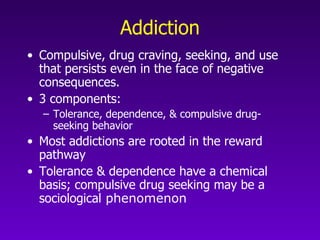
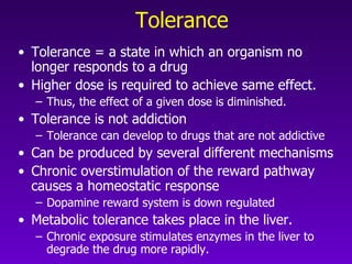
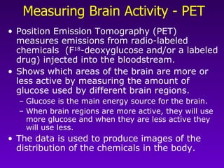
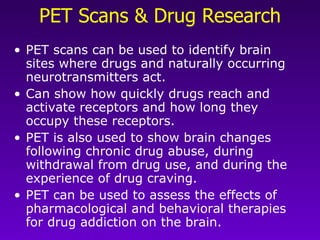
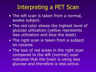
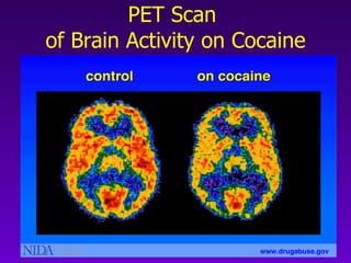
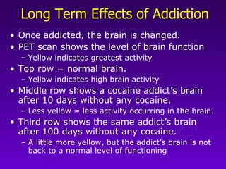
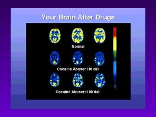
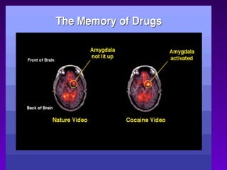
Drugs and the Brain Part 2 Addiction
Addiction Compulsive, drug craving, seeking, and use that persists even in the face of negative consequences.   3 components: Tolerance, dependence, & compulsive drug-seeking behavior Most addictions are rooted in the reward pathway Tolerance & dependence have a chemical basis; compulsive drug seeking may be a sociological  phenomenon
Tolerance Tolerance = a state in which an organism no longer responds to a drug Higher dose is required to achieve same effect.  Thus, the effect of a given dose is diminished.  Tolerance is not addiction Tolerance can develop to drugs that are not addictive Can be produced by several different mechanisms Chronic overstimulation of the reward pathway causes a homeostatic response Dopamine reward system is down regulated  Metabolic tolerance takes place in the liver. Chronic exposure stimulates enzymes in the liver to degrade the drug more rapidly.
Dependence A state in which the organism functions normally   only in the presence of the drug Manifests as physical disturbance when the drug is removed (withdrawal) Withdrawal symptoms are usually the opposite of the drug’s effect
Dependence & the Brain The development of dependence also involves specific areas of the brain. These are separate from the reward pathway.  The thalamus and brainstem are key areas in dependence.
The Reward Pathway The reward pathway plays a key role in addiction.  This pathway involves: the ventral tegmental area (VTA), the nucleus accumbens, and the prefrontal cortex.  When activated by a rewarding stimulus (e.g, food, water, sex), information travels from the VTA to the nucleus accumbens and then up to the prefrontal cortex. The neurons of the VTA contain dopamine which is released in the nucleus accumbens and in the prefrontal cortex.
The Reward Pathway Pictured
Understanding the Reward Pathway Research in rats led to understanding of the reward pathway  Rats were trained to press a lever for a tiny electrical jolt to certain parts of the brain. When an electrode is placed in the nucleus accumbens, the rat keeps pressing the lever to receive the electrical stimulus This stimulation is interpreted as pleasure Positive reinforcement.  When the electrode is placed near, but not in,  the nucleus accumbens, the rat will not press the lever because it does not activate the reward pathway.
Reward Pathway Research Activation of the reward pathway by an electrical stimulus.
Drugs Act in Different Parts of the Pathway Heroin & nicotine act on the VTA   Dopamine neurons from the VTA project through the lateral hypothalamus to the forebrain These dopaminergic neurons have both opiate and nicotinic acetylcholine receptors. Cocaine acts on the nucleus accumbens The nucleus accumbens is a target of the ascending dopaminergic axons in the forebrain
Rat Experiments Using Drugs to Stimulate the Reward Pathway
The Role of Dopamine Scientists can measure an increased release of dopamine in the reward pathway after the rat receives the reward.  If the dopamine release is prevented (either with a drug or by destroying the pathway), the rat won't press the bar for the electrical jolt.
Dopamine is the Link in Addiction Addictive drugs are biochemically quite different Activate different neurotransmitter systems Produce different psychoactive effects Heroin acts on the opiate system Nicotine acts on the cholinergic system Cocaine acts on dopaminergic & noradrenergic systems All either stimulate dopamine release (heroin, nicotine) or enhance dopamine action (cocaine) in the nucleus accumbens. Animals are motivated to perform behaviors that stimulate dopamine release in the nucleus accumbens & related structures
Measuring Brain Activity - PET Position Emission Tomography (PET) measures emissions from radio-labeled chemicals  (F 18 -deoxyglucose and/or a labeled drug) injected into the bloodstream.  Shows which areas of the brain are more or less active by measuring the amount of glucose used by different brain regions.  Glucose is the main energy source for the brain.  When brain regions are more active, they will use more glucose and when they are less active they will use less. The data is used to produce images of the distribution of the chemicals in the body.
Measuring Brain Activity
PET Scans & Drug Research PET scans can be used to identify brain sites where drugs and naturally occurring neurotransmitters act. Can show how quickly drugs reach and activate receptors and how long they occupy these receptors. PET is also used to show brain changes following chronic drug abuse, during withdrawal from drug use, and during the experience of drug craving.  PET can be used to assess the effects of pharmacological and behavioral therapies for drug addiction on the brain.
Interpreting a PET Scan The left scan is taken from a normal, awake subject.  The red color shows the highest level of glucose utilization (yellow represents less utilization and blue the least).  The right scan is taken from a subject on cocaine.  The loss of red areas in the right scan compared to the left (normal) scan indicates that the brain is using less glucose and therefore is less active.
PET Scan  of Brain Activity on Cocaine
Long Term Effects of Addiction Once addicted, the brain is changed.   PET scan shows the level of brain function Yellow indicates greatest activity Top row = normal brain.  Yellow indicates high brain activity Middle row shows a cocaine addict’s brain after 10 days without any cocaine. Less yellow = less activity occurring in the brain. Third row shows the same addict’s brain after 100 days without any cocaine.  A little more yellow, but the addict’s brain is not back to a normal level of functioning 
 
Memory of Drugs PET scan demonstrates how just the mention of items associated with drug use may cause an addict to “crave” or desire drugs.  Study compared recovering addicts, who had stopped using cocaine, with people who had no history of cocaine use.  Goal was to determine what parts of the brain are activated when drugs are craved.
 

Drugsandthe Brain Part2 Addiction

  • 1.
    Drugs and theBrain Part 2 Addiction
  • 2.
    Addiction Compulsive, drugcraving, seeking, and use that persists even in the face of negative consequences. 3 components: Tolerance, dependence, & compulsive drug-seeking behavior Most addictions are rooted in the reward pathway Tolerance & dependence have a chemical basis; compulsive drug seeking may be a sociological phenomenon
  • 3.
    Tolerance Tolerance =a state in which an organism no longer responds to a drug Higher dose is required to achieve same effect. Thus, the effect of a given dose is diminished. Tolerance is not addiction Tolerance can develop to drugs that are not addictive Can be produced by several different mechanisms Chronic overstimulation of the reward pathway causes a homeostatic response Dopamine reward system is down regulated Metabolic tolerance takes place in the liver. Chronic exposure stimulates enzymes in the liver to degrade the drug more rapidly.
  • 4.
    Dependence A statein which the organism functions normally only in the presence of the drug Manifests as physical disturbance when the drug is removed (withdrawal) Withdrawal symptoms are usually the opposite of the drug’s effect
  • 5.
    Dependence & theBrain The development of dependence also involves specific areas of the brain. These are separate from the reward pathway. The thalamus and brainstem are key areas in dependence.
  • 6.
    The Reward PathwayThe reward pathway plays a key role in addiction. This pathway involves: the ventral tegmental area (VTA), the nucleus accumbens, and the prefrontal cortex. When activated by a rewarding stimulus (e.g, food, water, sex), information travels from the VTA to the nucleus accumbens and then up to the prefrontal cortex. The neurons of the VTA contain dopamine which is released in the nucleus accumbens and in the prefrontal cortex.
  • 7.
  • 8.
    Understanding the RewardPathway Research in rats led to understanding of the reward pathway Rats were trained to press a lever for a tiny electrical jolt to certain parts of the brain. When an electrode is placed in the nucleus accumbens, the rat keeps pressing the lever to receive the electrical stimulus This stimulation is interpreted as pleasure Positive reinforcement. When the electrode is placed near, but not in, the nucleus accumbens, the rat will not press the lever because it does not activate the reward pathway.
  • 9.
    Reward Pathway ResearchActivation of the reward pathway by an electrical stimulus.
  • 10.
    Drugs Act inDifferent Parts of the Pathway Heroin & nicotine act on the VTA Dopamine neurons from the VTA project through the lateral hypothalamus to the forebrain These dopaminergic neurons have both opiate and nicotinic acetylcholine receptors. Cocaine acts on the nucleus accumbens The nucleus accumbens is a target of the ascending dopaminergic axons in the forebrain
  • 11.
    Rat Experiments UsingDrugs to Stimulate the Reward Pathway
  • 12.
    The Role ofDopamine Scientists can measure an increased release of dopamine in the reward pathway after the rat receives the reward. If the dopamine release is prevented (either with a drug or by destroying the pathway), the rat won't press the bar for the electrical jolt.
  • 13.
    Dopamine is theLink in Addiction Addictive drugs are biochemically quite different Activate different neurotransmitter systems Produce different psychoactive effects Heroin acts on the opiate system Nicotine acts on the cholinergic system Cocaine acts on dopaminergic & noradrenergic systems All either stimulate dopamine release (heroin, nicotine) or enhance dopamine action (cocaine) in the nucleus accumbens. Animals are motivated to perform behaviors that stimulate dopamine release in the nucleus accumbens & related structures
  • 14.
    Measuring Brain Activity- PET Position Emission Tomography (PET) measures emissions from radio-labeled chemicals (F 18 -deoxyglucose and/or a labeled drug) injected into the bloodstream. Shows which areas of the brain are more or less active by measuring the amount of glucose used by different brain regions. Glucose is the main energy source for the brain. When brain regions are more active, they will use more glucose and when they are less active they will use less. The data is used to produce images of the distribution of the chemicals in the body.
  • 15.
  • 16.
    PET Scans &Drug Research PET scans can be used to identify brain sites where drugs and naturally occurring neurotransmitters act. Can show how quickly drugs reach and activate receptors and how long they occupy these receptors. PET is also used to show brain changes following chronic drug abuse, during withdrawal from drug use, and during the experience of drug craving. PET can be used to assess the effects of pharmacological and behavioral therapies for drug addiction on the brain.
  • 17.
    Interpreting a PETScan The left scan is taken from a normal, awake subject. The red color shows the highest level of glucose utilization (yellow represents less utilization and blue the least). The right scan is taken from a subject on cocaine. The loss of red areas in the right scan compared to the left (normal) scan indicates that the brain is using less glucose and therefore is less active.
  • 18.
    PET Scan of Brain Activity on Cocaine
  • 19.
    Long Term Effectsof Addiction Once addicted, the brain is changed.   PET scan shows the level of brain function Yellow indicates greatest activity Top row = normal brain. Yellow indicates high brain activity Middle row shows a cocaine addict’s brain after 10 days without any cocaine. Less yellow = less activity occurring in the brain. Third row shows the same addict’s brain after 100 days without any cocaine. A little more yellow, but the addict’s brain is not back to a normal level of functioning 
  • 20.
  • 21.
    Memory of DrugsPET scan demonstrates how just the mention of items associated with drug use may cause an addict to “crave” or desire drugs. Study compared recovering addicts, who had stopped using cocaine, with people who had no history of cocaine use. Goal was to determine what parts of the brain are activated when drugs are craved.
  • 22.