Downloaded 324 times
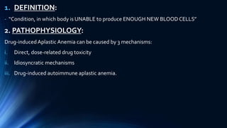
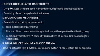
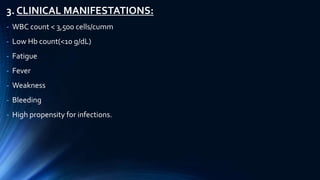


The document provides an overview of drug-induced hematological disorders, defining conditions such as aplastic anemia, agranulocytosis, hemolytic anemia, megaloblastic anemia, and thrombocytopenia, along with their pathophysiology, symptoms, causative drugs, and management strategies. It details the mechanisms by which these disorders occur and emphasizes the importance of recognizing and removing offending drugs, as well as supportive treatments and specific therapies like stem cell transplantation and immunosuppressive agents. The document serves as a comprehensive guide for understanding the implications of drug therapy on blood cell production and health.


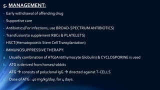
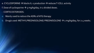
![PERI-PROSTHETIC FRACTURE NAIL-PLATE CONSTRUCT [NPC].pptx](https://cdn.slidesharecdn.com/ss_thumbnails/drarunkumardrmohamedashrafperiprostheticfrasturenail-plateconstructnpc-260209164459-7e9d15a1-thumbnail.jpg?width=640&height=640&fit=bounds)


