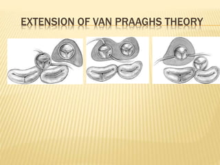
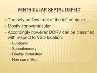
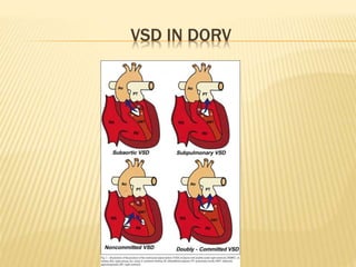
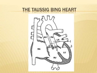
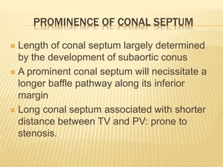
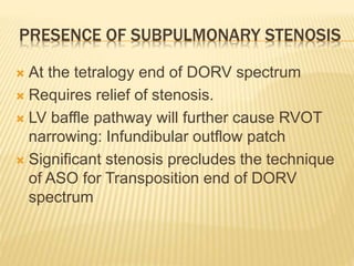
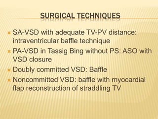
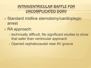
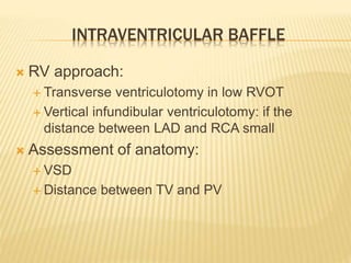
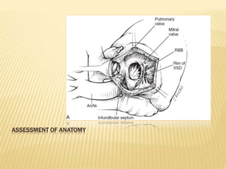
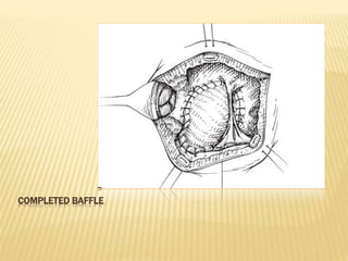
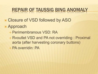
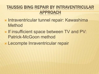
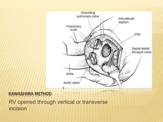
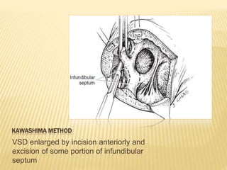
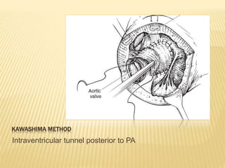
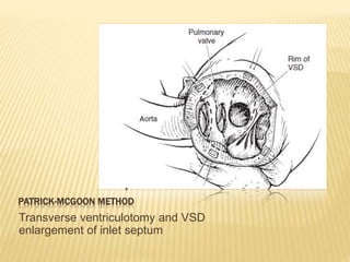
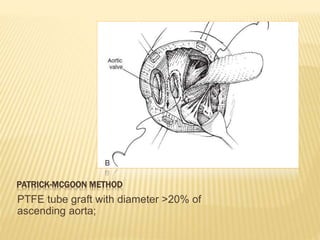
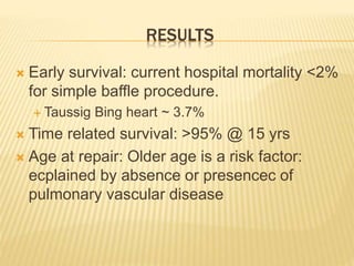
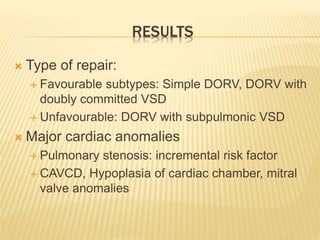
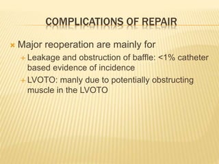
DOUBLE OUTLET RIGHT VENTRICLE
JOHN ABERNETHY
INTRODUCTION
 1793 - John Abernethy 1st described the
condition.
 1957 - Witham coined the term as DORV.
 1957 - 1st repair: Kirklin
DEFINITION
 When one great artery arises nearly or
wholely from RV and the other more than
50%.
 TOF with DORV is same as DORV with
pulmonary stenosis
 Taussig Bing Heart: variability in origin of
pulmonary artery
DORV
EMBRYOLOGY AND ANATOMY
 Conus: The muscle between semilunar and
AV valve.
 Lev’s theory of Conotruncal malseptation
 Van Praagh’s theory of Conal
underdevelopment
VAN PRAAGH THEORY OF CONAL
UNDERDEVELOPMENT
 “ the distal or semilunar part of infundibulum
or conus arteriosus performs an arterial
switch during cardiogenesis”
EXTENSION OF VAN PRAAGHS THEORY
VENTRICULAR SEPTAL DEFECT
 The only outflow tract of the left ventricle.
 Mostly conoventricular.
 Accordingly however DORV can be classified
with respect to VSD location.
 Subaortic
 Subpulmonary
 Doubly committed
 Non committed.
VSD IN DORV
CORONARY ARTERY ANATOMY
 Depends upon the great arteries
 At the transposition end of the spectrum:
LMCA and LAD pass anterior to PA
 TOF: LMCA and LAD pass posterior to PA.
HELLEN TAUSSIG AND RICHARD BING
THE TAUSSIG BING HEART
 Applied in physiological and anatomical
sense.
 Physiological
 Anatomical:
 Helen Taussig and Richard Bing (1949)
 Van praagh (1968) summarized anatomic
definition
THE TAUSSIG BING HEART
ASSOCIATED ANOMALIES
 Coarctation of aorta
 Arch hypoplasia
 Interrupted aortic arch
CLINICOPATHOLOGY AND HEMODYNAMICS
 Highly variable
 Large VSD with severe fixed pulmonary
stenosis: cyanotic child
 VSD with no pulmonary stenosis: features of
heart hailure with progression towards
pulmonary vascular disease.
STREAMING
 Determined by relationship of semilunar
valves to VSD and position and presence of
infundibular septum.
 Simple DORV: High Qp; heart failure; without
cyanosis
 Subpulmonary VSD: Taussig Bing heart:
SpaO2> SaO2. Infants present like TGA
CLINICAL FEATURES AND EVALUATION
 Type of symptoms and age of appearance
depends upon degree of pulmonary stenosis
 Echo:
 Size of VSD,
 Relation to semilunar valves
 Subvalvular conus
 AV valve abnormalities
 Position of great arteries
 Coronary artery anatomy
EVALUATION
 Cardiac catheterization: not in infants;
indicated in older children for
 PVR
 Ventricular end diastolic pressure
 MAPCA
 Cineangiography: Assessing complex
interrationships in DORV with superior-
inferior ventricles and criss cross hearts
MANAGEMENT: MEDICAL AND CATHETER BASED
 Decongestive medications: in cases of high
Qp heart failure; followed by surgery
 BAS: at the transposition end of DORV:
 Allows improved mixing
 Atrial septal defect allows decompression of left
side of heart: better surgical field
INDICATIONS AND TIMING OF SURGERY
 Diagnosis is sufficient for surgery
 Timing of surgery depends upon the subset
of the patients in question
 Simple DORV: electively by 3-6 months of age
 Sooner if symptoms of heart failure or failure to thrive
appear
 DORV with pulmonary stenosis: same as for
TOF
 Taussig Bing: during 1st month of age
 Operations including extracardiac conduit but
excluding ASO: 3-6 years of age.
SURGICAL MANAGEMENT
 1957 – Kirklin and 1958 – Barratt-Boyes: 1st
successful repair of DORV
ANATOMICAL DETERMINANTS OF METHOD OF
REPAIR
1. Separation of Pulmonary and tricuspid valve
2. Prominance of Conal septum
3. Presence of subpulmonic stenosis
SEPARATION OF PULMONARY AND TRICUSPID
VALVES
PROMINENCE OF CONAL SEPTUM
 Length of conal septum largely determined
by the development of subaortic conus
 A prominent conal septum will necissitate a
longer baffle pathway along its inferior
margin
 Long conal septum associated with shorter
distance between TV and PV: prone to
stenosis.
PRESENCE OF SUBPULMONARY STENOSIS
 At the tetralogy end of DORV spectrum
 Requires relief of stenosis.
 LV baffle pathway will further cause RVOT
narrowing: Infundibular outflow patch
 Significant stenosis precludes the technique
of ASO for Transposition end of DORV
spectrum
SURGICAL TECHNIQUES
 SA-VSD with adequate TV-PV distance:
intraventricular baffle technique
 PA-VSD in Tassig Bing without PS: ASO with
VSD closure
 Doubly committed VSD: Baffle
 Noncommitted VSD: baffle with myocardial
flap reconstruction of straddling TV
INTRAVENTRICULAR BAFFLE FOR
UNCOMPLICATED DORV
 Standard midline sternotomy/cardioplegic
arrest
 RA approach:
 technically difficult, No significant studies to show
that safer than ventricular approach
 Opened cephalocaudal near AV groove
INTRAVENTRICULAR BAFFLE
 RV approach:
 Transverse ventriculotomy in low RVOT
 Vertical infundibular ventriculotomy: if the
distance between LAD and RCA small
 Assessment of anatomy:
 VSD
 Distance between TV and PV
ASSESSMENT OF ANATOMY
INTRAVENTRICULAR REPAIR
 Enlargement of VSD: anteriorly,
 Risk of damaging LAD
 Risk of damaging MV
 VSD patch: Polyester tube graft diameter
>20% of Ascending aorta, Length: Anterior
edge of VSD to sub Ao conus
 Neonates and infants: standard pledgetted
interrupted suture technique
 Older: continuous
VSD PATCH IN INTRAVENTRICULAR BAFFLE
Flat patch : Knitted Daron Velour
Tube: PTFE
COMPLETED BAFFLE
REPAIR OF TAUSSIG BING ANOMALY
 Closure of VSD followed by ASO
 Approach
 Perimembranous VSD: RA
 Rvoutlet VSD and PA not overriding : Proximal
aorta (after harvesting coronary buttons)
 PA overridin: PA
TAUSSIG BING REPAIR BY INTRAVENTRICULAR
APPROACH
 Intraventricular tunnel repair: Kawashima
Method
 If insufficient space between TV and PV:
Patrick-McGoon method
 Lecompte Inraventricular repair
KAWASHIMA METHOD
RV opened through vertical or transverse
incision
KAWASHIMA METHOD
VSD enlarged by incision anteriorly and
excision of some portion of infundibular
septum
KAWASHIMA METHOD
Intraventricular tunnel posterior to PA
PATRICK-MCGOON METHOD
Transverse ventriculotomy and VSD
enlargement of inlet septum
PATRICK-MCGOON METHOD
PTFE tube graft with diameter >20% of
ascending aorta;
PATRICK-MCGOON METHOD
Continuous sutuiring of the tube graft
PATRICK-MCGOON METHOD
Completion of spiralled tube graft
RESULTS
 Early survival: current hospital mortality <2%
for simple baffle procedure.
 Taussig Bing heart ~ 3.7%
 Time related survival: >95% @ 15 yrs
 Age at repair: Older age is a risk factor:
ecplained by absence or presencec of
pulmonary vascular disease
RESULTS
 Type of repair:
 Favourable subtypes: Simple DORV, DORV with
doubly committed VSD
 Unfavourable: DORV with subpulmonic VSD
 Major cardiac anomalies
 Pulmonary stenosis: incremental risk factor
 CAVCD, Hypoplasia of cardiac chamber, mitral
valve anomalies
COMPLICATIONS OF REPAIR
 Major reoperation are mainly for
 Leakage and obstruction of baffle: <1% catheter
based evidence of incidence
 LVOTO: manly due to potentially obstructing
muscle in the LVOTO
THANK YOU

Dorv

  • 1.
  • 2.
  • 3.
    INTRODUCTION  1793 -John Abernethy 1st described the condition.  1957 - Witham coined the term as DORV.  1957 - 1st repair: Kirklin
  • 4.
    DEFINITION  When onegreat artery arises nearly or wholely from RV and the other more than 50%.  TOF with DORV is same as DORV with pulmonary stenosis  Taussig Bing Heart: variability in origin of pulmonary artery
  • 5.
  • 6.
    EMBRYOLOGY AND ANATOMY Conus: The muscle between semilunar and AV valve.  Lev’s theory of Conotruncal malseptation  Van Praagh’s theory of Conal underdevelopment
  • 7.
    VAN PRAAGH THEORYOF CONAL UNDERDEVELOPMENT  “ the distal or semilunar part of infundibulum or conus arteriosus performs an arterial switch during cardiogenesis”
  • 8.
    EXTENSION OF VANPRAAGHS THEORY
  • 9.
    VENTRICULAR SEPTAL DEFECT The only outflow tract of the left ventricle.  Mostly conoventricular.  Accordingly however DORV can be classified with respect to VSD location.  Subaortic  Subpulmonary  Doubly committed  Non committed.
  • 10.
  • 11.
    CORONARY ARTERY ANATOMY Depends upon the great arteries  At the transposition end of the spectrum: LMCA and LAD pass anterior to PA  TOF: LMCA and LAD pass posterior to PA.
  • 12.
    HELLEN TAUSSIG ANDRICHARD BING
  • 13.
    THE TAUSSIG BINGHEART  Applied in physiological and anatomical sense.  Physiological  Anatomical:  Helen Taussig and Richard Bing (1949)  Van praagh (1968) summarized anatomic definition
  • 14.
  • 15.
    ASSOCIATED ANOMALIES  Coarctationof aorta  Arch hypoplasia  Interrupted aortic arch
  • 16.
    CLINICOPATHOLOGY AND HEMODYNAMICS Highly variable  Large VSD with severe fixed pulmonary stenosis: cyanotic child  VSD with no pulmonary stenosis: features of heart hailure with progression towards pulmonary vascular disease.
  • 17.
    STREAMING  Determined byrelationship of semilunar valves to VSD and position and presence of infundibular septum.  Simple DORV: High Qp; heart failure; without cyanosis  Subpulmonary VSD: Taussig Bing heart: SpaO2> SaO2. Infants present like TGA
  • 18.
    CLINICAL FEATURES ANDEVALUATION  Type of symptoms and age of appearance depends upon degree of pulmonary stenosis  Echo:  Size of VSD,  Relation to semilunar valves  Subvalvular conus  AV valve abnormalities  Position of great arteries  Coronary artery anatomy
  • 19.
    EVALUATION  Cardiac catheterization:not in infants; indicated in older children for  PVR  Ventricular end diastolic pressure  MAPCA  Cineangiography: Assessing complex interrationships in DORV with superior- inferior ventricles and criss cross hearts
  • 20.
    MANAGEMENT: MEDICAL ANDCATHETER BASED  Decongestive medications: in cases of high Qp heart failure; followed by surgery  BAS: at the transposition end of DORV:  Allows improved mixing  Atrial septal defect allows decompression of left side of heart: better surgical field
  • 21.
    INDICATIONS AND TIMINGOF SURGERY  Diagnosis is sufficient for surgery  Timing of surgery depends upon the subset of the patients in question  Simple DORV: electively by 3-6 months of age  Sooner if symptoms of heart failure or failure to thrive appear  DORV with pulmonary stenosis: same as for TOF  Taussig Bing: during 1st month of age  Operations including extracardiac conduit but excluding ASO: 3-6 years of age.
  • 22.
    SURGICAL MANAGEMENT  1957– Kirklin and 1958 – Barratt-Boyes: 1st successful repair of DORV
  • 23.
    ANATOMICAL DETERMINANTS OFMETHOD OF REPAIR 1. Separation of Pulmonary and tricuspid valve 2. Prominance of Conal septum 3. Presence of subpulmonic stenosis
  • 24.
    SEPARATION OF PULMONARYAND TRICUSPID VALVES
  • 25.
    PROMINENCE OF CONALSEPTUM  Length of conal septum largely determined by the development of subaortic conus  A prominent conal septum will necissitate a longer baffle pathway along its inferior margin  Long conal septum associated with shorter distance between TV and PV: prone to stenosis.
  • 26.
    PRESENCE OF SUBPULMONARYSTENOSIS  At the tetralogy end of DORV spectrum  Requires relief of stenosis.  LV baffle pathway will further cause RVOT narrowing: Infundibular outflow patch  Significant stenosis precludes the technique of ASO for Transposition end of DORV spectrum
  • 27.
    SURGICAL TECHNIQUES  SA-VSDwith adequate TV-PV distance: intraventricular baffle technique  PA-VSD in Tassig Bing without PS: ASO with VSD closure  Doubly committed VSD: Baffle  Noncommitted VSD: baffle with myocardial flap reconstruction of straddling TV
  • 28.
    INTRAVENTRICULAR BAFFLE FOR UNCOMPLICATEDDORV  Standard midline sternotomy/cardioplegic arrest  RA approach:  technically difficult, No significant studies to show that safer than ventricular approach  Opened cephalocaudal near AV groove
  • 29.
    INTRAVENTRICULAR BAFFLE  RVapproach:  Transverse ventriculotomy in low RVOT  Vertical infundibular ventriculotomy: if the distance between LAD and RCA small  Assessment of anatomy:  VSD  Distance between TV and PV
  • 30.
  • 31.
    INTRAVENTRICULAR REPAIR  Enlargementof VSD: anteriorly,  Risk of damaging LAD  Risk of damaging MV  VSD patch: Polyester tube graft diameter >20% of Ascending aorta, Length: Anterior edge of VSD to sub Ao conus  Neonates and infants: standard pledgetted interrupted suture technique  Older: continuous
  • 32.
    VSD PATCH ININTRAVENTRICULAR BAFFLE Flat patch : Knitted Daron Velour Tube: PTFE
  • 33.
  • 34.
    REPAIR OF TAUSSIGBING ANOMALY  Closure of VSD followed by ASO  Approach  Perimembranous VSD: RA  Rvoutlet VSD and PA not overriding : Proximal aorta (after harvesting coronary buttons)  PA overridin: PA
  • 35.
    TAUSSIG BING REPAIRBY INTRAVENTRICULAR APPROACH  Intraventricular tunnel repair: Kawashima Method  If insufficient space between TV and PV: Patrick-McGoon method  Lecompte Inraventricular repair
  • 36.
    KAWASHIMA METHOD RV openedthrough vertical or transverse incision
  • 37.
    KAWASHIMA METHOD VSD enlargedby incision anteriorly and excision of some portion of infundibular septum
  • 38.
  • 39.
    PATRICK-MCGOON METHOD Transverse ventriculotomyand VSD enlargement of inlet septum
  • 40.
    PATRICK-MCGOON METHOD PTFE tubegraft with diameter >20% of ascending aorta;
  • 41.
  • 42.
  • 43.
    RESULTS  Early survival:current hospital mortality <2% for simple baffle procedure.  Taussig Bing heart ~ 3.7%  Time related survival: >95% @ 15 yrs  Age at repair: Older age is a risk factor: ecplained by absence or presencec of pulmonary vascular disease
  • 44.
    RESULTS  Type ofrepair:  Favourable subtypes: Simple DORV, DORV with doubly committed VSD  Unfavourable: DORV with subpulmonic VSD  Major cardiac anomalies  Pulmonary stenosis: incremental risk factor  CAVCD, Hypoplasia of cardiac chamber, mitral valve anomalies
  • 45.
    COMPLICATIONS OF REPAIR Major reoperation are mainly for  Leakage and obstruction of baffle: <1% catheter based evidence of incidence  LVOTO: manly due to potentially obstructing muscle in the LVOTO
  • 46.

Editor's Notes

  • #4 Diagnosis was correctly made at operation and the term DORV was coined in operating room.
  • #5 Artery is assigned to the ventricle it overrides more than 50%. aorta overrides >50% to RV in TOF. Taussig bing heart: PA arises wholely or nearly so from RV, Eaqually from right and left ventricles and >50% but not entirely from the Left ventricle When it arises entirely from LV then : TGS with VSD.
  • #7 Lev’s theory: Conotruncus which ultimately forms PA and Ao forms as spiral septation. If septum doesnot spiral in usual fashion at all: All great vessels in parallel: TGA If septum spiralling slightly abnormal: dextroposition of aorta relative to its usual location: TOF, Spiralling is more than TOF and less than TGA: DORV
  • #8 TOF: underdevelopment of pulmonary conus: AO is more anterior and to the right and superior, Conoventricular VSD with anterior malalignment of conal septum: RVOTO. Extension of Van Praagh:
  • #9 AV, TV, MV are in fibrous continuity PV is separate by sub pulmonary conus (infundibulum) DORV: Bilateral Coni: both AV and PV are lifted away from the AV valves TGA: No sup pulm conus and PV is in fibrous continuity with TV and MV, AoV is lifted away by the sub ao conus Greater digree of conal underdevelopment result in great arteries to lie side by side. Particularly if there is compensatory development of subaortic conus. No subpulmonary counus but aortic conus: TGA
  • #10 Conoventriccular: between the two limbs of TSM. Sub aortic: TOF end: no sub aortic conus: superior margin of VSD is the Ao valve itself. Sub pulmonary: TGA end: Progressive Subaortic conus; Ao Valve moves cephalad; and PV more intimately associated: Subpulmonary. Non committed: VSD not in conal septum or Jn of conal and interventricular septum(subaortic) : remote from aorta: difficult to direct LV flow to aorta:: inlet type Sub aortic VSD: VIF: posterior margin of VSD. Bundle away Subaortic Perimembranous VSD near TV : Posterio division of TSM is deficient: Bundle lies in poesterior inferior margin of VSD.
  • #12 LAD anterior excludes an intraventricular repair and suggests that the defect is closer to the transposition end of the spectrum and requires ASO.
  • #13 Richard John Bing (October 12, 1909 Nuremberg, Germany – November 8, 2010 La Cañada Flintridge, California) was a cardiologist who made significant contributions to his field of stud Helen Brooke Taussig (May 24, 1898 – May 20, 1986) was an American cardiologist, working in Baltimore and Boston who founded the field of pediatric cardiology. Notably, she is credited with developing the concept for a procedure that would extend the lives of children born with Tetralogy of Fallot (the most common cause of blue baby syndrome). This concept was applied in practice as a procedure known as the Blalock-Taussig shunt. The procedure was developed by Alfred Blalock and Vivien Thomas, who were Taussig's colleagues at the Johns Hopkins Hospital.
  • #14 Physiological: similar to transposition. Saturations higher in PA than in Aorta. Because of preferential blood flow from LV to PA.
  • #15 Both Subaortic and sub pulmonary conus present, separate both aortic and pulmonary valves from Atrioventricular valves. Semilunar valves lie side by side. And are at the same height Large subpulmonaryVSD present True DORV: Aorta arises entirely from RV and PA overrides the ventricular septum, but doesnot override the left ventricular cavity.
  • #18 Simple DORV: flow of highly oxygenated LV blood through VSD is directed prefrencially benieth infundibular septum to adjuscant aorta. Systemic venous blood to PA Taussig bing heart: LV blood into Adjascent PA by vertically positioned infundibular septum. Venous blood from RV going into Ao.
  • #19 Simple DORV: simple large VSD Also for DORV with doubly committed and non committed VSD Taussig bing: TGA DORV with pulm stenosis/ atresia: TOF with pulmonary stenosis/atresis
  • #23 1st repair was TOF type.
  • #25 Baffle from VSD to Aorta. RVOT passesaround the LV baffle but still within the RV. As AV moves superiorly and away from TV, PV moves closer to TV. Baffle pathway must pass between the PV and TV Adequate separation Separation less than the dia of Ao valve: likely to result in stenosis PV very close to TV: Precludes intraventricular repair.
  • #30 Assessment: whether VSD abutts or has muscular fold
  • #32 Continuous suture in infants cause unacceptable incidences of residual VSD
  • #33 Marking stitch: most anterior part of the repair 1st matress stitch: Atria->ventricle-> base of Tricuspid commissure between ATL and STL Flat patch whe VSD is closely related to Aorta Tube when baffle has to travel long.
  • #41 Initial sutures in usual mannar Preventing damage to the bundle of His
  • #42 Leftward arm of the continuous tuture continoud posterior then left margin of aortopulmonary trunk. Then finally along the anterior margin