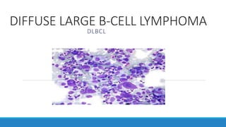
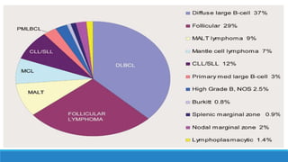
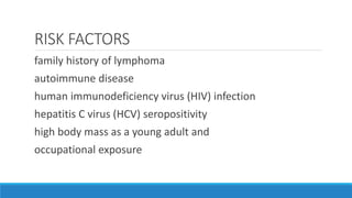
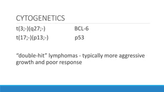
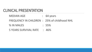
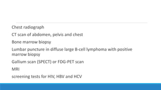
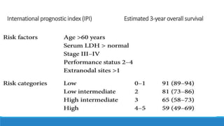
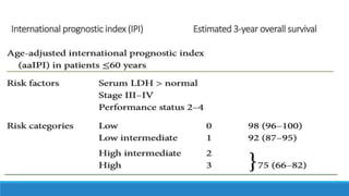
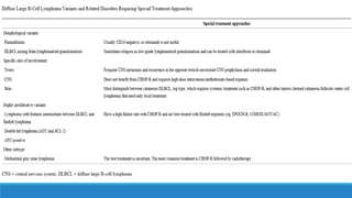
DIFFUSE LARGE B-CELL LYMPHOMA
DLBCL
*most common type of non-Hodgkin lymphoma (30-
40%)
* aggressive or intermediate-grade lymphoma
neoplastic cells are predominantly large Cells with vesicular
chromatin and prominent nucleoli.
RISK FACTORS
family history of lymphoma
autoimmune disease
human immunodeficiency virus (HIV) infection
hepatitis C virus (HCV) seropositivity
high body mass as a young adult and
occupational exposure
Epstein-Barr virus - Primary CNS diffuse large B cell
lymphoma
Human herpes virus 8 – primary effusion lymphoma
CYTOGENETICS
t(3;-)(q27;-) BCL-6
t(17;-)(p13;-) p53
“double-hit” lymphomas - typically more aggressive
growth and poor response
MEDIAN AGE : 64 years
FREQUENCY IN CHILDREN : 25% of childhood NHL
% IN MALES : 55%
5 YEARS SURVIVAL RATE : 46%
CLINICAL PRESENTATION
present as either primary lymph node disease or at extra nodal
sites (50%)
m/c extranodal site – GIT, BONE MARROW (15-20%)
OTHER SITES – PANCREAS , BRAIN,
OTHER UNUSUAL SUBTYPES : pleural effusion lymphoma ,
intravascular lymphoma
surgical excision biopsy
*allows assessment of nodal architecture and provides adequate material for
phenotypic and molecular studies.
*suggested immunohistochemical panel
CD20, CD79a, BCL6, CD10, MYC, BCL2, Ki67, IRF4, CyclinD1, CD5,
CD23, and EBER-1 staining
STAGING EVALUATION
*Physical examination
*Documentation of B symptoms
*Laboratory Evaluation
Complete blood counts
Liver function tests
Uric acid, Calcium, LDH
Serum protein
Electrophoresis
Serum ß2-microglobulin
Chest radiograph
CT scan of abdomen, pelvis and chest
Bone marrow biopsy
Lumbar puncture in diffuse large B-cell lymphoma with positive
marrow biopsy
Gallium scan (SPECT) or FDG-PET scan
MRI
screening tests for HIV, HBV and HCV
ANN ARBOR STAGING
Stage
I Involvement of a single lymphatic region (I) or localized involvement of
single extra lymphatic organ or site (IE)
II Involvement of two or more lymphatic regions on the same side of the
diaphragm (II) or localized involvement of a single extra lymphatic organ or site
and of one or more lymphatic regions on the same side of the diaphragm (IIE)
III Involvement of lymphatic regions on both sides of the diaphragm
IV Diffuse or disseminated involvement of one or more extra lymphatic
organs with or without lymphatic involvement
International prognostic index (IPI) Estimated 3-year overall survival
TREATMENT STRATEGIES
*young low-risk patients (aa-IPI=0)without bulky disease
R-CHOP21×6
*young low-intermediate-risk patients (aa-IPI=1) or IPI low risk (aa-IPI=0)
with bulky disease
(R)-CHOP 21×6 with IF-radiotherapy
OR
R-ACVBP with sequential consolidation
*young high-and high-intermediate-risk patients (aa-IPI≥2)
R-CHOP21×6–8
or
R-CHOP14×6 with 8 R
or
(R-CHOEP14×6 or R-CHOP or R-ACVBP plus HDCT with ASCT)
*patients aged 60–80years,fit
R-CHOP21×6–8 (R-CHOP21×6 for IPI low risk)
or
R-CHOP14×6 with 8R
*Unfit or frail or >60 years with cardiac dysfunction
R-C(X)OP21×6 (Doxorubicin substitution with gemcitabine, etoposide or liposomal doxorubicin)
or palliative care
- high-intermediate and high-risk IPI, especially those with more than
one extra nodal site or elevated LDH, are at higher risk of CNS
relapse.
- Testicular, renal and adrenal involvements have been validated as
additional risk factors.
- MYC gene rearrangement is associated with a high risk of CNS relapse
- Although widely used, intrathecal injections of methotrexate may
not be an optimal method. Intravenous high dose methotrexate has
been shown to be associated with efficient disease control.
Central nervous system (CNS) prophylaxis
post-treatment evaluation
* FDG-PET/CT - recommended for post-treatment assessment
follow-up
*Careful history and physical examination every 3 months for 1 year, every 6
months for 2 further years and then once a year.
* Blood count should be carried out at 3, 6, 12 and 24 months, then only as
needed for evaluation of suspicious symptoms or clinical findings in those
patients suitable for further therapy.
* Minimal radiological examinations at 6, 12 and 24 months after end of
treatment by CT scan is common practice.
Thank you……

DLBCL

  • 1.
    DIFFUSE LARGE B-CELLLYMPHOMA DLBCL
  • 2.
    *most common typeof non-Hodgkin lymphoma (30- 40%) * aggressive or intermediate-grade lymphoma
  • 4.
    neoplastic cells arepredominantly large Cells with vesicular chromatin and prominent nucleoli.
  • 5.
    RISK FACTORS family historyof lymphoma autoimmune disease human immunodeficiency virus (HIV) infection hepatitis C virus (HCV) seropositivity high body mass as a young adult and occupational exposure
  • 6.
    Epstein-Barr virus -Primary CNS diffuse large B cell lymphoma Human herpes virus 8 – primary effusion lymphoma
  • 7.
    CYTOGENETICS t(3;-)(q27;-) BCL-6 t(17;-)(p13;-) p53 “double-hit”lymphomas - typically more aggressive growth and poor response
  • 9.
    MEDIAN AGE :64 years FREQUENCY IN CHILDREN : 25% of childhood NHL % IN MALES : 55% 5 YEARS SURVIVAL RATE : 46% CLINICAL PRESENTATION
  • 10.
    present as eitherprimary lymph node disease or at extra nodal sites (50%) m/c extranodal site – GIT, BONE MARROW (15-20%) OTHER SITES – PANCREAS , BRAIN, OTHER UNUSUAL SUBTYPES : pleural effusion lymphoma , intravascular lymphoma
  • 12.
    surgical excision biopsy *allowsassessment of nodal architecture and provides adequate material for phenotypic and molecular studies. *suggested immunohistochemical panel CD20, CD79a, BCL6, CD10, MYC, BCL2, Ki67, IRF4, CyclinD1, CD5, CD23, and EBER-1 staining
  • 13.
    STAGING EVALUATION *Physical examination *Documentationof B symptoms *Laboratory Evaluation Complete blood counts Liver function tests Uric acid, Calcium, LDH Serum protein Electrophoresis Serum ß2-microglobulin
  • 14.
    Chest radiograph CT scanof abdomen, pelvis and chest Bone marrow biopsy Lumbar puncture in diffuse large B-cell lymphoma with positive marrow biopsy Gallium scan (SPECT) or FDG-PET scan MRI screening tests for HIV, HBV and HCV
  • 15.
    ANN ARBOR STAGING Stage IInvolvement of a single lymphatic region (I) or localized involvement of single extra lymphatic organ or site (IE) II Involvement of two or more lymphatic regions on the same side of the diaphragm (II) or localized involvement of a single extra lymphatic organ or site and of one or more lymphatic regions on the same side of the diaphragm (IIE) III Involvement of lymphatic regions on both sides of the diaphragm IV Diffuse or disseminated involvement of one or more extra lymphatic organs with or without lymphatic involvement
  • 16.
    International prognostic index(IPI) Estimated 3-year overall survival
  • 17.
    International prognostic index(IPI) Estimated 3-year overall survival
  • 18.
    TREATMENT STRATEGIES *young low-riskpatients (aa-IPI=0)without bulky disease R-CHOP21×6 *young low-intermediate-risk patients (aa-IPI=1) or IPI low risk (aa-IPI=0) with bulky disease (R)-CHOP 21×6 with IF-radiotherapy OR R-ACVBP with sequential consolidation *young high-and high-intermediate-risk patients (aa-IPI≥2) R-CHOP21×6–8 or R-CHOP14×6 with 8 R or (R-CHOEP14×6 or R-CHOP or R-ACVBP plus HDCT with ASCT)
  • 19.
    *patients aged 60–80years,fit R-CHOP21×6–8(R-CHOP21×6 for IPI low risk) or R-CHOP14×6 with 8R *Unfit or frail or >60 years with cardiac dysfunction R-C(X)OP21×6 (Doxorubicin substitution with gemcitabine, etoposide or liposomal doxorubicin) or palliative care
  • 20.
    - high-intermediate andhigh-risk IPI, especially those with more than one extra nodal site or elevated LDH, are at higher risk of CNS relapse. - Testicular, renal and adrenal involvements have been validated as additional risk factors. - MYC gene rearrangement is associated with a high risk of CNS relapse - Although widely used, intrathecal injections of methotrexate may not be an optimal method. Intravenous high dose methotrexate has been shown to be associated with efficient disease control. Central nervous system (CNS) prophylaxis
  • 21.
    post-treatment evaluation * FDG-PET/CT- recommended for post-treatment assessment
  • 24.
    follow-up *Careful history andphysical examination every 3 months for 1 year, every 6 months for 2 further years and then once a year. * Blood count should be carried out at 3, 6, 12 and 24 months, then only as needed for evaluation of suspicious symptoms or clinical findings in those patients suitable for further therapy. * Minimal radiological examinations at 6, 12 and 24 months after end of treatment by CT scan is common practice.
  • 26.

Editor's Notes

  • #8 molecular genetic studies are not necessary for diagnosis. that patients whose tumors overexpress the BCL-2 protein might be more likely to relapse than others
  • #12 ALK = anaplastic lymphoma kinase; CNS = central nervous system; EBV = Epstein-Barr virus; GCB = germinal center B-cell; HHV-8 = human herpesvirus 8.
  • #15 Focal bone marrow FDG uptake with or without increased diffuse uptake is more sensitive than bone marrow biopsy (BMB) for infiltration in DLBCL and is highly specifiC
  • #23  DHAP, cisplatin, cytarabine, dexamethasone,,, ICE, ifosfamide, carboplatin, etoposide,,,,, GDP, cisplatin, gemcitabine,dexa