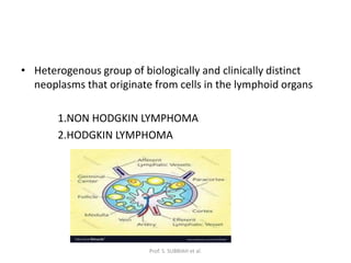
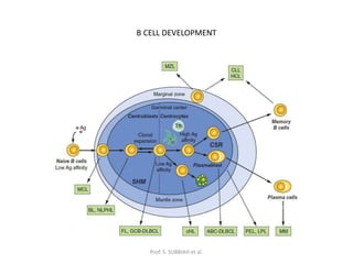
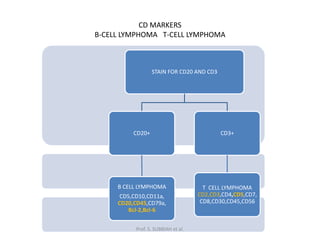
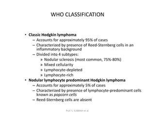
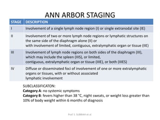
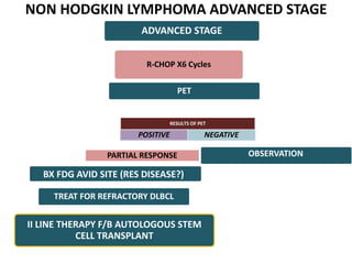
LYMPHOMA
DR.D.HEBER JOBSON
I YR RESIDENT-SURGICAL
ONCOLOGY
GOVERNMENT ROYAPETTAH
HOSPITAL
Prof. S. SUBBIAH et al.
• Heterogenous group of biologically and clinically distinct
neoplasms that originate from cells in the lymphoid organs
1.NON HODGKIN LYMPHOMA
2.HODGKIN LYMPHOMA
Prof. S. SUBBIAH et al.
B CELL DEVELOPMENT
Prof. S. SUBBIAH et al.
T CELL DEVELOPMENT
Prof. S. SUBBIAH et al.
CD MARKERS
B-CELL LYMPHOMA T-CELL LYMPHOMA
STAIN FOR CD20 AND CD3
CD20+
B CELL LYMPHOMA
CD5,CD10,CD11a,
CD20,CD45,CD79a,
Bcl-2,Bcl-6
CD3+
T CELL LYMPHOMA
CD2,CD3,CD4,CD5,CD7,
CD8,CD30,CD45,CD56
Prof. S. SUBBIAH et al.
HODGKIN LYMPHOMA
• Hodgkin lymphoma is an uncommon
lymphoproliferative malignancy of B
lymphocytes
Prof. S. SUBBIAH et al.
REED-STERNBERG CELL
Prof. S. SUBBIAH et al.
REED STERNBERG CELL
• Large cells with abundant cytoplasm and bilobed nuclei
• IHC : PAX 5 , CD 15 and CD 20 positive
• Constitute less than 1% of tumor cellularity, rest made of
inflammatory cells
Prof. S. SUBBIAH et al.
WHO CLASSIFICATION
• Classic Hodgkin lymphoma
– Accounts for approximately 95% of cases
– Characterized by presence of Reed-Sternberg cells in an
inflammatory background
– Divided into 4 subtypes:
» Nodular sclerosis (most common, 75%-80%)
» Mixed cellularity
» Lymphocyte-depleted
» Lymphocyte-rich
• Nodular lymphocyte predominant Hodgkin lymphoma
– Accounts for approximately 5% of cases
– Characterized by presence of lymphocyte-predominant cells
known as popcorn cells
– Reed-Sternberg cells are absent
Prof. S. SUBBIAH et al.
ANN ARBOR STAGING
STAGE DESCRIPTION
I Involvement of a single lymph node region (I) or single extranodal site (IE)
II Involvement of two or more lymph node regions or lymphatic structures on
the same side of the diaphragm alone (II) or
with involvement of limited, contiguous, extralymphatic organ or tissue (IIE)
III Involvement of lymph node regions on both sides of the diaphragm (III),
which may include the spleen (IIIS), or limited,
contiguous, extralymphatic organ or tissue (IIIE), or both (IIIES)
IV Diffuse or disseminated foci of involvement of one or more extralymphatic
organs or tissues, with or without associated
lymphatic involvement
SUBCLASSIFICATON:
Category A: no systemic symptoms
Category B: fevers higher than 38 °C, night sweats, or weight loss greater than
10% of body weight within 6 months of diagnosis
Prof. S. SUBBIAH et al.
• EARLY STAGE-I,II
FAVOURABLE/UNFAVOURABLE
• ADVANCED STAGE-III,IV
Prof. S. SUBBIAH et al.
UNFAVOURABLE PROGNOSTIC FACTORS
NCCN EORTC,GHSG
•Mediastinal lymphadenopathy with
ratio of maximum mass width to
maximum thoracic diameter of more
than 0.33
•Bulky lymphadenopathy >10 cm
•>3 nodal sites involved
•B symptoms (fever, night sweats,
and weight loss)
•ESR>50 mm/hr
Presence of extranodal contiguous
disease
>2 involved nodal sites
Age>50yrs
Prof. S. SUBBIAH et al.
INTERNATIONAL PROGNOSTIC INDEX-HL
MALE SEX
AGE>45
STAGE IV
SR. ALBUMIN<4
HB<10.5
WBC>15000
LYMPHOCYTE COUNT<600/MICROL OR <8% OF WBC
5 yr survival - 88% for no risk factor
- 62% for 4 or more risk factors
Prof. S. SUBBIAH et al.
RISK FACTORS
• Immunosuppression-acquired/EBV
• Increasing age (waning immunity)
• Previous history
• Family history
Prof. S. SUBBIAH et al.
CLINICAL PRESENTATION
• Lymphadenopathy-Painless/ Alcohol-pain
• B symptoms
• Hepatomegaly and splenomegaly
• Cough, dyspnea with bulky mediastinal
involvement.
• Aggressive/Indolent
Prof. S. SUBBIAH et al.
WORKUP
• Excisional or incisional biopsy is preferred.
• Image-guided core needle biopsies in patients
without peripheral adenopathy.
• FNA-not adequate for precise lymphoma
subclassification.
• Tissue biopsy with histologic,
immunophenotypic, and genetic studies
interpretation.
Prof. S. SUBBIAH et al.
LYMPH NODE BIOPSY
• A lymph node>1.5 × 1.5 cm, not associated
with a documented infection, persists longer
than 4 weeks.
• Patients with findings suggesting malignancy
(e.g., systemic complaints or B symptoms,
such as fever, night sweats, weight loss).
• Definitive diagnosis and histological
categorization.
Prof. S. SUBBIAH et al.
LYMPH NODE BIOPSY
• Peripheral Blood Smear-To rule out leukemia
• Core needle biopsy of lymph node is an
alternative in patients without accessible
lymph nodes, but not generally recommended
Prof. S. SUBBIAH et al.
BONE MARROW BIOPSY
Indicated in
Patients with B Symptoms
Stage III-IV
Recurrent Disease
• Not routine -PET scan demonstrates bone
marrow involvement, but indicated if
cytopenia is present and PET is negative.
Prof. S. SUBBIAH et al.
DIAGNOSTIC IMAGING
PET-CT scan
• Standard test for initial staging and
assessing response to therapy
• High FDG uptake should raise suspicion for
aggressive-histology lymphoma
• Diagnostic biopsy should be targeted to the
site of greatest FDG avidity .
Prof. S. SUBBIAH et al.
PET-CT
• FDG-PET scanning is highly sensitive for
detecting both nodal and extranodal
sites.
• The intensity of FDG avidity, or
Standardized Uptake Value, correlates
with histologic aggressiveness.
Prof. S. SUBBIAH et al.
• Detects actively metabolizing tumor in
residual masses following or during
chemotherapy.
• Persistent abnormal uptake predicts early
relapse and/or reduced survival.
• More accurate than the detection of a residual
mass on CT scans, which can often be a false
positive.
Prof. S. SUBBIAH et al.
PET-CT RESPONSE SCORE
SCORE UPTAKE
1 NO UPTAKE
2 <MEDIASTINUM
3 >MEDIASTINUM<LIVER
4 MODERATELY MORE THAN LIVER
5 MARKEDLY MORE THAN LIVER OR NEW SITE
Prof. S. SUBBIAH et al.
STAGE-IA/IIA(Non Bulky)/Favourable*
ABVD X 2Cycles
Restage with
INTERIM PET-CT
DEAUVILLE 1-2 DEAUVILLE 3 DEAUVILLE 4
ABVD X 2 CYCLES
or ISRT 20GY
ABD X 4 CYCLES or
ISRT 20GY or
ABVD X 1 CYCLE
ABVD - 2 Cycle
Restage with INTERIM PET/CT
DEAUVILLE 1-3 DEAUVILLE 4/5
BIOPSY – NEG-ISRT 30GY-
POS-Refractory disease
ISRT 30GY
*-Favourable Factors-ESR<50/<3 Nodal Sites/No Extra Nodal Disease
Unfavourable-B/Bulky->10CM
ABVD X 2Cycles
Restage with
INTERIM PET-CT
DEAUVILLE 1-3 DEAUVILLE 4/5
AVD X 4 CYCLES ESCALATED BEACOPP
STAGE III/IV
ABVD X 2Cycles
Restage with
INTERIM PET-CT
DEAUVILLE 1-3 DEAUVILLE 4/5
AVD X 4 CYCLES
ABVD-2
Cycles+ESCALATED
BEACOPP
REFRACTORY DISEASE
SECOND LINE AGENTS
(ICE/DHAP/GVD)
Restage with INTERIM
PET-CT
DEAUVILLE 1-3 DEAUVILLE 4/5
AUTOLOGOUS STEM
CELL TRANSPLANT
BRENTUXIMAB
VEDONTIN
BRENTUXIMAB
VEDONTIN
Prof. S. SUBBIAH et al.
WHO CLASSIFICATION
Prof. S. SUBBIAH et al.
INTERNATIONAL PROGNOSTIC INDEX
AGE OLDER THAN 60 YRS
LDH>UPPER LIMIT NORMAL
ECOG PERFORMANCE STATUS ≥2
ANN ARBOR STAGE III OR IV
NUMBER OF EXTRANODAL DISEASE SITES GREATER THAN ONE
Prof. S. SUBBIAH et al.
TREATMENT
• Cyclophosphamide-750 mg/m2 IV on DAY-1
• Doxorubicin-50mg/m2 IV on DAY-1
• Vincristine-1.4 mg/m2 IV on DAY-1
• Prednisone-100mg/ PO on DAYS 1-5
• Rituximab-375mg/m2 on DAY-1
CYCLE EVERY 21 DAYS
Prof. S. SUBBIAH et al.
NON HODGKIN LYMPHOMA STAGE I,II
LIMITED STAGE DLBCL (↑ LDH/ ECOG PS 2-4)
PARTIAL RESPONSE MORE EXTENSIVE DISEASE
RESULTS OF PET
POSITIVE NEGATIVE
R-CHOP X3 Cycles F/B RISK ADAPTED MANAGEMENT BASED ON
PET R-CHOP X4 Cycles
TREAT FOR REFRACTORY DLBCL
3 Addl CYCLES OF R-CHOP
(TOTAL 6 CYCLES) VS ISRT
30GY WITH 6-10 GY
BOOST TO FDG AVID
AREA (NO Addl R-CHOP)
3 Addl CYCLES OF R-CHOP (TOTAL6
CYCLES) VS ISRT 30GY (NO Addl R-
CHOP)
PET?
1 Addl CYCLE OF R-CHOP(TOTAL 4) VS ISRT 30
GY (NO ADDL R-CHOP)
BX FDG AVID SITE (RES DISEASE?)
NON HODGKIN LYMPHOMA STAGE I,II bulky
PARTIAL RESPONSE MORE EXTENSIVE DISEASE
RESULTS OF PET
POSITIVE NEGATIVE
R-CHOP X 6 Cycles
TREAT FOR REFRACTORY DLBCL
3 Addl CYCLES OF R-CHOP
(TOT 6 CYCLES) VS ISRT
30GY WITH 6-10 GY
BOOST TO FDG AVID
AREA (NO Addl R-CHOP)
ISRT 30 – 40
GY
PET?
ISRT 30 GY
BX FDG AVID SITE (RES DISEASE?)
NON HODGKIN LYMPHOMA ADVANCED STAGE
ADVANCED STAGE
PARTIAL RESPONSE
RESULTS OF PET
POSITIVE NEGATIVE
R-CHOP X6 Cycles
TREAT FOR REFRACTORY DLBCL
BX FDG AVID SITE (RES DISEASE?)
PET
OBSERVATION
II LINE THERAPY F/B AUTOLOGOUS STEM
CELL TRANSPLANT
THANK YOU
Prof. S. SUBBIAH et al.

LYMPHOMA.pptx

  • 1.
    LYMPHOMA DR.D.HEBER JOBSON I YRRESIDENT-SURGICAL ONCOLOGY GOVERNMENT ROYAPETTAH HOSPITAL Prof. S. SUBBIAH et al.
  • 2.
    • Heterogenous groupof biologically and clinically distinct neoplasms that originate from cells in the lymphoid organs 1.NON HODGKIN LYMPHOMA 2.HODGKIN LYMPHOMA Prof. S. SUBBIAH et al.
  • 3.
    B CELL DEVELOPMENT Prof.S. SUBBIAH et al.
  • 4.
    T CELL DEVELOPMENT Prof.S. SUBBIAH et al.
  • 5.
    CD MARKERS B-CELL LYMPHOMAT-CELL LYMPHOMA STAIN FOR CD20 AND CD3 CD20+ B CELL LYMPHOMA CD5,CD10,CD11a, CD20,CD45,CD79a, Bcl-2,Bcl-6 CD3+ T CELL LYMPHOMA CD2,CD3,CD4,CD5,CD7, CD8,CD30,CD45,CD56 Prof. S. SUBBIAH et al.
  • 6.
    HODGKIN LYMPHOMA • Hodgkinlymphoma is an uncommon lymphoproliferative malignancy of B lymphocytes Prof. S. SUBBIAH et al.
  • 7.
  • 8.
    REED STERNBERG CELL •Large cells with abundant cytoplasm and bilobed nuclei • IHC : PAX 5 , CD 15 and CD 20 positive • Constitute less than 1% of tumor cellularity, rest made of inflammatory cells Prof. S. SUBBIAH et al.
  • 9.
    WHO CLASSIFICATION • ClassicHodgkin lymphoma – Accounts for approximately 95% of cases – Characterized by presence of Reed-Sternberg cells in an inflammatory background – Divided into 4 subtypes: » Nodular sclerosis (most common, 75%-80%) » Mixed cellularity » Lymphocyte-depleted » Lymphocyte-rich • Nodular lymphocyte predominant Hodgkin lymphoma – Accounts for approximately 5% of cases – Characterized by presence of lymphocyte-predominant cells known as popcorn cells – Reed-Sternberg cells are absent Prof. S. SUBBIAH et al.
  • 10.
    ANN ARBOR STAGING STAGEDESCRIPTION I Involvement of a single lymph node region (I) or single extranodal site (IE) II Involvement of two or more lymph node regions or lymphatic structures on the same side of the diaphragm alone (II) or with involvement of limited, contiguous, extralymphatic organ or tissue (IIE) III Involvement of lymph node regions on both sides of the diaphragm (III), which may include the spleen (IIIS), or limited, contiguous, extralymphatic organ or tissue (IIIE), or both (IIIES) IV Diffuse or disseminated foci of involvement of one or more extralymphatic organs or tissues, with or without associated lymphatic involvement SUBCLASSIFICATON: Category A: no systemic symptoms Category B: fevers higher than 38 °C, night sweats, or weight loss greater than 10% of body weight within 6 months of diagnosis Prof. S. SUBBIAH et al.
  • 11.
    • EARLY STAGE-I,II FAVOURABLE/UNFAVOURABLE •ADVANCED STAGE-III,IV Prof. S. SUBBIAH et al.
  • 12.
    UNFAVOURABLE PROGNOSTIC FACTORS NCCNEORTC,GHSG •Mediastinal lymphadenopathy with ratio of maximum mass width to maximum thoracic diameter of more than 0.33 •Bulky lymphadenopathy >10 cm •>3 nodal sites involved •B symptoms (fever, night sweats, and weight loss) •ESR>50 mm/hr Presence of extranodal contiguous disease >2 involved nodal sites Age>50yrs Prof. S. SUBBIAH et al.
  • 13.
    INTERNATIONAL PROGNOSTIC INDEX-HL MALESEX AGE>45 STAGE IV SR. ALBUMIN<4 HB<10.5 WBC>15000 LYMPHOCYTE COUNT<600/MICROL OR <8% OF WBC 5 yr survival - 88% for no risk factor - 62% for 4 or more risk factors Prof. S. SUBBIAH et al.
  • 14.
    RISK FACTORS • Immunosuppression-acquired/EBV •Increasing age (waning immunity) • Previous history • Family history Prof. S. SUBBIAH et al.
  • 15.
    CLINICAL PRESENTATION • Lymphadenopathy-Painless/Alcohol-pain • B symptoms • Hepatomegaly and splenomegaly • Cough, dyspnea with bulky mediastinal involvement. • Aggressive/Indolent Prof. S. SUBBIAH et al.
  • 16.
    WORKUP • Excisional orincisional biopsy is preferred. • Image-guided core needle biopsies in patients without peripheral adenopathy. • FNA-not adequate for precise lymphoma subclassification. • Tissue biopsy with histologic, immunophenotypic, and genetic studies interpretation. Prof. S. SUBBIAH et al.
  • 17.
    LYMPH NODE BIOPSY •A lymph node>1.5 × 1.5 cm, not associated with a documented infection, persists longer than 4 weeks. • Patients with findings suggesting malignancy (e.g., systemic complaints or B symptoms, such as fever, night sweats, weight loss). • Definitive diagnosis and histological categorization. Prof. S. SUBBIAH et al.
  • 18.
    LYMPH NODE BIOPSY •Peripheral Blood Smear-To rule out leukemia • Core needle biopsy of lymph node is an alternative in patients without accessible lymph nodes, but not generally recommended Prof. S. SUBBIAH et al.
  • 19.
    BONE MARROW BIOPSY Indicatedin Patients with B Symptoms Stage III-IV Recurrent Disease • Not routine -PET scan demonstrates bone marrow involvement, but indicated if cytopenia is present and PET is negative. Prof. S. SUBBIAH et al.
  • 20.
    DIAGNOSTIC IMAGING PET-CT scan •Standard test for initial staging and assessing response to therapy • High FDG uptake should raise suspicion for aggressive-histology lymphoma • Diagnostic biopsy should be targeted to the site of greatest FDG avidity . Prof. S. SUBBIAH et al.
  • 21.
    PET-CT • FDG-PET scanningis highly sensitive for detecting both nodal and extranodal sites. • The intensity of FDG avidity, or Standardized Uptake Value, correlates with histologic aggressiveness. Prof. S. SUBBIAH et al.
  • 22.
    • Detects activelymetabolizing tumor in residual masses following or during chemotherapy. • Persistent abnormal uptake predicts early relapse and/or reduced survival. • More accurate than the detection of a residual mass on CT scans, which can often be a false positive. Prof. S. SUBBIAH et al.
  • 23.
    PET-CT RESPONSE SCORE SCOREUPTAKE 1 NO UPTAKE 2 <MEDIASTINUM 3 >MEDIASTINUM<LIVER 4 MODERATELY MORE THAN LIVER 5 MARKEDLY MORE THAN LIVER OR NEW SITE Prof. S. SUBBIAH et al.
  • 24.
  • 25.
    STAGE-IA/IIA(Non Bulky)/Favourable* ABVD X2Cycles Restage with INTERIM PET-CT DEAUVILLE 1-2 DEAUVILLE 3 DEAUVILLE 4 ABVD X 2 CYCLES or ISRT 20GY ABD X 4 CYCLES or ISRT 20GY or ABVD X 1 CYCLE ABVD - 2 Cycle Restage with INTERIM PET/CT DEAUVILLE 1-3 DEAUVILLE 4/5 BIOPSY – NEG-ISRT 30GY- POS-Refractory disease ISRT 30GY *-Favourable Factors-ESR<50/<3 Nodal Sites/No Extra Nodal Disease
  • 26.
    Unfavourable-B/Bulky->10CM ABVD X 2Cycles Restagewith INTERIM PET-CT DEAUVILLE 1-3 DEAUVILLE 4/5 AVD X 4 CYCLES ESCALATED BEACOPP
  • 27.
    STAGE III/IV ABVD X2Cycles Restage with INTERIM PET-CT DEAUVILLE 1-3 DEAUVILLE 4/5 AVD X 4 CYCLES ABVD-2 Cycles+ESCALATED BEACOPP
  • 28.
    REFRACTORY DISEASE SECOND LINEAGENTS (ICE/DHAP/GVD) Restage with INTERIM PET-CT DEAUVILLE 1-3 DEAUVILLE 4/5 AUTOLOGOUS STEM CELL TRANSPLANT BRENTUXIMAB VEDONTIN BRENTUXIMAB VEDONTIN
  • 29.
  • 30.
  • 31.
    INTERNATIONAL PROGNOSTIC INDEX AGEOLDER THAN 60 YRS LDH>UPPER LIMIT NORMAL ECOG PERFORMANCE STATUS ≥2 ANN ARBOR STAGE III OR IV NUMBER OF EXTRANODAL DISEASE SITES GREATER THAN ONE Prof. S. SUBBIAH et al.
  • 32.
    TREATMENT • Cyclophosphamide-750 mg/m2IV on DAY-1 • Doxorubicin-50mg/m2 IV on DAY-1 • Vincristine-1.4 mg/m2 IV on DAY-1 • Prednisone-100mg/ PO on DAYS 1-5 • Rituximab-375mg/m2 on DAY-1 CYCLE EVERY 21 DAYS Prof. S. SUBBIAH et al.
  • 33.
    NON HODGKIN LYMPHOMASTAGE I,II LIMITED STAGE DLBCL (↑ LDH/ ECOG PS 2-4) PARTIAL RESPONSE MORE EXTENSIVE DISEASE RESULTS OF PET POSITIVE NEGATIVE R-CHOP X3 Cycles F/B RISK ADAPTED MANAGEMENT BASED ON PET R-CHOP X4 Cycles TREAT FOR REFRACTORY DLBCL 3 Addl CYCLES OF R-CHOP (TOTAL 6 CYCLES) VS ISRT 30GY WITH 6-10 GY BOOST TO FDG AVID AREA (NO Addl R-CHOP) 3 Addl CYCLES OF R-CHOP (TOTAL6 CYCLES) VS ISRT 30GY (NO Addl R- CHOP) PET? 1 Addl CYCLE OF R-CHOP(TOTAL 4) VS ISRT 30 GY (NO ADDL R-CHOP) BX FDG AVID SITE (RES DISEASE?)
  • 34.
    NON HODGKIN LYMPHOMASTAGE I,II bulky PARTIAL RESPONSE MORE EXTENSIVE DISEASE RESULTS OF PET POSITIVE NEGATIVE R-CHOP X 6 Cycles TREAT FOR REFRACTORY DLBCL 3 Addl CYCLES OF R-CHOP (TOT 6 CYCLES) VS ISRT 30GY WITH 6-10 GY BOOST TO FDG AVID AREA (NO Addl R-CHOP) ISRT 30 – 40 GY PET? ISRT 30 GY BX FDG AVID SITE (RES DISEASE?)
  • 35.
    NON HODGKIN LYMPHOMAADVANCED STAGE ADVANCED STAGE PARTIAL RESPONSE RESULTS OF PET POSITIVE NEGATIVE R-CHOP X6 Cycles TREAT FOR REFRACTORY DLBCL BX FDG AVID SITE (RES DISEASE?) PET OBSERVATION II LINE THERAPY F/B AUTOLOGOUS STEM CELL TRANSPLANT
  • 36.
    THANK YOU Prof. S.SUBBIAH et al.

Editor's Notes

  • #9 PAX 5-Transcription factor BSAP-B CELL LINEAGE SPECIFIC ACTIVATOR PROTEIN
  • #10 MULTILOBATED OR EXTREMELY FOLDED NUCLEUS-L&H
  • #11 NUMBER,LOCATION,LYMPHATIC ORGAN
  • #16 Aggressive lymphomas-acute or subacute presentation with increasing size of the mass and B symptoms. Indolent lymphomas-chronic course, with asymptomatic lymphadenopathy and/or slowly progressive cytopenias.
  • #17 CBC,LDH
  • #20 Less commonly in practice Staging and Prognostic purposes depending on the disease histology. Overall involvement in Hodgkin is less than 5%
  • #25 BLEOMYCIN 10MG/M2 ETOPOSIDE 200MG/M2 DOXORUBICIN 35MG/M2 CYCLOPHOSPHAMIDE 1250MG/M2 VINCRISTINE 1.4MG/M2 PROCARBAZINE 100 MG/M2 PO PREDNISONE 40 MG/M2 PO
  • #29 HDT/ASCT-BV HDT/RT-BV
  • #33 Serum protein electrophoresis-pres of monoclonal paraproteins b2 microglobulin and LDH
  • #37 DEXAMETHASONE/CISPLATIN/CYTARABINE+/-RITUXIMAB DEXAMETHASONE/CYTARABINE/OXALIPLATIN+/-RITUXIMAB TUMOR LYSIS SYNDROME In the first 12 to 72 hrs of treatment VIRAL REACTIVATION RCHOP FOR 6 CYCLES NO ROLE OF INTERIM PET R-POLA CHP VS RCHOP II LINE THERAPY ICE/DHAP CAR T CELL THERAPY AXICABTAGENE CILOLEUCEL