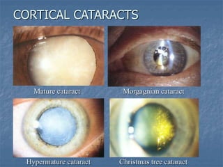
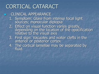
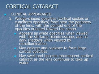
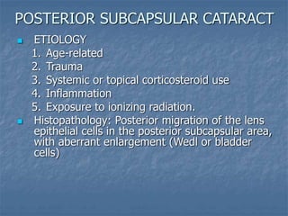
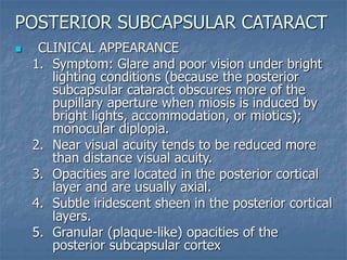
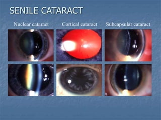
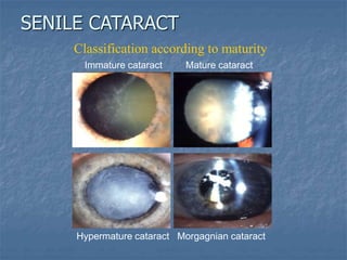
ACQUIRED CATARACTS
Sengdy Chandra Chauhari
ACQUIRED CATARACTS
1. Aging changes
2. Trauma
3. Metabolic cataracts
4. Cataracts associated with uveitis
5. Exfoliation syndromes
6. Cataract and atopic dermatitis
7. Lens-induced uveitis
8. Lens-induced glaucoma
9. Ischemia
10. Cataracts associated with degenerative ocular
disorders
AGE-RELATED CHANGES in the LENS
1. Change of crystallins (lens proteins) by chemical
modification and aggregation into high-
molecular-weight protein.
2. Compression and hardening of lens nucleus
(nuclear sclerosis)
3. Decreased concentrations of glutathione and
potassium
4. Increased concentrations of sodium and calcium
5. Increased hydration
6. Progressive pigmentation (yellow or brownish
hue)
AGING CHANGES
SENILE CATARACTS
1. Nuclear cataracts
2. Cortical cataracts
3. Posterior subcapsular cataracts (cupuliform)
AGING CHANGES
 Definition: Central lens opacity caused by
excessive amount of sclerosis and yellowing of
the lens nucleus
 Natural course
1. Usually bilateral but asymmetric
2. Tend to progress slowly
 Evaluation of the degree of sclerosis, yellowing
and opacification
1. Slit-lamp biomicroscope
2. Examining the red-reflex with pupil dilated
 Histopathology: Homogeneity of lens nucleus
with loss of cellular laminations
NUCLEAR CATARACTS
 CLINICAL APPEARANCE
1. Typically cause greater impairment of distance
vision than of near vision
2. In the early stages, the progressive hardening
of the lens nucleus commonly causes an
increase in the refractive index of the lens and
thus a myopic shift in refraction, sometimes
known as lenticular myopia.
3. In some cases, the myopic shift transiently
enables otherwise presbyopic individuals to
read without spectacles (second sight)
NUCLEAR CATARACTS
 CLINICAL APPEARANCE
4. Occasionally, the abrupt change in refractive
index between the sclerotic nucleus and the
lens cortex can cause monocular diplopia.
5. Progressive yellowing of the lens causes poor
hue discrimination, especially at the blue end
of the visible light spectrum.
6. Decreased photopic retinal function (in
advanced cases)
7. Opacification of lens nucleus in very
advanced cases (brunescent cataract)
NUCLEAR CATARACTS
 Pathogenesis: Changes in the ionic composition
of the lens cortex and subsequent changes in
hydration of the lens fibers
 Natural course
1. Usually bilateral but asymmetric
2. Vary greatly in rate of progression; some
remain unchanged for prolonged periods,
while others progress rapidly.
 Histopathology: Hydropic swelling of the lens
fibers
CORTICAL CATARACTS
 Classification according to maturation
1. Mature cataract occurs when the entire
cortex from the capsule to the nucleus
becomes white and opaque
2. Hypermature cataract occurs when
degenerated cortical material leaks through
the lens capsule, leaving the capsule wrinkled
and shrunken
3. Morgagnian cataract occurs when further
liquefaction of the cortex allows free
movement of the nucleus within the capsular
bag
CORTICAL CATARACTS
CORTICAL CATARACTS
Mature cataract Morgagnian cataract
Hypermature cataract Christmas tree cataract
 CLINICAL APPEARANCE
1. Symptom: Glare from intense focal light
sources; monocular diplopia
2. Effect on visual function varies greatly,
depending on the location of the opacification
relative to the visual axis
3. First sign: Vacuoles and water clefts in the
anterior or posterior cortex
4. The cortical lamellae may be separated by
fluid.
CORTICAL CATARACT
 CLINICAL APPEARANCE
5. Wedge-shaped opacities (cortical spokes or
cuneiform opacities) form near the periphery
of the lens, with the pointed end of the
opacities oriented toward the center
 Appears as white opacities when viewed
with the slit-lamp biomicroscope, and as
dark shadows when viewed by
retroillumination
 May enlarge and coalesce to form large
cortical opacities
 May swell and become intumescent cortical
cataract as the lens continues to take up
water
CORTICAL CATARACT
 ETIOLOGY
1. Age-related
2. Trauma
3. Systemic or topical corticosteroid use
4. Inflammation
5. Exposure to ionizing radiation.
 Histopathology: Posterior migration of the lens
epithelial cells in the posterior subcapsular area,
with aberrant enlargement (Wedl or bladder
cells)
POSTERIOR SUBCAPSULAR CATARACT
 CLINICAL APPEARANCE
1. Symptom: Glare and poor vision under bright
lighting conditions (because the posterior
subcapsular cataract obscures more of the
pupillary aperture when miosis is induced by
bright lights, accommodation, or miotics);
monocular diplopia.
2. Near visual acuity tends to be reduced more
than distance visual acuity.
3. Opacities are located in the posterior cortical
layer and are usually axial.
4. Subtle iridescent sheen in the posterior cortical
layers.
5. Granular (plaque-like) opacities of the
posterior subcapsular cortex
POSTERIOR SUBCAPSULAR CATARACT
Nuclear cataract Cortical cataract Subcapsular cataract
SENILE CATARACT
Immature cataract Mature cataract
Hypermature cataract Morgagnian cataract
Classification according to maturity
SENILE CATARACT
HARDNESS of the NUCLEUS
Grade Color Type of cataract Red reflex
1 Transparent or pale
gray
Cortical or recent
subcapsular
High
2 Gray or gray-yellow Subcapsular posterior Marked
3 Yellow or yellow-
gray
Nuclear, cortico-nuclear Good
4 Yellow-amber or
amber
Cortico-nuclear, dense Poor
5 Dark brown or black Totally dense Absent
 CORTICOSTEROIDS
 Posterior subcapsular
cataracts may be induced
depending on:
1. Dose and duration of
corticosteroid treatment
2. Individual susceptibility to
corticosteroid
 PHENOTHIAZINES
 Pigmented deposits in the
anterior lens epithelium in
an axial configuration →
affected by both dose and
duration of medication
DRUG-INDUCED LENS CHANGES
 MIOTICS (ANTICHOLINESTERASES)
(echothiopate and demecarium Br)
Cataracts may be induced depending on dose
and duration of treatment.
1. First appear as small vacuoles within and
posterior to the anterior lens capsule and
epithelium (by retroillumination).
2. May progress to posterior cortical and
nuclear lens changes.
 AMIODARONE
 Stellate anterior axial pigment deposition
DRUG-INDUCED LENS CHANGES
Traumatic lens damage may be caused by:
1. Mechanical injury
2. Physical forces (radiation, electrical current,
chemicals)
3. Osmotic influences (diabetes mellitus)
TRAUMA
TRAUMA to the LENS
1. Contusion
2. Perforating and penetrating injury
3. Radiation-induced cataracts
4. Chemical injuries
5. Electrical injury
6. Intralenticular foreign bodies
7. Metallosis
TRAUMA
VOSSIUS RING
1. Imprinting of pigment from the pupillary ruff
onto the anterior surface
2. Indicator of prior blunt trauma
3. Visually insignificant; resolves gradually with
time
CONTUSION
TRAUMATIC CATARACT
1. Lens opacification may occur as an acute event
or as late sequela and may involve only a
portion of the lens or the entire lens.
 Common initial manifestation: Stellate or
rosette-shaped opacification that is axial in
location, involves the posterior lens
capsule and may progress to opacification
of the entire lens
2. Lens dislocation
CONTUSION
DISLOCATION and SUBLUXATION
1. Pathogenesis: Compression of the globe →
rapid expansion in an equatorial plane →
disruption of zonular fibers → dislocation or
subluxation.
2. Traumatic lens subluxation
CONTUSION
 Fluctuation of vision,
impaired accommodation,
monocular diplopia, and
high astigmatism
 Iridodonesis or
phacodonesis
 Zonular disruption
IONIZING RADIATION
1. The lens is extremely sensitive to ionizing
radiation
2. Dose of radiation: Ionizing radiation in the x-ray
range (0.001-10 nm wavelength) can cause
cataracts in dosages as low as 200 rads in one
fraction → A routine chest x-ray equals 0.1 rads
exposure to the thorax
3. Patient’s age: Younger patients are more
susceptible because of more actively growing
lens cells.
4. First appear as punctate opacities within the
posterior capsule and feathery anterior
subcapsular opacities that radiate toward the
equator of the lens and may progress to
complete opacification of the lens.
RADIATION-INDUCED CATARACT
INFRARED RADIATION
(GLASSBLOWER’S CATARACT)
 May cause the outer layers of the anterior lens
capsule to peel off a single layer.
ULTRAVIOLET RADIATION
 Long-term exposure of UV-B (290-320 nm) from
sun exposure → increased risk of cortical and
posterior subcapsular cataracts
MICROWAVE RADIATION
1. Non-ionizing radiation with wavelengths
between infrared and shortwave on the
electromagnetic spectrum
2. No evidence that microwaves cause cataracts in
humans. The only biological effect of
microwaves is thermal.
RADIATION-INDUCED CATARACT
ALKALI INJURIES to the ocular surface
1. Often result in cortical cataract as an acute
event or as late sequela
2. Alkali compounds penetrate the eye readily,
causing an increase in aqueous pH and a
decrease in the level of aqueous glucose and
ascorbate.
ACID INJURIES to ocular surface
 Less likely to result in cataract formation
because acid tends to penetrate the eye less
easily than alkali.
CHEMICAL INJURIES
1. Can cause protein coagulation and cataract
formation. This cataract may regress, remain
stationary, or mature to complete cataract over
months or years
2. Lens manifestations are more likely when the
transmission of current involves the patient’s
head
3. Initially, lens vacuoles appear in the anterior
midperiphery of the lens, followed by linear
opacities in the anterior subcapsular cortex
ELECTRICAL INJURY
 Opacification of the cortex at the site of the
rupture that usually progresses rapidly to
complete opacification
PERFORATING and
PENETRATING INJURY
INTRALENTICULAR FOREIGN BODIES
1. May cause cataract formation but do not
always lead to lens opacification.
2. Foreign body is sometimes retained within the
lens if the foreign body is not composed of a
ferric or cupric material, or the anterior lens
capsule seals the perforation site.
PERFORATING and
PENETRATING INJURY
SIDEROSIS BULBI
1. Deposition of iron molecules in the trabecular
meshwork, lens epithelium, iris and retina
2. The epithelium and cortical fibers of the
affected lens at first show a yellowish tinge,
followed later by a rusty brown discoloration
METALLOSIS
CHALCOSIS
1. Occurs when an intraocular copper-containing
foreign body deposits copper in Descemet’s
membrane, the anterior lens capsule, and other
intraocular basement membranes.
2. Sunflower cataract (petal-shaped deposition of
yellow or brown pigmentation in the lens
capsule that radiates from the anterior axial
pole of the lens to its equator) usually causes
no significant loss of visual acuity
METALLOSIS
1. Diabetes mellitus
2. Galactosemia
3. Hypocalcemic cataract (tetanic cataract)
4. Wilson disease (hepatolenticular degeneration)
5. Myotonic dystrophy
METABOLIC CATARACT
PATHOGENESIS
1. As the blood sugar level increases, so also does
the glucose content in the aqueous humor.
Because glucose enters the lens by diffusion,
glucose content in the lens will be increased.
2. Some of the glucose is converted by aldose
reductase to sorbitol, which is not metabolized
but remains in the lens. Subsequently, osmotic
pressure causes an influx of water into the lens,
which leads to swelling of the lens fibers.
 Change of refractive power of the lens
(most commonly myopic).
 Decreased amplitude of accommodation
 Presence of presbyopia at a younger age
DIABETES MELLITUS
DIABETIC CATARACT (SNOWFLAKE CATARACT)
1. Bilateral, widespread subcapsular lens changes
of abrupt onset and acute progression, typically
in young people with uncontrolled diabetes
mellitus
2. Multiple gray-white subcapsular opacities that
have a snowflake appearance are seen initially in
the superficial anterior and posterior lens cortex
3. Vacuoles in the lens capsule; clefts in the
underlying cortex
4. Intumescence and maturity of the cortical
cataract follow shortly thereafter
 Any rapidly maturing bilateral cortical cataracts
in a child or young adult should alert the clinician
to the possibility of diabetes mellitus.
DIABETES MELLITUS
SENESCENT CATARACT
1. Accumulation of sorbitol within the lens
2. Subsequent hydration changes
3. Increased glycosylation of proteins in the
diabetic lens
→ Increased risk of age-related lens changes which
tend to occur at a younger age
DIABETES MELLITUS
DEFINITION
Autosomal recessive inherited inability to
convert galactose to glucose. Consequently,
excessive galactose accumulates in body
tissues, with further metabolic conversion of
galactose to galactitol (dulcitol)
ETIOLOGY
Defects in one of three enzymes – galactose-1-
phosphate uridyl transferase, galactokinase, or
UDP-galactose-4-epimerase
GALACTOSEMIA
CLASSIC GALACTOSEMIA
(caused by a defect in transferase)
1. Malnutrition, hepatomegaly, jaundice, and
mental deficiency present within the first few
weeks of life
2. Cataract develops in 75% of cases, usually
within the first few weeks of life
3. Oil-droplet bilateral cataract (opacification of
the nucleus and deep cortex) that may
progress to total opacification of the lens
TREATMENT
Elimination of milk and milk products from the
diet
GALACTOSEMIA
1. Usually bilateral punctate iridescent opacities
in the anterior and posterior cortex that lie
beneath lens capsule and separated from it by
a zone of clear lens.
2. May either remain stable or mature into
complete cortical cataract
HYPOCALCEMIC CATARACT
(TETANIC CATARACT)
DEFINITION
Autosomal recessive inherited disorder of
copper metabolism
CLINICAL APPEARANCE
1. Kayser-Fleischer ring (golden brown
discoloration of Descemet’s membrane around
the periphery of the cornea)
2. Characteristic sunflower cataract – Deposition
of reddish brown pigment (cuprous oxide) in
the anterior lens capsule and subcapsular
cortex in a stellate shape → usually does not
produce serious visual impairment
WILSON DISEASE
(HEPATOLENTICULAR DEGENERATION)
DEFINITION
Autosomal dominant inherited
condition characterized by delayed
relaxation of contracted muscles,
ptosis, facial musculature
weakness, cardiac conduction
defects, and prominent frontal
balding in affected male patients.
OCULAR MANIFESTATIONS
Polychromatic iridescent crystals
(whorls of plasma-lemma from the
lens fibers ultrastructurally) in the
cortex, with sequential posterior
subcapsular cataract progressing
to complete cortical opacification.
MYOTONIC DYSTROPHY
CLINICAL APPEARANCE
1. Posterior subcapsular cataract that may
progress to a mature cataract
2. Posterior synechiae formation, often associated
with thickening of the anterior lens capsule and
development of a fibrovascular membrane
across it and the pupil (pupillary membrane)
CATARACT associated with UVEITIS
3. Calcium deposits on the
anterior capsule or within
the lens substance
TRUE EXFOLIATION
1. Pathogenesis: Intense exposure to infrared
radiation and heat causes the superficial lens
capsule to delaminate and peel off in scrolls
2. Occurs primarily in glassblowers and blast
furnace operators
EXFOLIATION SYNDROME
EXFOLIATION SYNDROME (PSEUDOEXFOLIATION)
CLINICAL APPEARANCE
1. Unilateral or bilateral disorder; onset often occurs
in the seventh decade
2. Basement membrane-like fibrillogranular white
material is deposited on the lens, cornea, iris,
anterior hyaloid face, ciliary processes, zonular
fibers, and trabecular meshwork. These deposits
arise from basement membranes within the eye,
and appear as grayish white flecks that are
prominent at the pupillary margin and on the lens
capsule
3. Atrophy of the iris at the pupillary margin
4. Deposition of pigment on the anterior surface of
the iris
EXFOLIATION SYNDROME
EXFOLIATION SYNDROME (PSEUDOEXFOLIATION)
CLINICAL APPEARANCE
5. Poorly dilating pupil
6. Increased pigmentation of the trabecular
meshwork
7. Capsular fragility
8. Zonular weakness, spontaneous lens subluxation,
phacodonesis
9. Open-angle glaucoma
EXFOLIATION SYNDROME
Cataract may develop in up to 25% of cases
1. Usually bilateral (70%)
2. Onset occurs in the second to third decade
3. Typically anterior subcapsular opacities in the
pupillary area that resemble shieldlike plaques.
CATARACT and ATOPIC DERMATITIS
PHACOANTIGENIC (PHACOANAPHYLACTIC UVEITIS)
DEFINITION: Immune-mediated granulomatous
inflammation initiated by lens proteins released
through a ruptured lens capsule
PATHOGENESIS: Liberation of a large amount of lens
protein into the anterior chamber disrupts the
normal immunologic tolerance and may trigger a
severe inflammatory reaction
CLINICAL APPEARANCE
1. Usually occurs days to weeks following traumatic
rupture of the lens capsule or following cataract
surgery when cortical material is retained within
the eye
2. Secondary glaucoma
LENS-INDUCED UVEITIS
PHACOANTIGENIC (PHACOANAPHYLACTIC UVEITIS)
HISTOPATHOLOGY: Zonal granulomatous
inflammation surrounding a breach of the lens
capsule.
COMPLICATION
Cyclitic membrane, synechiae formation, phthisis
bulbi.
TREATMENT: Lens extraction
LENS-INDUCED UVEITIS
1. Glaukomflecken
2. Phacolytic glaucoma
3. Lens-particle glaucoma
4. Phacomorphic glaucoma
LENS-INDUCED GLAUCOMA
GLAUKOMFLECKEN
1. Gray-white epithelial and anterior cortical lens
opacities that occur following an episode of
markedly elevated IOP, as in acute ACG.
2. Histopathology: Necrotic lens epithelial cells,
degenerated subepithelial cortex
LENS-INDUCED GLAUCOMA
PATHOGENESIS
1. Complication of a mature or hypermature
cataract
2. Denatured, liquefied high-molecular-weight lens
proteins leak through an intact but permeable
lens capsule. An immune response is not elicited;
rather, macrophages ingest these lens proteins.
The trabecular meshwork can become clogged
with both the lens proteins and the engorged
macrophages.
PHACOLYTIC GLAUCOMA
CLINICAL APPEARANCE
1. Abrupt onset in a cataractous eye that has had
poor vision for some time
2. White flocculent material in the anterior
chamber and adheres to lens capsule
3. Open angle glaucoma
TREATMENT
1. Initial treatment: Control of IOP with
antiglaucoma medications, and of the
inflammation with topical corticosteroids.
2. Definitive treatment: Surgical removal of the
lens
PHACOLYTIC GLAUCOMA
PATHOGENESIS
Liberation of lens cortex into the anterior
chamber following penetrating lens injury, ECCE
with retained cortical material, or Nd:YAG
capsulotomy → obstruction of aqueous outflow
through trabecular meshwork
CLINICAL APPEARANCE
1. Usually occurs days or weeks after the surgical
event or lens injury
2. White, fluffy, cortical lens material in the
anterior chamber
3. Open angle glaucoma
LENS-PARTICLE GLAUCOMA
TREATMENT
1. Medical therapy to lower IOP and to reduce
intraocular inflammation
2. Surgical removal of the retained lens material
LENS-PARTICLE GLAUCOMA
PATHOGENESIS
Intumescent cataractous lens → pupillary block
and shallowing of the anterior chamber →
secondary ACG.
CLINICAL APPEARANCE
1. History of decreased vision (cataract formation
prior to acute event)
2. Angle-closure glaucoma
TREATMENT
1. Initial treatment: Medical therapy to lower IOP;
laser iridotomy
2. Definitive treatment: Cataract extraction
PHACOMORPHIC GLAUCOMA
1. Takayasu arteritis
2. Thromboangiitis obliterans
3. Anterior segment necrosis
Posterior subcapsular cataract may develop and
may progress rapidly to total opacification of the
lens
ISCHEMIA
 Retinitis pigmentosa
 Essential iris atrophy
 Chronic hypotony
 Absolute glaucoma
Usually begins as posterior subcapsular cataracts
and may progress to total lens opacification.
CATARACTS associated with
DEGENERATIVE OCULAR DISORDER
1. Anterior
capsulotomy
3. Expression of
nucleus
5. Care not to aspirate
posterior capsule
accidentally
2. Completion of
incision
4. Cortical cleanup
6. Polishing of posterior
capsule, if appropriate
Extracapsular cataract extraction
CATARACT EXTRACTION
Extracapsular cataract extraction ( cont. )
7. Injection of
viscoelastic
substance
9. Insertion of inferior
haptic and optic
11. Placement of haptics
into capsular bag
and not into ciliary
sulcus
8. Grasping of IOL and
coating with viscoelastic
substance
10. Insertion of superior
haptic
12. Dialing of IOL into
horizontal position
CATARACT EXTRACTION
Phacoemulsification
1. Capsulorrhexis
3. Sculpting of nucleus
5. Emulsification of
each quadrant
2. Hydrodissection
4. Cracking of nucleus
6. Cortical clean-up and
insertion of IOL
CATARACT EXTRACTION

Acquired cataracts.ppt

  • 1.
  • 2.
    ACQUIRED CATARACTS 1. Agingchanges 2. Trauma 3. Metabolic cataracts 4. Cataracts associated with uveitis 5. Exfoliation syndromes 6. Cataract and atopic dermatitis 7. Lens-induced uveitis 8. Lens-induced glaucoma 9. Ischemia 10. Cataracts associated with degenerative ocular disorders
  • 3.
    AGE-RELATED CHANGES inthe LENS 1. Change of crystallins (lens proteins) by chemical modification and aggregation into high- molecular-weight protein. 2. Compression and hardening of lens nucleus (nuclear sclerosis) 3. Decreased concentrations of glutathione and potassium 4. Increased concentrations of sodium and calcium 5. Increased hydration 6. Progressive pigmentation (yellow or brownish hue) AGING CHANGES
  • 4.
    SENILE CATARACTS 1. Nuclearcataracts 2. Cortical cataracts 3. Posterior subcapsular cataracts (cupuliform) AGING CHANGES
  • 5.
     Definition: Centrallens opacity caused by excessive amount of sclerosis and yellowing of the lens nucleus  Natural course 1. Usually bilateral but asymmetric 2. Tend to progress slowly  Evaluation of the degree of sclerosis, yellowing and opacification 1. Slit-lamp biomicroscope 2. Examining the red-reflex with pupil dilated  Histopathology: Homogeneity of lens nucleus with loss of cellular laminations NUCLEAR CATARACTS
  • 6.
     CLINICAL APPEARANCE 1.Typically cause greater impairment of distance vision than of near vision 2. In the early stages, the progressive hardening of the lens nucleus commonly causes an increase in the refractive index of the lens and thus a myopic shift in refraction, sometimes known as lenticular myopia. 3. In some cases, the myopic shift transiently enables otherwise presbyopic individuals to read without spectacles (second sight) NUCLEAR CATARACTS
  • 7.
     CLINICAL APPEARANCE 4.Occasionally, the abrupt change in refractive index between the sclerotic nucleus and the lens cortex can cause monocular diplopia. 5. Progressive yellowing of the lens causes poor hue discrimination, especially at the blue end of the visible light spectrum. 6. Decreased photopic retinal function (in advanced cases) 7. Opacification of lens nucleus in very advanced cases (brunescent cataract) NUCLEAR CATARACTS
  • 8.
     Pathogenesis: Changesin the ionic composition of the lens cortex and subsequent changes in hydration of the lens fibers  Natural course 1. Usually bilateral but asymmetric 2. Vary greatly in rate of progression; some remain unchanged for prolonged periods, while others progress rapidly.  Histopathology: Hydropic swelling of the lens fibers CORTICAL CATARACTS
  • 9.
     Classification accordingto maturation 1. Mature cataract occurs when the entire cortex from the capsule to the nucleus becomes white and opaque 2. Hypermature cataract occurs when degenerated cortical material leaks through the lens capsule, leaving the capsule wrinkled and shrunken 3. Morgagnian cataract occurs when further liquefaction of the cortex allows free movement of the nucleus within the capsular bag CORTICAL CATARACTS
  • 10.
    CORTICAL CATARACTS Mature cataractMorgagnian cataract Hypermature cataract Christmas tree cataract
  • 11.
     CLINICAL APPEARANCE 1.Symptom: Glare from intense focal light sources; monocular diplopia 2. Effect on visual function varies greatly, depending on the location of the opacification relative to the visual axis 3. First sign: Vacuoles and water clefts in the anterior or posterior cortex 4. The cortical lamellae may be separated by fluid. CORTICAL CATARACT
  • 12.
     CLINICAL APPEARANCE 5.Wedge-shaped opacities (cortical spokes or cuneiform opacities) form near the periphery of the lens, with the pointed end of the opacities oriented toward the center  Appears as white opacities when viewed with the slit-lamp biomicroscope, and as dark shadows when viewed by retroillumination  May enlarge and coalesce to form large cortical opacities  May swell and become intumescent cortical cataract as the lens continues to take up water CORTICAL CATARACT
  • 13.
     ETIOLOGY 1. Age-related 2.Trauma 3. Systemic or topical corticosteroid use 4. Inflammation 5. Exposure to ionizing radiation.  Histopathology: Posterior migration of the lens epithelial cells in the posterior subcapsular area, with aberrant enlargement (Wedl or bladder cells) POSTERIOR SUBCAPSULAR CATARACT
  • 14.
     CLINICAL APPEARANCE 1.Symptom: Glare and poor vision under bright lighting conditions (because the posterior subcapsular cataract obscures more of the pupillary aperture when miosis is induced by bright lights, accommodation, or miotics); monocular diplopia. 2. Near visual acuity tends to be reduced more than distance visual acuity. 3. Opacities are located in the posterior cortical layer and are usually axial. 4. Subtle iridescent sheen in the posterior cortical layers. 5. Granular (plaque-like) opacities of the posterior subcapsular cortex POSTERIOR SUBCAPSULAR CATARACT
  • 15.
    Nuclear cataract Corticalcataract Subcapsular cataract SENILE CATARACT
  • 16.
    Immature cataract Maturecataract Hypermature cataract Morgagnian cataract Classification according to maturity SENILE CATARACT
  • 17.
    HARDNESS of theNUCLEUS Grade Color Type of cataract Red reflex 1 Transparent or pale gray Cortical or recent subcapsular High 2 Gray or gray-yellow Subcapsular posterior Marked 3 Yellow or yellow- gray Nuclear, cortico-nuclear Good 4 Yellow-amber or amber Cortico-nuclear, dense Poor 5 Dark brown or black Totally dense Absent
  • 18.
     CORTICOSTEROIDS  Posteriorsubcapsular cataracts may be induced depending on: 1. Dose and duration of corticosteroid treatment 2. Individual susceptibility to corticosteroid  PHENOTHIAZINES  Pigmented deposits in the anterior lens epithelium in an axial configuration → affected by both dose and duration of medication DRUG-INDUCED LENS CHANGES
  • 19.
     MIOTICS (ANTICHOLINESTERASES) (echothiopateand demecarium Br) Cataracts may be induced depending on dose and duration of treatment. 1. First appear as small vacuoles within and posterior to the anterior lens capsule and epithelium (by retroillumination). 2. May progress to posterior cortical and nuclear lens changes.  AMIODARONE  Stellate anterior axial pigment deposition DRUG-INDUCED LENS CHANGES
  • 20.
    Traumatic lens damagemay be caused by: 1. Mechanical injury 2. Physical forces (radiation, electrical current, chemicals) 3. Osmotic influences (diabetes mellitus) TRAUMA
  • 21.
    TRAUMA to theLENS 1. Contusion 2. Perforating and penetrating injury 3. Radiation-induced cataracts 4. Chemical injuries 5. Electrical injury 6. Intralenticular foreign bodies 7. Metallosis TRAUMA
  • 22.
    VOSSIUS RING 1. Imprintingof pigment from the pupillary ruff onto the anterior surface 2. Indicator of prior blunt trauma 3. Visually insignificant; resolves gradually with time CONTUSION
  • 23.
    TRAUMATIC CATARACT 1. Lensopacification may occur as an acute event or as late sequela and may involve only a portion of the lens or the entire lens.  Common initial manifestation: Stellate or rosette-shaped opacification that is axial in location, involves the posterior lens capsule and may progress to opacification of the entire lens 2. Lens dislocation CONTUSION
  • 24.
    DISLOCATION and SUBLUXATION 1.Pathogenesis: Compression of the globe → rapid expansion in an equatorial plane → disruption of zonular fibers → dislocation or subluxation. 2. Traumatic lens subluxation CONTUSION  Fluctuation of vision, impaired accommodation, monocular diplopia, and high astigmatism  Iridodonesis or phacodonesis  Zonular disruption
  • 25.
    IONIZING RADIATION 1. Thelens is extremely sensitive to ionizing radiation 2. Dose of radiation: Ionizing radiation in the x-ray range (0.001-10 nm wavelength) can cause cataracts in dosages as low as 200 rads in one fraction → A routine chest x-ray equals 0.1 rads exposure to the thorax 3. Patient’s age: Younger patients are more susceptible because of more actively growing lens cells. 4. First appear as punctate opacities within the posterior capsule and feathery anterior subcapsular opacities that radiate toward the equator of the lens and may progress to complete opacification of the lens. RADIATION-INDUCED CATARACT
  • 26.
    INFRARED RADIATION (GLASSBLOWER’S CATARACT) May cause the outer layers of the anterior lens capsule to peel off a single layer. ULTRAVIOLET RADIATION  Long-term exposure of UV-B (290-320 nm) from sun exposure → increased risk of cortical and posterior subcapsular cataracts MICROWAVE RADIATION 1. Non-ionizing radiation with wavelengths between infrared and shortwave on the electromagnetic spectrum 2. No evidence that microwaves cause cataracts in humans. The only biological effect of microwaves is thermal. RADIATION-INDUCED CATARACT
  • 27.
    ALKALI INJURIES tothe ocular surface 1. Often result in cortical cataract as an acute event or as late sequela 2. Alkali compounds penetrate the eye readily, causing an increase in aqueous pH and a decrease in the level of aqueous glucose and ascorbate. ACID INJURIES to ocular surface  Less likely to result in cataract formation because acid tends to penetrate the eye less easily than alkali. CHEMICAL INJURIES
  • 28.
    1. Can causeprotein coagulation and cataract formation. This cataract may regress, remain stationary, or mature to complete cataract over months or years 2. Lens manifestations are more likely when the transmission of current involves the patient’s head 3. Initially, lens vacuoles appear in the anterior midperiphery of the lens, followed by linear opacities in the anterior subcapsular cortex ELECTRICAL INJURY
  • 29.
     Opacification ofthe cortex at the site of the rupture that usually progresses rapidly to complete opacification PERFORATING and PENETRATING INJURY
  • 30.
    INTRALENTICULAR FOREIGN BODIES 1.May cause cataract formation but do not always lead to lens opacification. 2. Foreign body is sometimes retained within the lens if the foreign body is not composed of a ferric or cupric material, or the anterior lens capsule seals the perforation site. PERFORATING and PENETRATING INJURY
  • 31.
    SIDEROSIS BULBI 1. Depositionof iron molecules in the trabecular meshwork, lens epithelium, iris and retina 2. The epithelium and cortical fibers of the affected lens at first show a yellowish tinge, followed later by a rusty brown discoloration METALLOSIS
  • 32.
    CHALCOSIS 1. Occurs whenan intraocular copper-containing foreign body deposits copper in Descemet’s membrane, the anterior lens capsule, and other intraocular basement membranes. 2. Sunflower cataract (petal-shaped deposition of yellow or brown pigmentation in the lens capsule that radiates from the anterior axial pole of the lens to its equator) usually causes no significant loss of visual acuity METALLOSIS
  • 33.
    1. Diabetes mellitus 2.Galactosemia 3. Hypocalcemic cataract (tetanic cataract) 4. Wilson disease (hepatolenticular degeneration) 5. Myotonic dystrophy METABOLIC CATARACT
  • 34.
    PATHOGENESIS 1. As theblood sugar level increases, so also does the glucose content in the aqueous humor. Because glucose enters the lens by diffusion, glucose content in the lens will be increased. 2. Some of the glucose is converted by aldose reductase to sorbitol, which is not metabolized but remains in the lens. Subsequently, osmotic pressure causes an influx of water into the lens, which leads to swelling of the lens fibers.  Change of refractive power of the lens (most commonly myopic).  Decreased amplitude of accommodation  Presence of presbyopia at a younger age DIABETES MELLITUS
  • 35.
    DIABETIC CATARACT (SNOWFLAKECATARACT) 1. Bilateral, widespread subcapsular lens changes of abrupt onset and acute progression, typically in young people with uncontrolled diabetes mellitus 2. Multiple gray-white subcapsular opacities that have a snowflake appearance are seen initially in the superficial anterior and posterior lens cortex 3. Vacuoles in the lens capsule; clefts in the underlying cortex 4. Intumescence and maturity of the cortical cataract follow shortly thereafter  Any rapidly maturing bilateral cortical cataracts in a child or young adult should alert the clinician to the possibility of diabetes mellitus. DIABETES MELLITUS
  • 36.
    SENESCENT CATARACT 1. Accumulationof sorbitol within the lens 2. Subsequent hydration changes 3. Increased glycosylation of proteins in the diabetic lens → Increased risk of age-related lens changes which tend to occur at a younger age DIABETES MELLITUS
  • 37.
    DEFINITION Autosomal recessive inheritedinability to convert galactose to glucose. Consequently, excessive galactose accumulates in body tissues, with further metabolic conversion of galactose to galactitol (dulcitol) ETIOLOGY Defects in one of three enzymes – galactose-1- phosphate uridyl transferase, galactokinase, or UDP-galactose-4-epimerase GALACTOSEMIA
  • 38.
    CLASSIC GALACTOSEMIA (caused bya defect in transferase) 1. Malnutrition, hepatomegaly, jaundice, and mental deficiency present within the first few weeks of life 2. Cataract develops in 75% of cases, usually within the first few weeks of life 3. Oil-droplet bilateral cataract (opacification of the nucleus and deep cortex) that may progress to total opacification of the lens TREATMENT Elimination of milk and milk products from the diet GALACTOSEMIA
  • 39.
    1. Usually bilateralpunctate iridescent opacities in the anterior and posterior cortex that lie beneath lens capsule and separated from it by a zone of clear lens. 2. May either remain stable or mature into complete cortical cataract HYPOCALCEMIC CATARACT (TETANIC CATARACT)
  • 40.
    DEFINITION Autosomal recessive inheriteddisorder of copper metabolism CLINICAL APPEARANCE 1. Kayser-Fleischer ring (golden brown discoloration of Descemet’s membrane around the periphery of the cornea) 2. Characteristic sunflower cataract – Deposition of reddish brown pigment (cuprous oxide) in the anterior lens capsule and subcapsular cortex in a stellate shape → usually does not produce serious visual impairment WILSON DISEASE (HEPATOLENTICULAR DEGENERATION)
  • 41.
    DEFINITION Autosomal dominant inherited conditioncharacterized by delayed relaxation of contracted muscles, ptosis, facial musculature weakness, cardiac conduction defects, and prominent frontal balding in affected male patients. OCULAR MANIFESTATIONS Polychromatic iridescent crystals (whorls of plasma-lemma from the lens fibers ultrastructurally) in the cortex, with sequential posterior subcapsular cataract progressing to complete cortical opacification. MYOTONIC DYSTROPHY
  • 42.
    CLINICAL APPEARANCE 1. Posteriorsubcapsular cataract that may progress to a mature cataract 2. Posterior synechiae formation, often associated with thickening of the anterior lens capsule and development of a fibrovascular membrane across it and the pupil (pupillary membrane) CATARACT associated with UVEITIS 3. Calcium deposits on the anterior capsule or within the lens substance
  • 43.
    TRUE EXFOLIATION 1. Pathogenesis:Intense exposure to infrared radiation and heat causes the superficial lens capsule to delaminate and peel off in scrolls 2. Occurs primarily in glassblowers and blast furnace operators EXFOLIATION SYNDROME
  • 44.
    EXFOLIATION SYNDROME (PSEUDOEXFOLIATION) CLINICALAPPEARANCE 1. Unilateral or bilateral disorder; onset often occurs in the seventh decade 2. Basement membrane-like fibrillogranular white material is deposited on the lens, cornea, iris, anterior hyaloid face, ciliary processes, zonular fibers, and trabecular meshwork. These deposits arise from basement membranes within the eye, and appear as grayish white flecks that are prominent at the pupillary margin and on the lens capsule 3. Atrophy of the iris at the pupillary margin 4. Deposition of pigment on the anterior surface of the iris EXFOLIATION SYNDROME
  • 45.
    EXFOLIATION SYNDROME (PSEUDOEXFOLIATION) CLINICALAPPEARANCE 5. Poorly dilating pupil 6. Increased pigmentation of the trabecular meshwork 7. Capsular fragility 8. Zonular weakness, spontaneous lens subluxation, phacodonesis 9. Open-angle glaucoma EXFOLIATION SYNDROME
  • 46.
    Cataract may developin up to 25% of cases 1. Usually bilateral (70%) 2. Onset occurs in the second to third decade 3. Typically anterior subcapsular opacities in the pupillary area that resemble shieldlike plaques. CATARACT and ATOPIC DERMATITIS
  • 47.
    PHACOANTIGENIC (PHACOANAPHYLACTIC UVEITIS) DEFINITION:Immune-mediated granulomatous inflammation initiated by lens proteins released through a ruptured lens capsule PATHOGENESIS: Liberation of a large amount of lens protein into the anterior chamber disrupts the normal immunologic tolerance and may trigger a severe inflammatory reaction CLINICAL APPEARANCE 1. Usually occurs days to weeks following traumatic rupture of the lens capsule or following cataract surgery when cortical material is retained within the eye 2. Secondary glaucoma LENS-INDUCED UVEITIS
  • 48.
    PHACOANTIGENIC (PHACOANAPHYLACTIC UVEITIS) HISTOPATHOLOGY:Zonal granulomatous inflammation surrounding a breach of the lens capsule. COMPLICATION Cyclitic membrane, synechiae formation, phthisis bulbi. TREATMENT: Lens extraction LENS-INDUCED UVEITIS
  • 49.
    1. Glaukomflecken 2. Phacolyticglaucoma 3. Lens-particle glaucoma 4. Phacomorphic glaucoma LENS-INDUCED GLAUCOMA
  • 50.
    GLAUKOMFLECKEN 1. Gray-white epithelialand anterior cortical lens opacities that occur following an episode of markedly elevated IOP, as in acute ACG. 2. Histopathology: Necrotic lens epithelial cells, degenerated subepithelial cortex LENS-INDUCED GLAUCOMA
  • 51.
    PATHOGENESIS 1. Complication ofa mature or hypermature cataract 2. Denatured, liquefied high-molecular-weight lens proteins leak through an intact but permeable lens capsule. An immune response is not elicited; rather, macrophages ingest these lens proteins. The trabecular meshwork can become clogged with both the lens proteins and the engorged macrophages. PHACOLYTIC GLAUCOMA
  • 52.
    CLINICAL APPEARANCE 1. Abruptonset in a cataractous eye that has had poor vision for some time 2. White flocculent material in the anterior chamber and adheres to lens capsule 3. Open angle glaucoma TREATMENT 1. Initial treatment: Control of IOP with antiglaucoma medications, and of the inflammation with topical corticosteroids. 2. Definitive treatment: Surgical removal of the lens PHACOLYTIC GLAUCOMA
  • 53.
    PATHOGENESIS Liberation of lenscortex into the anterior chamber following penetrating lens injury, ECCE with retained cortical material, or Nd:YAG capsulotomy → obstruction of aqueous outflow through trabecular meshwork CLINICAL APPEARANCE 1. Usually occurs days or weeks after the surgical event or lens injury 2. White, fluffy, cortical lens material in the anterior chamber 3. Open angle glaucoma LENS-PARTICLE GLAUCOMA
  • 54.
    TREATMENT 1. Medical therapyto lower IOP and to reduce intraocular inflammation 2. Surgical removal of the retained lens material LENS-PARTICLE GLAUCOMA
  • 55.
    PATHOGENESIS Intumescent cataractous lens→ pupillary block and shallowing of the anterior chamber → secondary ACG. CLINICAL APPEARANCE 1. History of decreased vision (cataract formation prior to acute event) 2. Angle-closure glaucoma TREATMENT 1. Initial treatment: Medical therapy to lower IOP; laser iridotomy 2. Definitive treatment: Cataract extraction PHACOMORPHIC GLAUCOMA
  • 56.
    1. Takayasu arteritis 2.Thromboangiitis obliterans 3. Anterior segment necrosis Posterior subcapsular cataract may develop and may progress rapidly to total opacification of the lens ISCHEMIA
  • 57.
     Retinitis pigmentosa Essential iris atrophy  Chronic hypotony  Absolute glaucoma Usually begins as posterior subcapsular cataracts and may progress to total lens opacification. CATARACTS associated with DEGENERATIVE OCULAR DISORDER
  • 58.
    1. Anterior capsulotomy 3. Expressionof nucleus 5. Care not to aspirate posterior capsule accidentally 2. Completion of incision 4. Cortical cleanup 6. Polishing of posterior capsule, if appropriate Extracapsular cataract extraction CATARACT EXTRACTION
  • 59.
    Extracapsular cataract extraction( cont. ) 7. Injection of viscoelastic substance 9. Insertion of inferior haptic and optic 11. Placement of haptics into capsular bag and not into ciliary sulcus 8. Grasping of IOL and coating with viscoelastic substance 10. Insertion of superior haptic 12. Dialing of IOL into horizontal position CATARACT EXTRACTION
  • 60.
    Phacoemulsification 1. Capsulorrhexis 3. Sculptingof nucleus 5. Emulsification of each quadrant 2. Hydrodissection 4. Cracking of nucleus 6. Cortical clean-up and insertion of IOL CATARACT EXTRACTION