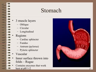
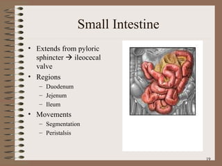
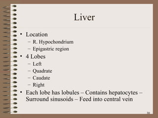
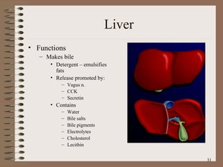
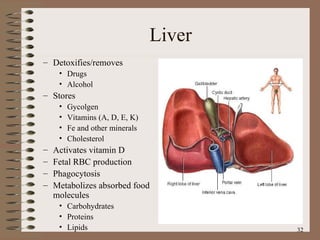
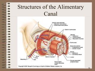
The Digestive System
Digestion Processing of food Types Mechanical (physical)‏ Chew Tear Grind Mash Mix Chemical Catabolic reactions Enzymatic hydrolysis Carbohydrate Protein Lipid
Digestion Phases Ingestion Movement Digestion Absorption Further digestion
Digestive System Organization Gastrointestinal (Gl) tract (Alimentary canal)‏ Tube within a tube Direct link/path between organs Structures Mouth Oral Cavity Pharynx Esophagus Stomach Duedenum  Jejenum Ileum Cecum Ascending colon Transverse colon
Digestive System Organization Descending colon Sigmoid colon Rectum Anus Accessory structures Not in tube path Organs Teeth Tongue Salivary glands Liver Gall bladder Pancreas
Anatomy of the Mouth and Throat
Human Deciduous and Permanent Teeth
Dorsal Surface of the Tongue
The Major Salivary Glands
Deglutition (swallowing)‏ Sequence Voluntary stage Push food to back of mouth Pharyngeal stage Raise Soft palate Larynx + hyoid Tongue to soft palate Esophageal stage Contract pharyngeal muscles Open esophagus Start peristalsis
Deglutition (swallowing)‏ Control Nerves Glossopharyngeal Vagus Accessory Brain stem Deglutition center  Medulla oblongata Pons Disorders Dysphagia Aphagia
Esophagus Usually collapsed (closed)‏ 3 constrictions Aortic arch Left primary bronchus Diaphragm  Surrounded by SNS plexus Blood vessels Functions Secrete mucous Transport food
Peristalsis and Segmentation
Esophagus Sphincters Upper Lower Abnormalities Achalasia Atresia Hernia Barret’s esophagus Esophageal varices
Stomach Usually “J” shaped Left side, anterior to the spleen Mucous membrane G cells – make gastrin Goblet cells – make mucous Gastric pit – Oxyntic gland – Parietal cells – Make HCl Chief cells – Zymogenic cells Pepsin Gastric lipase
Anatomy of the Stomach
Stomach 3 muscle layers Oblique Circular Longitudinal Regions Cardiac sphincter Fundus Antrum (pylorus)‏ Pyloric sphincter Vascular Inner surface thrown into folds – Rugae Contains enzymes that work best at pH 1-2
Stomach Functions Mix food Reservoir Start digestion of Protein Nucleic acids Fats Activates some enzymes Destroy some bacteria Makes intrinsic factor – B 12 absorption Destroys some bacteria Absorbs Alcohol Water Lipophilic acid B 12
Small Intestine Extends from pyloric sphincter    ileocecal valve Regions Duodenum Jejenum Ileum Movements Segmentation Peristalsis
Small Intestine Histology Intestinal glands – Intestinal enzymes Duodenal glands – Alkaline mucous Paneth cells – Lysozyme Microvilli Lacteals Plica circularis Smooth muscle Lymphatic tissue – GALT Vascular
Small Intestine Absorbs 80% ingested water Electrolytes Vitamins Minerals Carbonates Active/facilitated transport Monosaccharides Proteins Di-/tripeptides Amino acids Lipids Monoglycerides Fatty acids Micelles Chylomicrons
Structure of the Villi in the Small Intestine
Small Intestine Secretes digestive enzymes Peptidases Amino- Di- Tri- Sucrases Maltase Lactase Saccharidases Di- Tri- Lipase Nucleases
Small Intestine Control Requires pancreatic enzymes & bile to complete digestion
Large Intestine Extends from ileocecal valve to anus Regions Cecum – Appendix Colon Ascending Transverse Descending Rectum Anal canal
Anatomy of the Large Intestine
Large Intestine Histology No villi No permanent circular folds Smooth muscle Taeniae coli Haustra Epiploic appendages Otherwise like rest of Gl tract
Large Intestine Functions Mechanical digestion Haustral churning Peristalsis Reflexes  Gastroileal Gastrocolic Chemical digestion – Bacterial digestion Ferment carbohydrates Protein/amino acid breakdown Absorbs More water Vitamins  B K Concentrate/eliminate wastes
Feces Formation and Defecation Chyme dehydrated to form feces Feces composition Water Inorganic salts Epithelial cells Bacteria Byproducts of digestion Defecation Peristalsis pushes feces into rectum Rectal walls stretch Control Parasympathetic Voluntary
Liver Location R. Hypochondrium Epigastric region 4 Lobes Left  Quadrate Caudate Right Each lobe has lobules – Contains hepatocytes – Surround sinusoids – Feed into central vein
Liver Functions Makes bile Detergent – emulsifies fats Release promoted by: Vagus n. CCK Secretin Contains Water Bile salts Bile pigments Electrolytes Cholesterol Lecithin
Liver Detoxifies/removes Drugs Alcohol Stores Gycolgen Vitamins (A, D, E, K)‏ Fe and other minerals Cholesterol Activates vitamin D Fetal RBC production Phagocytosis Metabolizes absorbed food molecules Carbohydrates Proteins Lipids
Liver Dual blood supply Hepatic portal vein Direct input from small intestine Hepatic artery/vein Direct links to heart
The Duodenum and Related Organs
The Organs and Positions in the Abdominal Cavity
Structures of the Alimentary Canal

Digestivesystem

  • 1.
  • 2.
    Digestion Processing offood Types Mechanical (physical)‏ Chew Tear Grind Mash Mix Chemical Catabolic reactions Enzymatic hydrolysis Carbohydrate Protein Lipid
  • 3.
    Digestion Phases IngestionMovement Digestion Absorption Further digestion
  • 4.
    Digestive System OrganizationGastrointestinal (Gl) tract (Alimentary canal)‏ Tube within a tube Direct link/path between organs Structures Mouth Oral Cavity Pharynx Esophagus Stomach Duedenum Jejenum Ileum Cecum Ascending colon Transverse colon
  • 5.
    Digestive System OrganizationDescending colon Sigmoid colon Rectum Anus Accessory structures Not in tube path Organs Teeth Tongue Salivary glands Liver Gall bladder Pancreas
  • 6.
    Anatomy of theMouth and Throat
  • 7.
    Human Deciduous andPermanent Teeth
  • 8.
  • 9.
  • 10.
    Deglutition (swallowing)‏ SequenceVoluntary stage Push food to back of mouth Pharyngeal stage Raise Soft palate Larynx + hyoid Tongue to soft palate Esophageal stage Contract pharyngeal muscles Open esophagus Start peristalsis
  • 11.
    Deglutition (swallowing)‏ ControlNerves Glossopharyngeal Vagus Accessory Brain stem Deglutition center Medulla oblongata Pons Disorders Dysphagia Aphagia
  • 12.
    Esophagus Usually collapsed(closed)‏ 3 constrictions Aortic arch Left primary bronchus Diaphragm Surrounded by SNS plexus Blood vessels Functions Secrete mucous Transport food
  • 13.
  • 14.
    Esophagus Sphincters UpperLower Abnormalities Achalasia Atresia Hernia Barret’s esophagus Esophageal varices
  • 15.
    Stomach Usually “J”shaped Left side, anterior to the spleen Mucous membrane G cells – make gastrin Goblet cells – make mucous Gastric pit – Oxyntic gland – Parietal cells – Make HCl Chief cells – Zymogenic cells Pepsin Gastric lipase
  • 16.
  • 17.
    Stomach 3 musclelayers Oblique Circular Longitudinal Regions Cardiac sphincter Fundus Antrum (pylorus)‏ Pyloric sphincter Vascular Inner surface thrown into folds – Rugae Contains enzymes that work best at pH 1-2
  • 18.
    Stomach Functions Mixfood Reservoir Start digestion of Protein Nucleic acids Fats Activates some enzymes Destroy some bacteria Makes intrinsic factor – B 12 absorption Destroys some bacteria Absorbs Alcohol Water Lipophilic acid B 12
  • 19.
    Small Intestine Extendsfrom pyloric sphincter  ileocecal valve Regions Duodenum Jejenum Ileum Movements Segmentation Peristalsis
  • 20.
    Small Intestine HistologyIntestinal glands – Intestinal enzymes Duodenal glands – Alkaline mucous Paneth cells – Lysozyme Microvilli Lacteals Plica circularis Smooth muscle Lymphatic tissue – GALT Vascular
  • 21.
    Small Intestine Absorbs80% ingested water Electrolytes Vitamins Minerals Carbonates Active/facilitated transport Monosaccharides Proteins Di-/tripeptides Amino acids Lipids Monoglycerides Fatty acids Micelles Chylomicrons
  • 22.
    Structure of theVilli in the Small Intestine
  • 23.
    Small Intestine Secretesdigestive enzymes Peptidases Amino- Di- Tri- Sucrases Maltase Lactase Saccharidases Di- Tri- Lipase Nucleases
  • 24.
    Small Intestine ControlRequires pancreatic enzymes & bile to complete digestion
  • 25.
    Large Intestine Extendsfrom ileocecal valve to anus Regions Cecum – Appendix Colon Ascending Transverse Descending Rectum Anal canal
  • 26.
    Anatomy of theLarge Intestine
  • 27.
    Large Intestine HistologyNo villi No permanent circular folds Smooth muscle Taeniae coli Haustra Epiploic appendages Otherwise like rest of Gl tract
  • 28.
    Large Intestine FunctionsMechanical digestion Haustral churning Peristalsis Reflexes Gastroileal Gastrocolic Chemical digestion – Bacterial digestion Ferment carbohydrates Protein/amino acid breakdown Absorbs More water Vitamins B K Concentrate/eliminate wastes
  • 29.
    Feces Formation andDefecation Chyme dehydrated to form feces Feces composition Water Inorganic salts Epithelial cells Bacteria Byproducts of digestion Defecation Peristalsis pushes feces into rectum Rectal walls stretch Control Parasympathetic Voluntary
  • 30.
    Liver Location R.Hypochondrium Epigastric region 4 Lobes Left Quadrate Caudate Right Each lobe has lobules – Contains hepatocytes – Surround sinusoids – Feed into central vein
  • 31.
    Liver Functions Makesbile Detergent – emulsifies fats Release promoted by: Vagus n. CCK Secretin Contains Water Bile salts Bile pigments Electrolytes Cholesterol Lecithin
  • 32.
    Liver Detoxifies/removes DrugsAlcohol Stores Gycolgen Vitamins (A, D, E, K)‏ Fe and other minerals Cholesterol Activates vitamin D Fetal RBC production Phagocytosis Metabolizes absorbed food molecules Carbohydrates Proteins Lipids
  • 33.
    Liver Dual bloodsupply Hepatic portal vein Direct input from small intestine Hepatic artery/vein Direct links to heart
  • 34.
    The Duodenum andRelated Organs
  • 35.
    The Organs andPositions in the Abdominal Cavity
  • 36.
    Structures of theAlimentary Canal