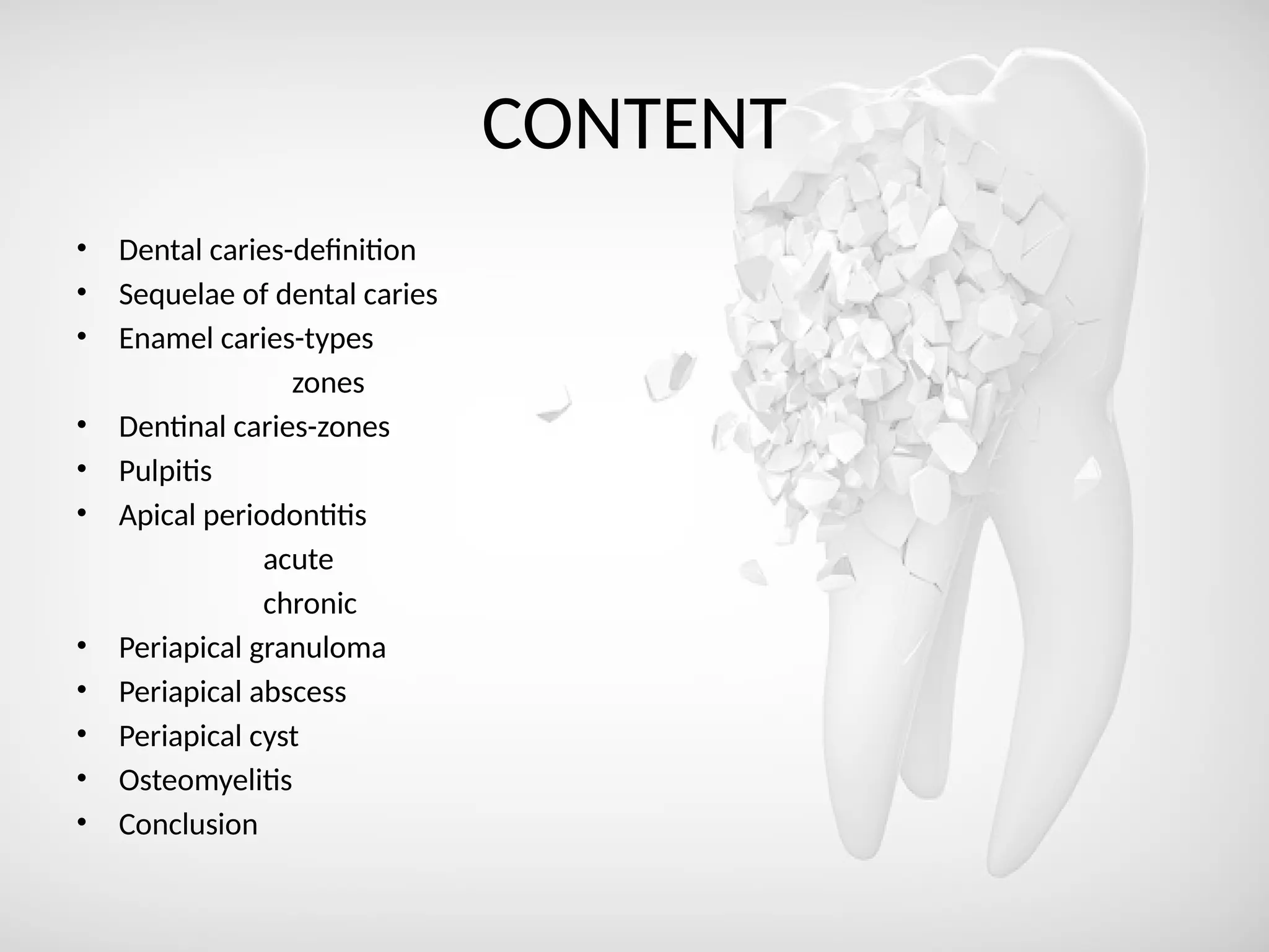
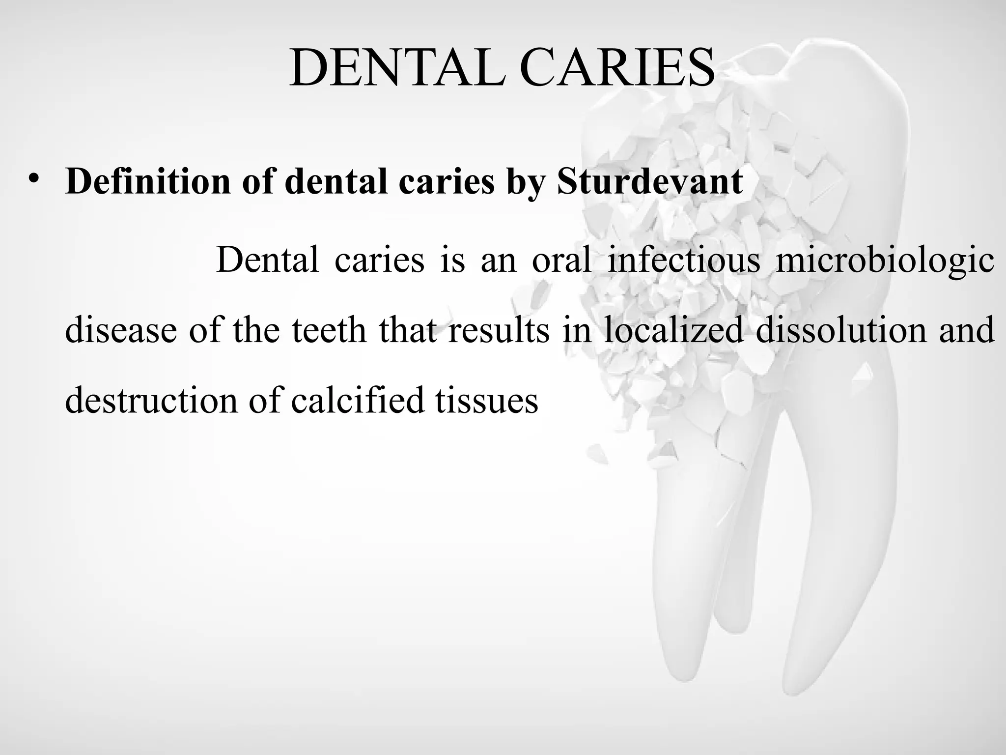
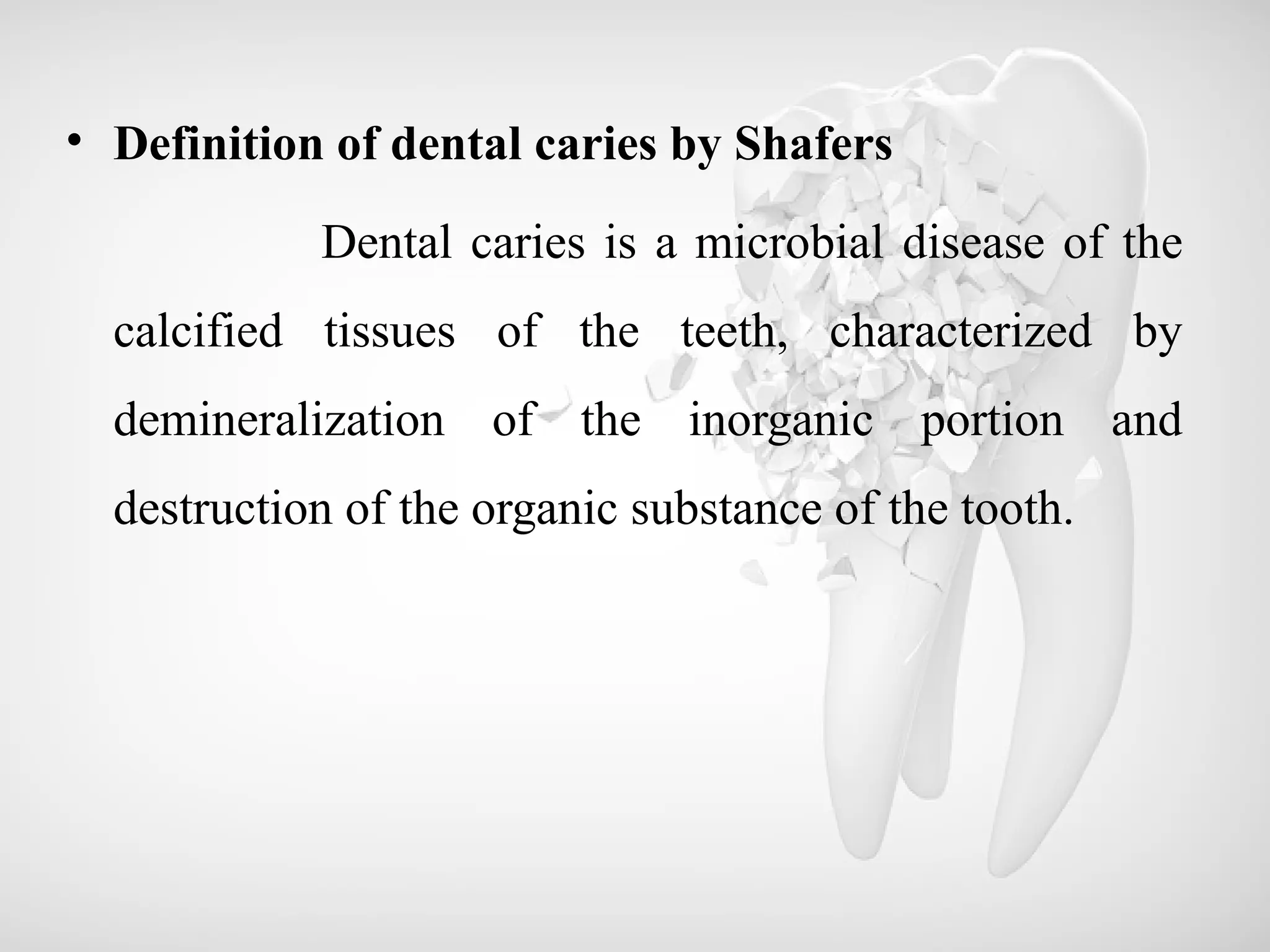
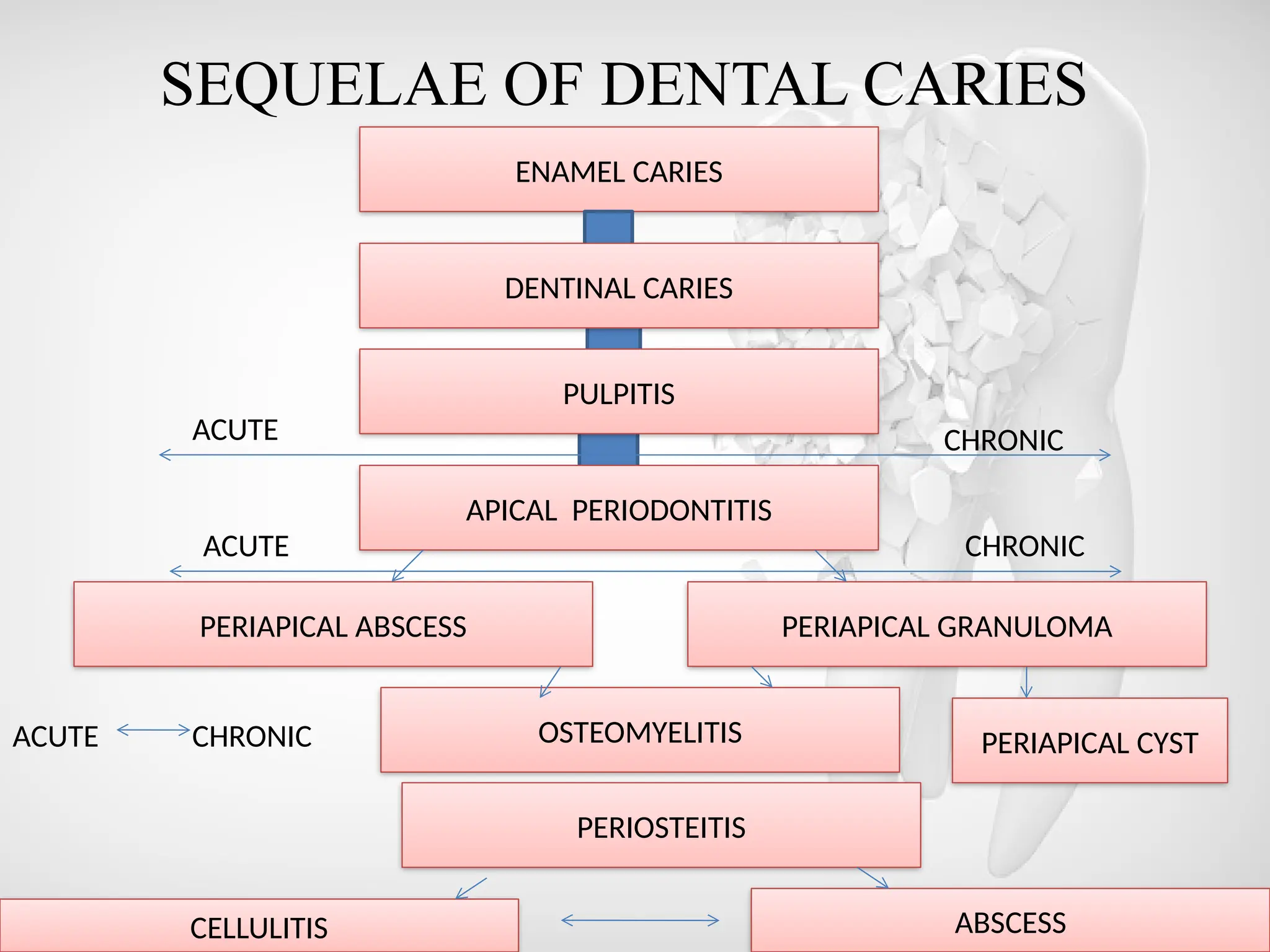
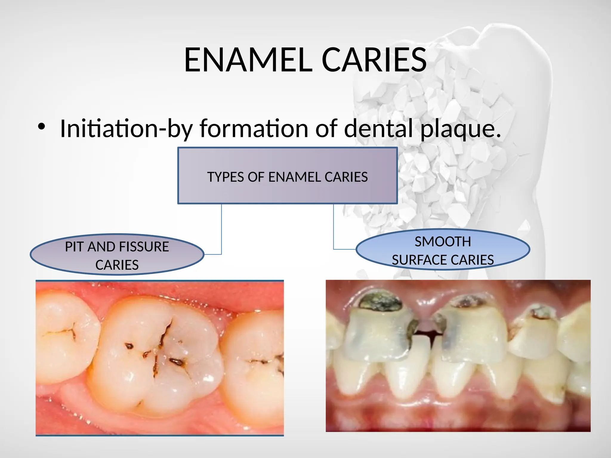
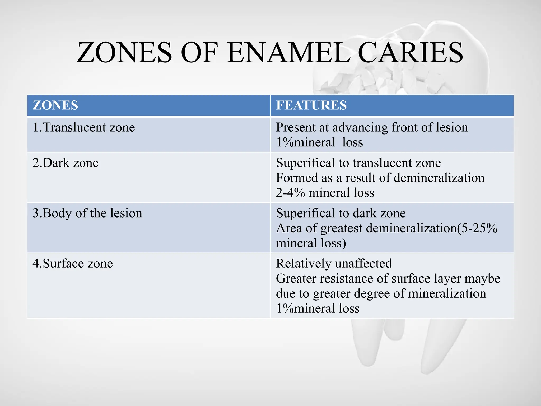
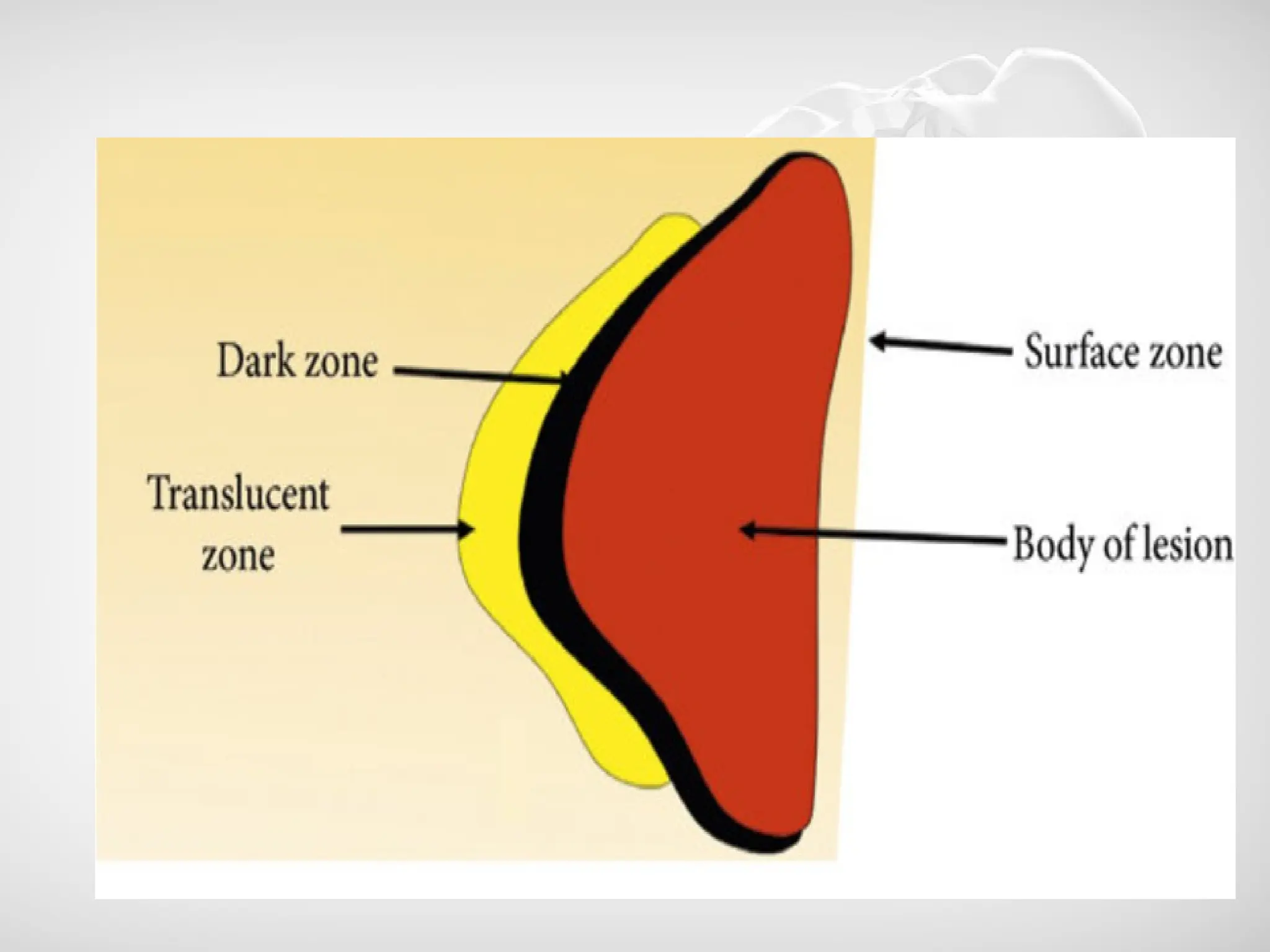
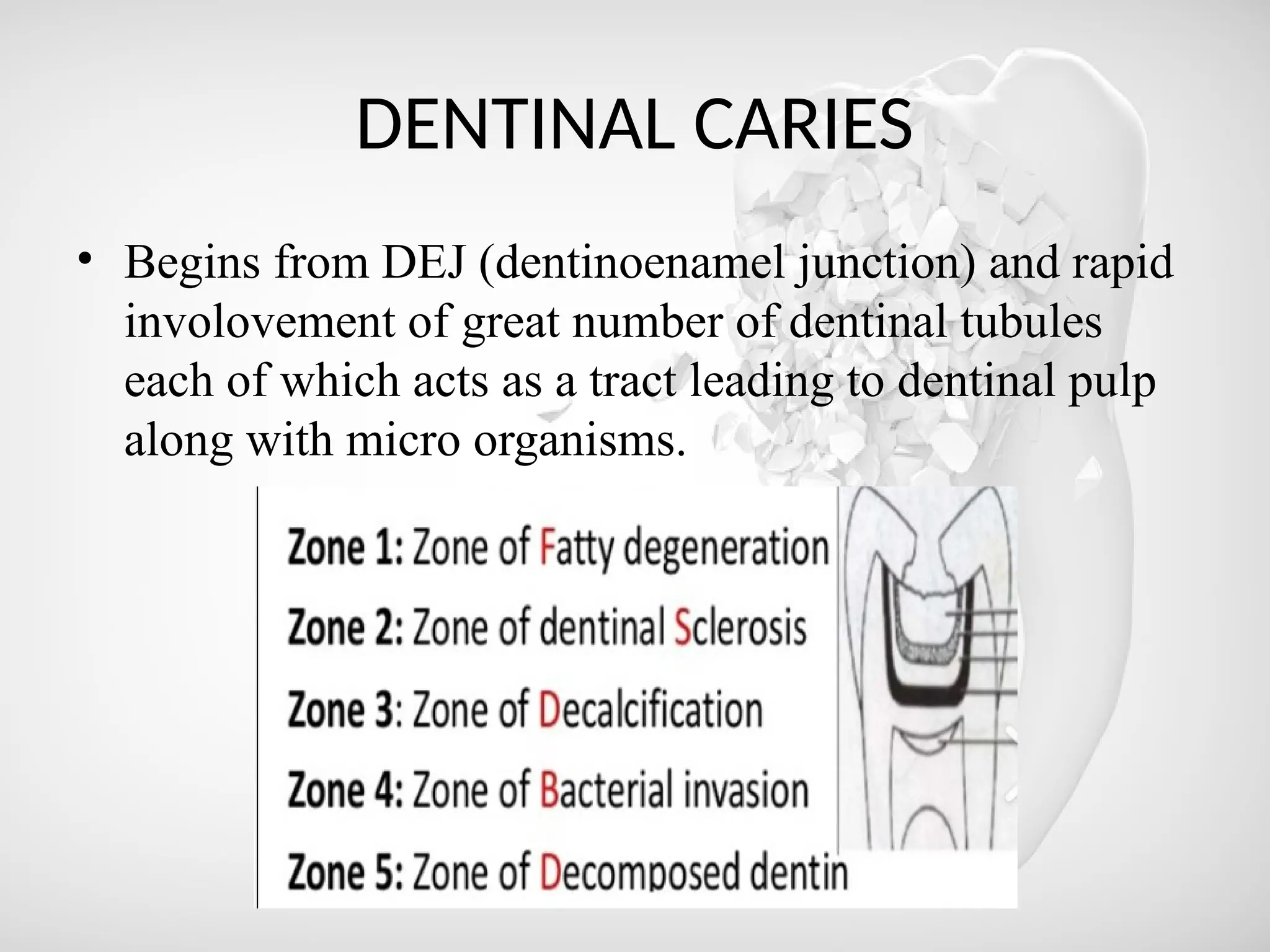
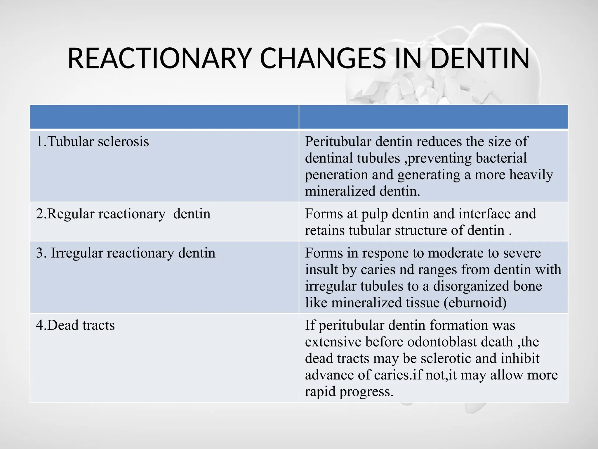
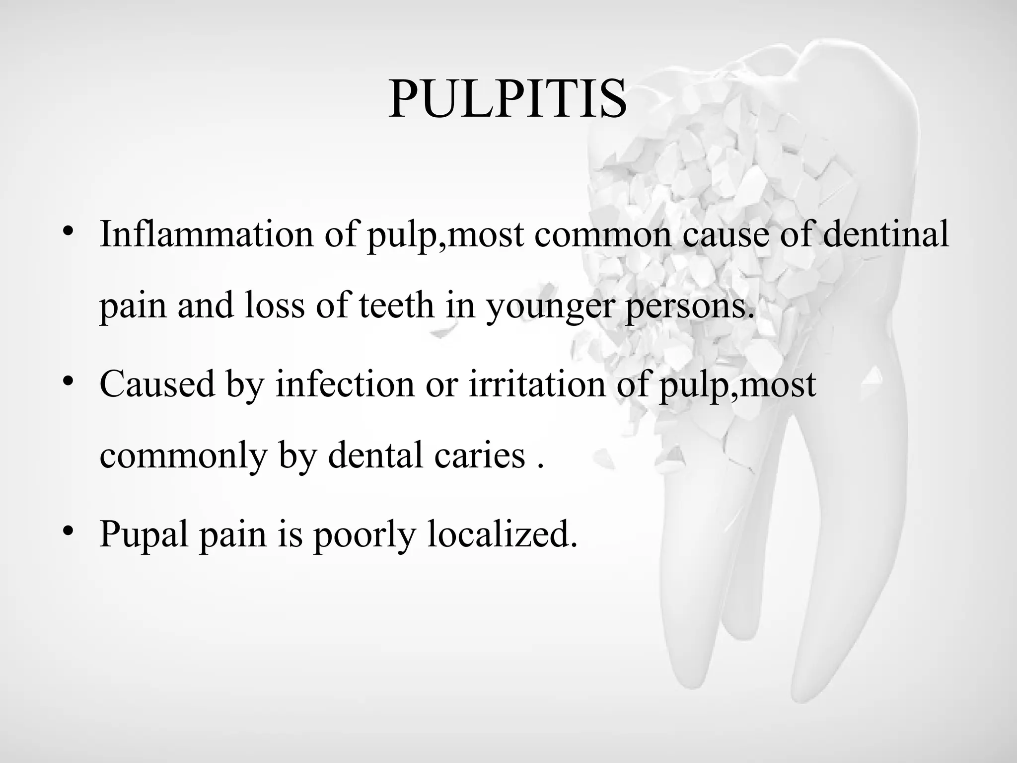
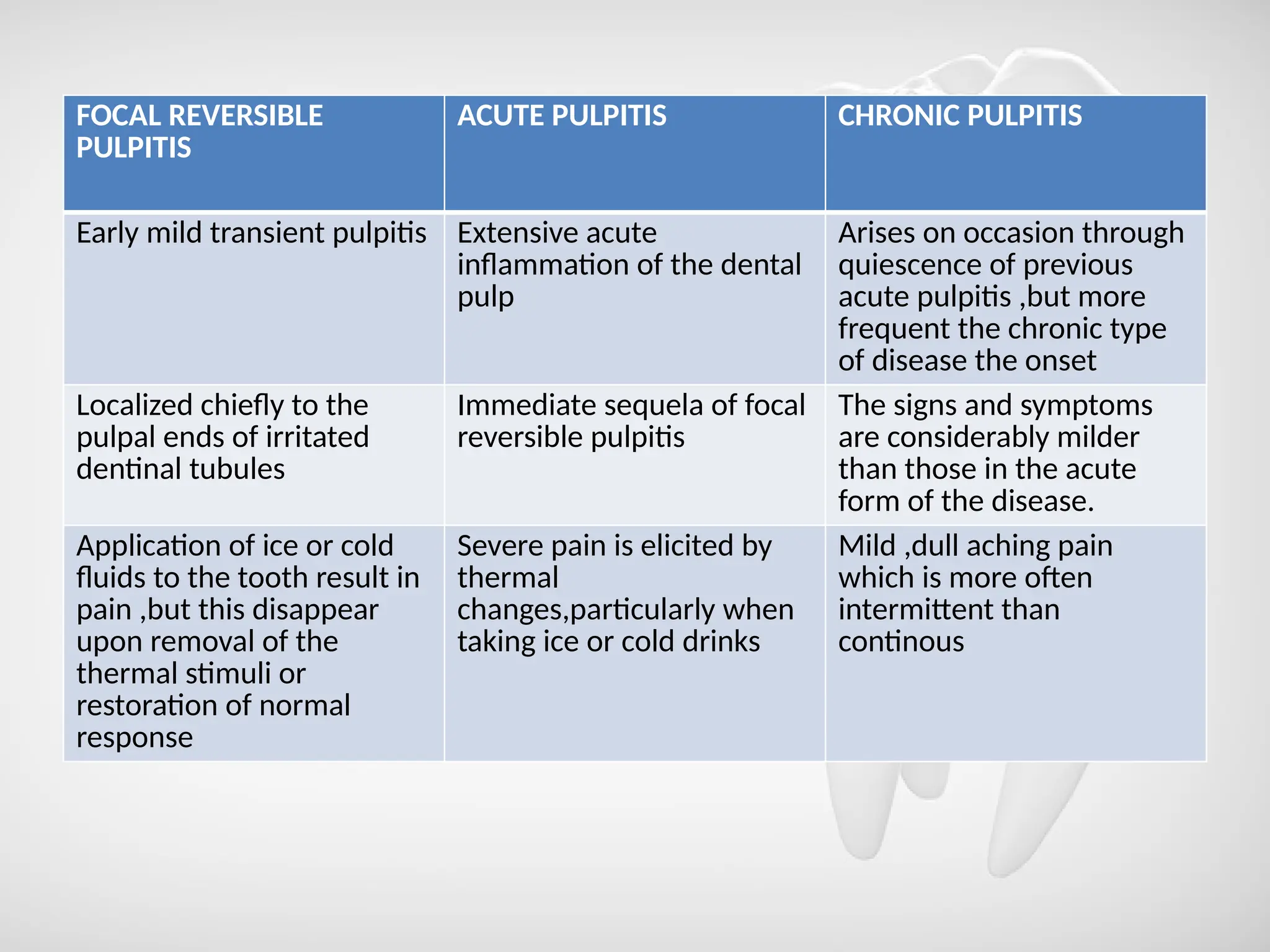
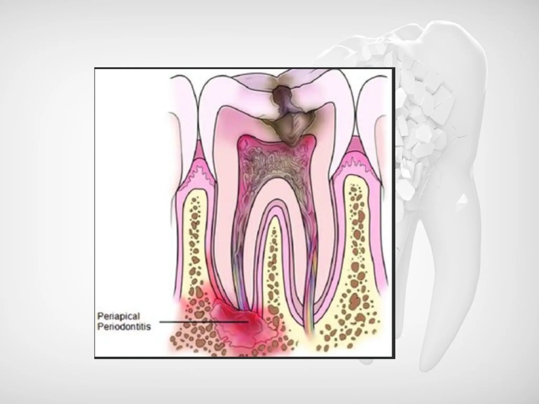
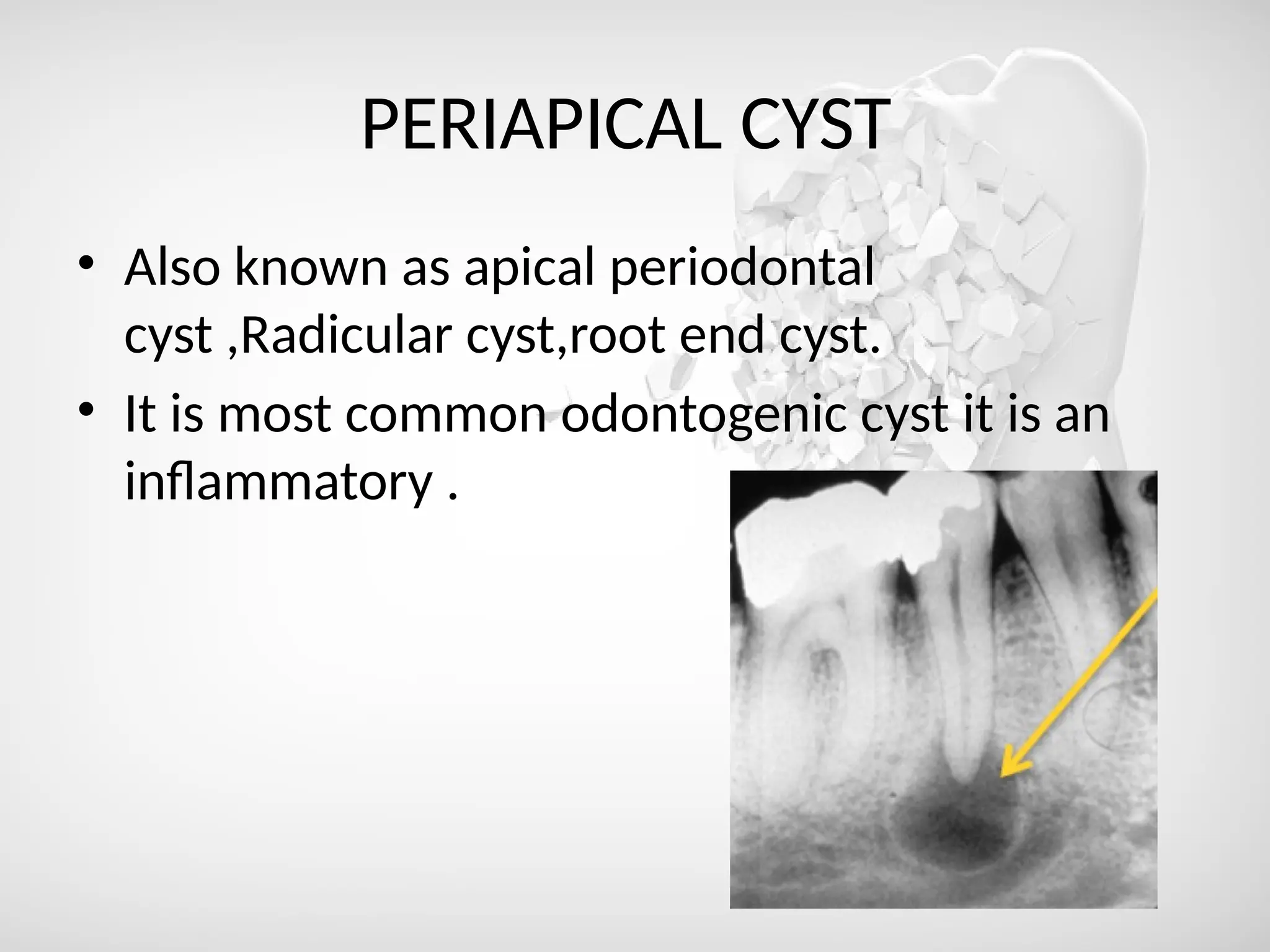
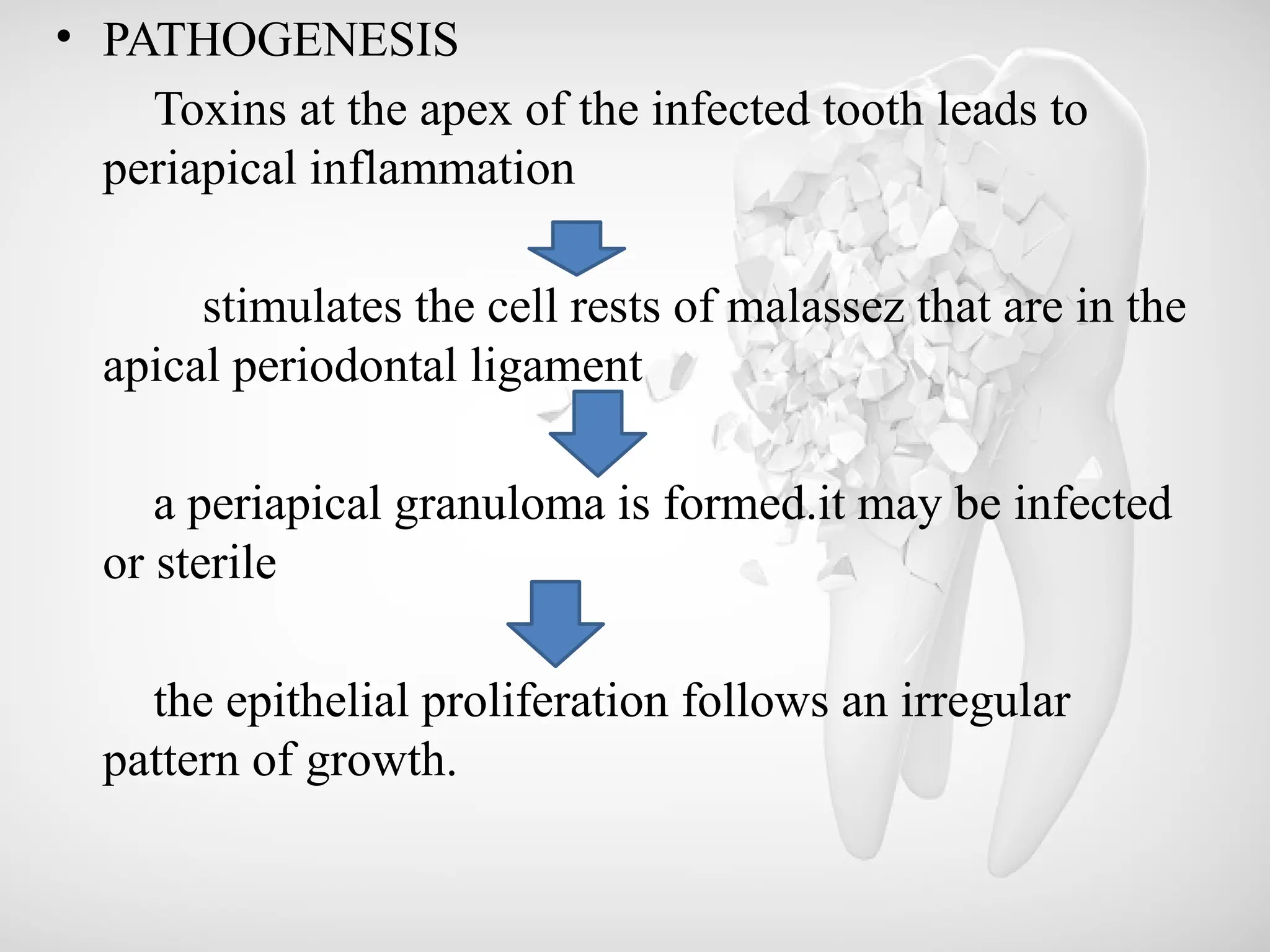
SEQUELAE OF DENTAL CARIES
-DR.SUGUMARI V
MDS FIRST YEAR
CONTENT
• Dental caries-definition
• Sequelae of dental caries
• Enamel caries-types
zones
• Dentinal caries-zones
• Pulpitis
• Apical periodontitis
acute
chronic
• Periapical granuloma
• Periapical abscess
• Periapical cyst
• Osteomyelitis
• Conclusion
DENTAL CARIES
• Definition of dental caries by Sturdevant
Dental caries is an oral infectious microbiologic
disease of the teeth that results in localized dissolution and
destruction of calcified tissues
• Definition of dental caries by Shafers
Dental caries is a microbial disease of the
calcified tissues of the teeth, characterized by
demineralization of the inorganic portion and
destruction of the organic substance of the tooth.
SEQUELAE OF DENTAL CARIES
ENAMEL CARIES
DENTINAL CARIES
PULPITIS
APICAL PERIODONTITIS
PERIAPICAL ABSCESS PERIAPICAL GRANULOMA
OSTEOMYELITIS
PERIOSTEITIS
PERIAPICAL CYST
ABSCESS
CELLULITIS
ACUTE
CHRONIC
ACUTE
CHRONIC
ACUTE CHRONIC
ENAMEL CARIES
• Initiation-by formation of dental plaque.
TYPES OF ENAMEL CARIES
SMOOTH
SURFACE CARIES
PIT AND FISSURE
CARIES
ZONES OF ENAMEL CARIES
ZONES FEATURES
1.Translucent zone Present at advancing front of lesion
1%mineral loss
2.Dark zone Superifical to translucent zone
Formed as a result of demineralization
2-4% mineral loss
3.Body of the lesion Superifical to dark zone
Area of greatest demineralization(5-25%
mineral loss)
4.Surface zone Relatively unaffected
Greater resistance of surface layer maybe
due to greater degree of mineralization
1%mineral loss
DENTINAL CARIES
• Begins from DEJ (dentinoenamel junction) and rapid
involovement of great number of dentinal tubules
each of which acts as a tract leading to dentinal pulp
along with micro organisms.
ZONES OF DENTINAL CARIES
Zones of
dentinal
caries
Zone of fatty
degeneration of
tomes s fibres
Zone of
decomposed
dentin Zone of
bacterial
invasion
Zone of
decalcified
dentin
Zone of
dentinal
sclerosis
REACTIONARY CHANGES IN DENTIN
1.Tubular sclerosis Peritubular dentin reduces the size of
dentinal tubules ,preventing bacterial
peneration and generating a more heavily
mineralized dentin.
2.Regular reactionary dentin Forms at pulp dentin and interface and
retains tubular structure of dentin .
3. Irregular reactionary dentin Forms in respone to moderate to severe
insult by caries nd ranges from dentin with
irregular tubules to a disorganized bone
like mineralized tissue (eburnoid)
4.Dead tracts If peritubular dentin formation was
extensive before odontoblast death ,the
dead tracts may be sclerotic and inhibit
advance of caries.if not,it may allow more
rapid progress.
PULPITIS
• Inflammation of pulp,most common cause of dentinal
pain and loss of teeth in younger persons.
• Caused by infection or irritation of pulp,most
commonly by dental caries .
• Pupal pain is poorly localized.
FOCAL REVERSIBLE
PULPITIS
ACUTE PULPITIS CHRONIC PULPITIS
Early mild transient pulpitis Extensive acute
inflammation of the dental
pulp
Arises on occasion through
quiescence of previous
acute pulpitis ,but more
frequent the chronic type
of disease the onset
Localized chiefly to the
pulpal ends of irritated
dentinal tubules
Immediate sequela of focal
reversible pulpitis
The signs and symptoms
are considerably milder
than those in the acute
form of the disease.
Application of ice or cold
fluids to the tooth result in
pain ,but this disappear
upon removal of the
thermal stimuli or
restoration of normal
response
Severe pain is elicited by
thermal
changes,particularly when
taking ice or cold drinks
Mild ,dull aching pain
which is more often
intermittent than
continous
APICAL PERIODONTITIS
• Apical periodontitis is the inflammation of the
periodontal ligament around the root apex.
• This process may be acute or chronic depending upon
the virulence of the microorganisms involved, the
type and severity of the physical or chemical irritants,
and host resistance.
• The common causes of apical periodontitis include
spread of infection following pulp necrosis, occlusal
trauma from a high restoration or biting suddenly on a
hard object, inadvertent endodontic procedures such
as over instrumentation, pushing the infected material
into the apical portion or chemical irritation from root
canal medicaments.
ACUTE APICAL PERIODONTITIS
• Give the history of previous pulpitis.
• Thermal change does not induce pain as in pulpitis.
• Due to the collection of inflammatory edema in the
periodontal ligament, the tooth is slightly elevated in
its socket and causes tenderness while biting or even
to mere touch.
• The external pressure on the tooth forces the edema
fluid against already sensitized nerve endings and
results in severe pain.
• RAGIOLOGIC FEATURES:
Radiographic appearance is essentially normal at
this stage except for a slight widening of periodontal
ligament space.
HISTOLOGIC FEATURES :
• The periodontal ligament shows signs of
inflammation characterized by vascular dilatation and
infiltration with polymorphonuclear leukocytes.
• Initially, these changes are localized around the root
apex, as this area is richly vascular.
• The inflammation is transient if it is caused by acute
trauma.
TREATMENT AND PROGNOSIS:
• If the inflammation is caused by occlusal trauma, it
should be relieved by selective occlusal grinding.
• If the periapical periodontitis occurs due to
the spread of pulpal infection, the tooth should be
extracted.
CHRONIC APICAL PERIODONTITIS
• Chronic apical periodontitis, also known as periapical
granuloma, is a low-grade infection and one of the
most common of all sequelae of pulpitis or acute
periapical periodontitis.
• If the acute process is left untreated, it is
incompletely resolved and becomes chronic.
• The acute inflammatory process is an exudative
response whereas the chronic one is proliferative.
• Periapical granuloma is essentially a localized mass
of chronic granulation tissue formed in response to
the infection.
Clinical features:
• The involved tooth is usually nonvital and may be
slightly tender to percussion, and percussion may
produce a dull sound instead of a normal metallic
sound because of the presence of granulation tissue
around the root apex.
• Patients may complain of mild pain on biting or
chewing on solid food.
Histologic features
• The typical periapical granuloma shows the delicate
fibrillar stroma with intense lymphocytic and plasma cell
infiltration and sometimes polymorphonuclear
leukocytes as well as many small capillaries
• collections of macrophages that are often filled with
lipoid material and cholesterol slits in the tissue The
typical granuloma is usually surrounded by a connective
tissue ‘capsule’
Treatment and Prognosis
• The treatment of the periapical granuloma consists in
extraction of the involved teeth, or under certain
conditions, root canal therapy with or without
subsequent apicoectomy.
• If left untreated, the periapical granuloma may
ultimately undergo transformation into an apical
periodontal cyst.
PERIAPICAL CYST
• Also known as apical periodontal
cyst ,Radicular cyst,root end cyst.
• It is most common odontogenic cyst it is an
inflammatory .
• PATHOGENESIS
Toxins at the apex of the infected tooth leads to
periapical inflammation
stimulates the cell rests of malassez that are in the
apical periodontal ligament
a periapical granuloma is formed.it may be infected
or sterile
the epithelial proliferation follows an irregular
pattern of growth.
the epithelial mass increases in size if by the division of
the cells in the periphery corresponding to the basal layer
of the cell in the surface epithelium.
cells in the centre becomes separated from their source of
nutrition
cell in the centre degenerates become necrotic and liquefy
This creates an epithelium lined cavity filled with fluid the
apica;l periodontal cyst
CLINICAL FEATURES:
• mostly asymptomatic
• Most commonly involved tooth maxillary anteriors
followed by premolars and molars.
• Expansion of cortical plate are uncommon
TREATMENT
• Root canal therapy of the involved teeth with periapical
surgery or extraction of involved tooth followed by
curretage.
• The cyst do not recur if the surgical removal in
thorough.
PERIAPICAL ABSCESS
• Dentoalveolar abscess, alveolar abscess)
• Periapical abscess is an acute or chronic suppurative
process of the dental periapical region.
• It may develop either from acute periapical
periodontitis or more commonly from a periapical
granuloma.
• Acute exacerbation of a chronic periapical lesion is known as
Phoenix abscess.
• It usually arises as a result of infection following carious
involvement of the tooth and pulp infection, but it also does
occur after traumatic injury to the teeth, resulting in necrosis of
the pulp, and in cases of irritation of the periapical tissues either
by mechanical manipulation or by the application of chemicals
in endodontic procedures.
• It is a mixed infection with the culture of pus yielding to a wide
range of different bacterial species.
Clinical Features
• The acute periapical abscess presents the features of an
acute inflammation of the apical periodontium.
• The initial stages produce tenderness of the tooth,
which is often relieved by application of pressure. In
time, the tooth is extremely painful and is slightly
extruded from its socket
• regional lymphadenitis and fever may be present.
• The chronic periapical abscess generally presents no
clinical features, since it is essentially a mild,well-
circumscribed area of suppuration that shows little
tendency to spread from the local area.
Radiographic Features:
• The acute periapical abscess is such a rapidly
progressive lesion that, except for slight thickening of
the periodontal ligament space, there is usually no
radiographic evidence of its presence.
• The chronic abscess, developing in a periapical
granuloma, presents the radiolucent area at the apex of
the tooth described previously or the radiolucency may
be ill-defined.
Histologic Features:
• The area of suppuration is composed chiefly of a central
area of disintegrating polymorphonuclear leukocytes
surrounded by viable leukocytes, occasional
lymphocytes, cellular debris, necrotic materials and
bacterial colonies.
• There is dilatation of the blood vessels in the periodontal
ligament and adjacent marrow spaces of the bone.
• These marrow spaces also show an inflammatory cell
infiltrate.
• The tissue around the area of suppuration contains a
serous exudate
Treatment and Prognosis:
• The principle of treatment of the periapical abscess is the
same as for any abscess, drainage must be established.
• This can be accomplished by either opening the pulp
chamber or extracting the tooth.
• Under some circumstances, the tooth may be retained and
root canal therapy is carried out if the lesion can be
sterilized.
• If the periapical abscess is not treated, it may lead to
serious complications through the spread of the
infection.
• These include osteomyelitis, cellulitis, and bacteremia
and the ultimate formation of the fistulous tract
opening on the skin or oral mucosa.
• Cavernous sinus thrombosis has also been reported.
OSTEOMYELITIS
• Osteomyelitis is defined as the inflammation of bone
and its marrow contents.
• Changes in the calcified tissue are secondary to
inflammation of the soft tissue component of the
bone.
• Though the pathologic changes of periapical abscess
can even be considered as osteomyelitis as there is
involvement of bone, the term ‘osteomyelitis’ is
reserved for infections which spread through the bone
to a larger extent.
• Predisposing factors include fractures due to trauma and
road traffic accidents; gunshot wounds, radiation
damage, Paget’s disease, and osteopetrosis.
• Systemic conditions like malnutrition, acute leukemia,
uncontrolled diabetes mellitus, sickle cell anemia, and
chronic alcoholism may also predispose to osteomyelitis.
• The disease may be acute, subacute, or chronic and
presents a different clinical course, depending upon its
nature.
ACUTE SUPPURATIVE OSTEOMYELITIS
Acute suppurative osteomyelitis of the jaw
is a serious sequela of periapical infection that often
results in a diffuse spread of infection throughout the
medullary spaces, with subsequent necrosis of a
variable amount of bone.
Clinical features :
• this form of osteomyelitis, which arises from a dental
infection, are the same as those present after infection
due to a fracture of the jaw, a gunshot wound, or even
hematogenous spread.
• Dental infection is the most frequent cause of acute
osteomyelitis of the jaws. It may be a rather well-
localized infection of one involving a great volume of
bone.
• A periapical infection (usually an abscess), if it is a
particularly virulent one and not walled off, may
spread spontaneously throughout the bone.
• In other instances, a chronic periapical infection such
as a granuloma, or even a cyst that is walled off, may
undergo an acute exacerbation, especially if the area is
traumatized or surgically disturbed without
establishing and maintaining drainage.
Clinical Features:
• Acute or subacute suppurative osteomyelitis may
involve either the maxilla or the mandible.
• In the maxilla the disease usually remains fairly
well localized to the area of initial infection.
• In the mandible, bone involvement tends to be
more diffuse and widespread. The disease may
occur at any age.
• A particular form of acute osteomyelitis, referred to as
neonatal maxillitis in infants and young children, is a
well recognized entity that is fortunately becoming
extremely uncommon nowadays because of antibiotic
drugs.
• In some instances, this osteomyelitis of infants is of
hematogenous origin, but at other times it seems to be a
result of local oral infection following some minor
injury or abrasion.
Radiographic Features:
• Acute osteomyelitis progresses rapidly and demonstrates
little radiographic evidence of its presence until the
disease has developed for at least one to two weeks.
• At this time diffuse lytic changes in the bone begin to
appear.
• Individual trabeculae become fuzzy and indistinct, and
radiolucent areas begin to appear .
Histologic Features:
• The medullary spaces are filled with inflammatory exudates .
• The inflammatory cells are chiefly polymorphonuclear
leukocytes, but may show occasional lymphocytes and plasma
cells.
• The osteoblasts bordering the bony trabeculae are generally
destroyed, and depending upon the duration of the process, the
trabeculae may lose their viability and begin to undergo slow
resorption.
Treatment and Prognosis:
• General principles of management includes
debridement, drainage, and antimicrobial therapy.
• When the intensity of the disease becomes attenuated,
either spontaneously or under treatment, the
sequestrum begins to separate from the living bone.
• If the sequestrum is small, it gradually exfoliates
through the mucosa.
• If large, surgical removal may be necessary, since
its removal by normal processes of bone resorption would
be extremely slow.
• Sometimes an involucrum forms when the sequestrum
becomes surrounded by new living bone.
• Unless proper treatment is instituted, acute suppurative
osteomyelitis may proceed to the development of
periostitis, soft-tissue abscess, or cellulitis .
CHRONIC SUPPURATIVE OSTEOMYELITIS
• Chronic suppurative osteomyelitis may develop in
inadequately treated acute osteomyelitis or may arise
from a dental infection without a preceding acute
stage.
Clinical features
• Similar to those of acute osteomyelitis except that all
signs and symptoms are milder.
• The pain is less severe; the temperature is still
elevated, but only mildly; and the leukocytosis is only
slightly greater than normal.
• Teeth may not be loose or sore, so that mastication is
at least possible even though the jaw may not be
perfectly comfortable.
• Acute exacerbations of the chronic stage may occur
periodically, and these present all features of acute
suppurative osteomyelitis.
• The suppuration may perforate the bone and
overlying skin or mucosa to form a fistulous tract and
empty on the surface
CHRONIC FOCAL SCLEROSING OSTEOMYELITIS
(CONDENSING OSTEITIS)
• Chronic focal sclerosing osteomyelitis is an unusual
reaction of the bone to infection: a reaction to mild
bacterial infection entering
• In either case, the radiopacity stands out in distinct
contrast to the trabeculation of the normal bone.
Histologic Features:
• Histologic examination reveals only a dense mass of bony
trabeculae with little interstitial marrow tissue .
• The osteocytic lacunae appear empty.
• The bony trabeculae exhibit many reversal and resting lines
giving pagetoid appearance.
• If interstitial soft tissue is present, it is generally fibrotic and
infiltrated only by small numbers of lymphocytes.
• Osteoblastic activity may have completely subsided at the time
of microscopic study.
Garres chronic nonsuppurative sclerosing osteitis, periostitis
ossificans:
• This is a distinctive type of chronic osteomyelitis in which
there is focal gross thickening of the periosteum, with
peripheral reactive bone formation resulting from mild
irritation or infection.
• It is essentially a periosteal osteosclerosis analogous to the
endosteal sclerosis of chronic focal and diffuse
sclerosingosteomyelitis.
CELULITIS
• Diffuse inflammation of soft tissue.
• Tends to spread through tissue space and
facial planes.
• Caused by organisms producing hyaluronidase
and fibrinolysins like streptococci.
THANK YOU

sequele of dental caries-1.pptx sequeale

  • 1.
    SEQUELAE OF DENTALCARIES -DR.SUGUMARI V MDS FIRST YEAR
  • 2.
    CONTENT • Dental caries-definition •Sequelae of dental caries • Enamel caries-types zones • Dentinal caries-zones • Pulpitis • Apical periodontitis acute chronic • Periapical granuloma • Periapical abscess • Periapical cyst • Osteomyelitis • Conclusion
  • 3.
    DENTAL CARIES • Definitionof dental caries by Sturdevant Dental caries is an oral infectious microbiologic disease of the teeth that results in localized dissolution and destruction of calcified tissues
  • 4.
    • Definition ofdental caries by Shafers Dental caries is a microbial disease of the calcified tissues of the teeth, characterized by demineralization of the inorganic portion and destruction of the organic substance of the tooth.
  • 5.
    SEQUELAE OF DENTALCARIES ENAMEL CARIES DENTINAL CARIES PULPITIS APICAL PERIODONTITIS PERIAPICAL ABSCESS PERIAPICAL GRANULOMA OSTEOMYELITIS PERIOSTEITIS PERIAPICAL CYST ABSCESS CELLULITIS ACUTE CHRONIC ACUTE CHRONIC ACUTE CHRONIC
  • 6.
    ENAMEL CARIES • Initiation-byformation of dental plaque. TYPES OF ENAMEL CARIES SMOOTH SURFACE CARIES PIT AND FISSURE CARIES
  • 7.
    ZONES OF ENAMELCARIES ZONES FEATURES 1.Translucent zone Present at advancing front of lesion 1%mineral loss 2.Dark zone Superifical to translucent zone Formed as a result of demineralization 2-4% mineral loss 3.Body of the lesion Superifical to dark zone Area of greatest demineralization(5-25% mineral loss) 4.Surface zone Relatively unaffected Greater resistance of surface layer maybe due to greater degree of mineralization 1%mineral loss
  • 9.
    DENTINAL CARIES • Beginsfrom DEJ (dentinoenamel junction) and rapid involovement of great number of dentinal tubules each of which acts as a tract leading to dentinal pulp along with micro organisms.
  • 10.
    ZONES OF DENTINALCARIES Zones of dentinal caries Zone of fatty degeneration of tomes s fibres Zone of decomposed dentin Zone of bacterial invasion Zone of decalcified dentin Zone of dentinal sclerosis
  • 11.
    REACTIONARY CHANGES INDENTIN 1.Tubular sclerosis Peritubular dentin reduces the size of dentinal tubules ,preventing bacterial peneration and generating a more heavily mineralized dentin. 2.Regular reactionary dentin Forms at pulp dentin and interface and retains tubular structure of dentin . 3. Irregular reactionary dentin Forms in respone to moderate to severe insult by caries nd ranges from dentin with irregular tubules to a disorganized bone like mineralized tissue (eburnoid) 4.Dead tracts If peritubular dentin formation was extensive before odontoblast death ,the dead tracts may be sclerotic and inhibit advance of caries.if not,it may allow more rapid progress.
  • 12.
    PULPITIS • Inflammation ofpulp,most common cause of dentinal pain and loss of teeth in younger persons. • Caused by infection or irritation of pulp,most commonly by dental caries . • Pupal pain is poorly localized.
  • 13.
    FOCAL REVERSIBLE PULPITIS ACUTE PULPITISCHRONIC PULPITIS Early mild transient pulpitis Extensive acute inflammation of the dental pulp Arises on occasion through quiescence of previous acute pulpitis ,but more frequent the chronic type of disease the onset Localized chiefly to the pulpal ends of irritated dentinal tubules Immediate sequela of focal reversible pulpitis The signs and symptoms are considerably milder than those in the acute form of the disease. Application of ice or cold fluids to the tooth result in pain ,but this disappear upon removal of the thermal stimuli or restoration of normal response Severe pain is elicited by thermal changes,particularly when taking ice or cold drinks Mild ,dull aching pain which is more often intermittent than continous
  • 14.
    APICAL PERIODONTITIS • Apicalperiodontitis is the inflammation of the periodontal ligament around the root apex. • This process may be acute or chronic depending upon the virulence of the microorganisms involved, the type and severity of the physical or chemical irritants, and host resistance.
  • 15.
    • The commoncauses of apical periodontitis include spread of infection following pulp necrosis, occlusal trauma from a high restoration or biting suddenly on a hard object, inadvertent endodontic procedures such as over instrumentation, pushing the infected material into the apical portion or chemical irritation from root canal medicaments.
  • 17.
    ACUTE APICAL PERIODONTITIS •Give the history of previous pulpitis. • Thermal change does not induce pain as in pulpitis. • Due to the collection of inflammatory edema in the periodontal ligament, the tooth is slightly elevated in its socket and causes tenderness while biting or even to mere touch.
  • 18.
    • The externalpressure on the tooth forces the edema fluid against already sensitized nerve endings and results in severe pain.
  • 19.
    • RAGIOLOGIC FEATURES: Radiographicappearance is essentially normal at this stage except for a slight widening of periodontal ligament space.
  • 20.
    HISTOLOGIC FEATURES : •The periodontal ligament shows signs of inflammation characterized by vascular dilatation and infiltration with polymorphonuclear leukocytes. • Initially, these changes are localized around the root apex, as this area is richly vascular. • The inflammation is transient if it is caused by acute trauma.
  • 21.
    TREATMENT AND PROGNOSIS: •If the inflammation is caused by occlusal trauma, it should be relieved by selective occlusal grinding. • If the periapical periodontitis occurs due to the spread of pulpal infection, the tooth should be extracted.
  • 22.
    CHRONIC APICAL PERIODONTITIS •Chronic apical periodontitis, also known as periapical granuloma, is a low-grade infection and one of the most common of all sequelae of pulpitis or acute periapical periodontitis. • If the acute process is left untreated, it is incompletely resolved and becomes chronic.
  • 23.
    • The acuteinflammatory process is an exudative response whereas the chronic one is proliferative. • Periapical granuloma is essentially a localized mass of chronic granulation tissue formed in response to the infection.
  • 24.
    Clinical features: • Theinvolved tooth is usually nonvital and may be slightly tender to percussion, and percussion may produce a dull sound instead of a normal metallic sound because of the presence of granulation tissue around the root apex. • Patients may complain of mild pain on biting or chewing on solid food.
  • 25.
    Histologic features • Thetypical periapical granuloma shows the delicate fibrillar stroma with intense lymphocytic and plasma cell infiltration and sometimes polymorphonuclear leukocytes as well as many small capillaries • collections of macrophages that are often filled with lipoid material and cholesterol slits in the tissue The typical granuloma is usually surrounded by a connective tissue ‘capsule’
  • 26.
    Treatment and Prognosis •The treatment of the periapical granuloma consists in extraction of the involved teeth, or under certain conditions, root canal therapy with or without subsequent apicoectomy. • If left untreated, the periapical granuloma may ultimately undergo transformation into an apical periodontal cyst.
  • 27.
    PERIAPICAL CYST • Alsoknown as apical periodontal cyst ,Radicular cyst,root end cyst. • It is most common odontogenic cyst it is an inflammatory .
  • 28.
    • PATHOGENESIS Toxins atthe apex of the infected tooth leads to periapical inflammation stimulates the cell rests of malassez that are in the apical periodontal ligament a periapical granuloma is formed.it may be infected or sterile the epithelial proliferation follows an irregular pattern of growth.
  • 29.
    the epithelial massincreases in size if by the division of the cells in the periphery corresponding to the basal layer of the cell in the surface epithelium. cells in the centre becomes separated from their source of nutrition cell in the centre degenerates become necrotic and liquefy This creates an epithelium lined cavity filled with fluid the apica;l periodontal cyst
  • 30.
    CLINICAL FEATURES: • mostlyasymptomatic • Most commonly involved tooth maxillary anteriors followed by premolars and molars. • Expansion of cortical plate are uncommon
  • 31.
    TREATMENT • Root canaltherapy of the involved teeth with periapical surgery or extraction of involved tooth followed by curretage. • The cyst do not recur if the surgical removal in thorough.
  • 32.
    PERIAPICAL ABSCESS • Dentoalveolarabscess, alveolar abscess) • Periapical abscess is an acute or chronic suppurative process of the dental periapical region. • It may develop either from acute periapical periodontitis or more commonly from a periapical granuloma.
  • 34.
    • Acute exacerbationof a chronic periapical lesion is known as Phoenix abscess. • It usually arises as a result of infection following carious involvement of the tooth and pulp infection, but it also does occur after traumatic injury to the teeth, resulting in necrosis of the pulp, and in cases of irritation of the periapical tissues either by mechanical manipulation or by the application of chemicals in endodontic procedures. • It is a mixed infection with the culture of pus yielding to a wide range of different bacterial species.
  • 35.
    Clinical Features • Theacute periapical abscess presents the features of an acute inflammation of the apical periodontium. • The initial stages produce tenderness of the tooth, which is often relieved by application of pressure. In time, the tooth is extremely painful and is slightly extruded from its socket • regional lymphadenitis and fever may be present.
  • 36.
    • The chronicperiapical abscess generally presents no clinical features, since it is essentially a mild,well- circumscribed area of suppuration that shows little tendency to spread from the local area.
  • 37.
    Radiographic Features: • Theacute periapical abscess is such a rapidly progressive lesion that, except for slight thickening of the periodontal ligament space, there is usually no radiographic evidence of its presence. • The chronic abscess, developing in a periapical granuloma, presents the radiolucent area at the apex of the tooth described previously or the radiolucency may be ill-defined.
  • 38.
    Histologic Features: • Thearea of suppuration is composed chiefly of a central area of disintegrating polymorphonuclear leukocytes surrounded by viable leukocytes, occasional lymphocytes, cellular debris, necrotic materials and bacterial colonies. • There is dilatation of the blood vessels in the periodontal ligament and adjacent marrow spaces of the bone.
  • 39.
    • These marrowspaces also show an inflammatory cell infiltrate. • The tissue around the area of suppuration contains a serous exudate
  • 40.
    Treatment and Prognosis: •The principle of treatment of the periapical abscess is the same as for any abscess, drainage must be established. • This can be accomplished by either opening the pulp chamber or extracting the tooth. • Under some circumstances, the tooth may be retained and root canal therapy is carried out if the lesion can be sterilized.
  • 41.
    • If theperiapical abscess is not treated, it may lead to serious complications through the spread of the infection. • These include osteomyelitis, cellulitis, and bacteremia and the ultimate formation of the fistulous tract opening on the skin or oral mucosa. • Cavernous sinus thrombosis has also been reported.
  • 42.
    OSTEOMYELITIS • Osteomyelitis isdefined as the inflammation of bone and its marrow contents. • Changes in the calcified tissue are secondary to inflammation of the soft tissue component of the bone.
  • 43.
    • Though thepathologic changes of periapical abscess can even be considered as osteomyelitis as there is involvement of bone, the term ‘osteomyelitis’ is reserved for infections which spread through the bone to a larger extent.
  • 44.
    • Predisposing factorsinclude fractures due to trauma and road traffic accidents; gunshot wounds, radiation damage, Paget’s disease, and osteopetrosis. • Systemic conditions like malnutrition, acute leukemia, uncontrolled diabetes mellitus, sickle cell anemia, and chronic alcoholism may also predispose to osteomyelitis. • The disease may be acute, subacute, or chronic and presents a different clinical course, depending upon its nature.
  • 45.
    ACUTE SUPPURATIVE OSTEOMYELITIS Acutesuppurative osteomyelitis of the jaw is a serious sequela of periapical infection that often results in a diffuse spread of infection throughout the medullary spaces, with subsequent necrosis of a variable amount of bone.
  • 46.
    Clinical features : •this form of osteomyelitis, which arises from a dental infection, are the same as those present after infection due to a fracture of the jaw, a gunshot wound, or even hematogenous spread. • Dental infection is the most frequent cause of acute osteomyelitis of the jaws. It may be a rather well- localized infection of one involving a great volume of bone.
  • 47.
    • A periapicalinfection (usually an abscess), if it is a particularly virulent one and not walled off, may spread spontaneously throughout the bone. • In other instances, a chronic periapical infection such as a granuloma, or even a cyst that is walled off, may undergo an acute exacerbation, especially if the area is traumatized or surgically disturbed without establishing and maintaining drainage.
  • 48.
    Clinical Features: • Acuteor subacute suppurative osteomyelitis may involve either the maxilla or the mandible. • In the maxilla the disease usually remains fairly well localized to the area of initial infection. • In the mandible, bone involvement tends to be more diffuse and widespread. The disease may occur at any age.
  • 49.
    • A particularform of acute osteomyelitis, referred to as neonatal maxillitis in infants and young children, is a well recognized entity that is fortunately becoming extremely uncommon nowadays because of antibiotic drugs. • In some instances, this osteomyelitis of infants is of hematogenous origin, but at other times it seems to be a result of local oral infection following some minor injury or abrasion.
  • 50.
    Radiographic Features: • Acuteosteomyelitis progresses rapidly and demonstrates little radiographic evidence of its presence until the disease has developed for at least one to two weeks. • At this time diffuse lytic changes in the bone begin to appear. • Individual trabeculae become fuzzy and indistinct, and radiolucent areas begin to appear .
  • 51.
    Histologic Features: • Themedullary spaces are filled with inflammatory exudates . • The inflammatory cells are chiefly polymorphonuclear leukocytes, but may show occasional lymphocytes and plasma cells. • The osteoblasts bordering the bony trabeculae are generally destroyed, and depending upon the duration of the process, the trabeculae may lose their viability and begin to undergo slow resorption.
  • 52.
    Treatment and Prognosis: •General principles of management includes debridement, drainage, and antimicrobial therapy. • When the intensity of the disease becomes attenuated, either spontaneously or under treatment, the sequestrum begins to separate from the living bone. • If the sequestrum is small, it gradually exfoliates through the mucosa.
  • 53.
    • If large,surgical removal may be necessary, since its removal by normal processes of bone resorption would be extremely slow. • Sometimes an involucrum forms when the sequestrum becomes surrounded by new living bone. • Unless proper treatment is instituted, acute suppurative osteomyelitis may proceed to the development of periostitis, soft-tissue abscess, or cellulitis .
  • 54.
    CHRONIC SUPPURATIVE OSTEOMYELITIS •Chronic suppurative osteomyelitis may develop in inadequately treated acute osteomyelitis or may arise from a dental infection without a preceding acute stage.
  • 55.
    Clinical features • Similarto those of acute osteomyelitis except that all signs and symptoms are milder. • The pain is less severe; the temperature is still elevated, but only mildly; and the leukocytosis is only slightly greater than normal.
  • 56.
    • Teeth maynot be loose or sore, so that mastication is at least possible even though the jaw may not be perfectly comfortable. • Acute exacerbations of the chronic stage may occur periodically, and these present all features of acute suppurative osteomyelitis. • The suppuration may perforate the bone and overlying skin or mucosa to form a fistulous tract and empty on the surface
  • 57.
    CHRONIC FOCAL SCLEROSINGOSTEOMYELITIS (CONDENSING OSTEITIS) • Chronic focal sclerosing osteomyelitis is an unusual reaction of the bone to infection: a reaction to mild bacterial infection entering • In either case, the radiopacity stands out in distinct contrast to the trabeculation of the normal bone.
  • 58.
    Histologic Features: • Histologicexamination reveals only a dense mass of bony trabeculae with little interstitial marrow tissue . • The osteocytic lacunae appear empty. • The bony trabeculae exhibit many reversal and resting lines giving pagetoid appearance. • If interstitial soft tissue is present, it is generally fibrotic and infiltrated only by small numbers of lymphocytes. • Osteoblastic activity may have completely subsided at the time of microscopic study.
  • 59.
    Garres chronic nonsuppurativesclerosing osteitis, periostitis ossificans: • This is a distinctive type of chronic osteomyelitis in which there is focal gross thickening of the periosteum, with peripheral reactive bone formation resulting from mild irritation or infection. • It is essentially a periosteal osteosclerosis analogous to the endosteal sclerosis of chronic focal and diffuse sclerosingosteomyelitis.
  • 60.
    CELULITIS • Diffuse inflammationof soft tissue. • Tends to spread through tissue space and facial planes. • Caused by organisms producing hyaluronidase and fibrinolysins like streptococci.
  • 61.