SlideShare a Scribd company logo
ANATOMIC PERIAPICAL
RADIOLUCENCIES(FALSE)
TRUE PERIAPICAL
RADIOLUCENCIES
Marrow spaces and
hematopoietic defect of the
jaws
Sequelae of pulpitis
Granuloma
Dental papilla Radicular cyst
Maxillary sinus Scar
Incisive foramina and canals Chronic and acute
dentoalveolar abscess
Nasolacrimal duct Surgical defect
Naris Cholesteatoma
Greater palatine foramen Osteomyelitis
Mental foramen Dentigerous cyst
Submandibular fossa Periapical cementoma
Mandibular canal Periodontal disease
Traumatic bone cyst
Nonradicular cyst
Malignant tumors
 TRUE PERIAPICAL RADIOLUCENCIES represents
lesions that are truly located in contact with apex of a
tooth
 Their shadow cannot be shifted from the periapex by
taking additional radiographs at different angles
• FALSE PERIAPICAL RADIOLUCENCIES Are produced
by anatomic cavities or lytic bony lesions that donot
contact the apex of a tooth
 The radiolucent shadows may be shifted from the
periapex by taking additional radiograph at different
angles
INCISIVE
FORAMEN
MENTAL FORAMEN
MAXILLARY SINUS
SUBMANDIBULAR GLAND FOSSA
GREATER PALATINE FORAMEN
MANDIBULAR CANAL
NASAL FOSSA
 Most common type
 Result of a successful attempt by the periapical tissues to
neutralize and confine the irritating toxic
products(prostaglandins,kinins,lysosomes and endotoxins)that
are escaping from the root canal
 Low grade inflammation
 Microstructure consists of proliferating endothelial
cells,capillaries,young fibroblasts,a minimal amount of collagen and
chronic inflammatory cells
 FOUR TYPES ON BASIS OF HISTOPATHOLOGY
 Exudative
 Granulomatous
 Granulofibrous
 fibrous
 Radiographically,lesion is a rather well circumscribed
radiolucency somewhat rounded in shape and surrounding
the apex of the tooth
 May or may not have a thin
radiopaque(hyperostotic)border.
 Radiographs reveal presence of deep restorations,extensive
caries,fractures or a narrower pulp canal
 Periapical radiolucency of diameter less than 1.6cm is
considered as periapical granuloma
 Electrical and thermal pulp testing will usually indicate
that pulp is non-vital
Tooth is completely asymptomatic
Absence of sensitivity to percussion
Crown may have darker colour than of its neighbours
because of blood pigments that have diffused into
empty dentinal tubules
Swelling and expansion of cortical plate is unusual
since they rarely reach a size to produce such an effect
GRANULOMA
Small round or oval
radiolucency <1.6cm in
diameter surrounding apex
of tooth with or without
well-circumscribed borders
 Second most common
 Inflammatory cyst
 Origin from cell rest of Malassez
 Orginate in pre-existing periapical granuloma
 As the masses of proliferating epithelial nests increase in size,
the central cells start to degenerate and liquefy
 Leads to liquid-filled cavity lined with epithelium
 Cysts continues to grow because of a combination of factors
1. The products from the cell lysis are probably irritating and may
provide growth stimulus
2. Epilthelial cells discharged into the cyst lumen increase the
protein content and hence the osmotic pressure of cyst
fluid leading to more water diffusion into lumen
3. The pressure exerted by the enlarging cyst on the alveolar
bone induces osteoclastic action and resorption of the
periapical bone
 FEATURES
 The more pronounced the hyperostotic border of the
lesion, the more likely is the lesion to be a cyst
 Should measure atleast 1.6cm in diameter
 An untreated cyst may slowly enlarge and cause
expansion of cortical plates,observed clinically as dome
like swelling on the alveolus over the periapical region of
involved tooth
The swelling may develop on either the buccal or lingual side
of alveolar process and will be covered with normal-appearing
Mucosa
Initially,bony hard to palpation,but later it may demonstrate
A crackling sound(crepitus) as cortical plates become thinned
Usually,the cortical plates remain intact
Large cysts may involve a complete quadrant with some of the
Teeth occasionally mobile and some of the pulps non-vital
Root resorption is also seen
the causative tooth and the alveolar swelling are painless in
case of sterile cysts,however when infected develops pain
RADICULAR CYST
Round or oval well-
defined radiolucency
>1.6cm in diameter
 Well defined radiolucency at apex of untreated
asymptomatic tooth with nonvital or diseased pulp:90%
cases dental granuloma or radicular cyst(1.6cm or
more=radicular cyst)
 PERIAPICAL SCAR: if well sealed, persistent
asymptomatic nonenlarging radiolucency associated with
teeth which received non-surgical endodontic treatment
for granuloma and cyst
 Asymptomatic radiolucency persisting after root resection
can be either a scar or surgical defect
 PERIAPICAL CEMENTOMA: pulp is vital and mostly
involve lower teeth ,mostly incisors
 TRAUMATIC BONE CYST :pulp is vital and 90% occurs in
mandible(molar,premolar,incisor region)
 Extraction
 Conservative root canal therapy
 Cyst respond well to nonsurgical endodontic treatment:2
theories
1. Instrumentation beyond apex permits the cyst to drain into
canal
2. When root canal is sealed the irritating products from
gangrenous pulp are no longer present and inflammation
subsides
 Biopsy via buccal window to determine the nature of lesion
 Apical curettage or root resection procedure if 2cm or
larger(radicular cyst)
 Retrograde filling and periapical curettage if root canal is
nonnegotiable
For large radicular cysts with considerable bone loss
Surgical enucleation
Restoration of defect with graft,preferably autogenous
bone
Marsupialization
Decompression
Decompression with delayed enucleation
Creation of common chamber with maxillary sinus or
nasal cavity
Sequential post surgical radiographs to ensure
regression of defect
 Composed of dense fibrous tissue
 Situated at periapex of a pulpless tooth in which
rootcanals have been filled
 Represented by granuloma,cyst or abscess whose healing
has stopped in the formation of dense scar tissue(cicatrix)
rather than bone
 In some instances,granulation tissue during resolution
slowly organizes with production of more collagen fibers
leading to dense collagenous connective tissue scar
:permanent and radiolucent
 Microscopically : few spindle shaped fibroblasts scattered
throughout dense collagen bundles
 Scarring periapical granuloma: relatively young ,less dense
scar experiencing intermittent inflammation
Well circumscribed radiolucency that is more or less
round resembling granuloma and cyst radiographically
but usually smaller
Asymptomatic
Occurs mostly in anterior region of maxilla
Majority of involved teeth have been endodontically
treated
MANAGEMENT
No treatment required
SCAR
Well defined borders
Remain constant in size
over years
asymptomatic
 Primary or neoteric abscesses: associated with teeth that have
not developed apparent radiolucent lesion
• Acute apical periodontitis/acute periapical abscess
 Secondary or recrudescent abscesses: develop in a previously
existing asymptomatic periapical radiolucent lesion
PRIMARY ABSCESS:
 Radiographically normal periapical region
 Occurs due to rapid spread of virulent bacteria from canal to
periapical tissues
 Very sensitive tooth and perhaps alveolar swelling
 No resorption of bone due to sudden onset and course
 Frequent swelling of the periodontal ligament which force the
tooth out slightly from its socket leading to an increased
periodontal ligament space around entire root
Chronic or acute type depending on
1. Number and virulence of invading organism
2. Resistance of host
3. Type and timing of treatment
 MICROORGANISMS: Streptococci,Staphylococci,
Bacteroides, Peptococcus, Peptostreptococci,
Actinomyces, Eubacterium, Fusobacterium
 RADIOGRAPHIC
 periapical radiolucency is a feature of secondary abscess
 Radiolucency may vary in size
 Depending on duration ,acuteness and chronicity,margins
may be well defined with hyperostotic border to poorly
defined in chronic cases
 Sometimes present as blurred area of lessened density than
surrounding bone
 Deep restorations, caries, narrowed pulp chamber or canals
: suggestive of non-vital pulp
 Roots may show resorption of apex
APICAL ABSCESS
Diffuse
radiolucency in
periapical region of
affected tooth
MICROSCOPICALLY
Central region of necrosis with dense
accumulation of PMN leukocytes surrounded by
inflamed connective tissue wall of varying thickness
CLINICALLY
Very painful to percussion
No response to electric pulp tests
Ice application relieves pain while heat aggravates
the pain
Progression leads to penetration of cortical plates,
formation of space infection and sinus tracts
Warmth of overlying skin or mucosa
Elevation of systemic temperature
A small proliferation of granulomatous tissue
often forms on the surface referred to as parules
pain subsides when drainage is established
COMPLICATIONS
Osteomyelitis
Septicemia
Septic emboli
Ludwig’s angina
Cavernous sinus thrombosis
 Drainage established by opening pulp chamber and passing file
through canal into periapical region
 If not possible,TREPHINATION procedure done:opening made
through mucosa and bone to abscess at apex
 A THROUGH AND THROUGH DRAIN may be placed in abscess
in case of vestibular and lingual space infection along with
frequent irrigation with H2O2 solution and saline
 sample of pus for culture and sensitivity test
 Penicillin therapy(not less than 500mg qid for atleast 5
days)/erythromycin
 Antibiotics before extraction
 In case of draining sinus,exact location of the abscess can be
located by inserting a gutta-percha cone to the extent of sinus
and making radiograph of that area
 Occurs in patients with periapical abscess with underlying
systemic disease or who have received large doses of
radiation therapy
 Defined as an infection of bone that involves all 3
components: periosteum, cortex, and marrow
 Osteitis: localized ; Osteomyelitis: diffuse
 ACUTE OSTEOMYELITIS: Similar to acute primary
alveolar abscess, rapid onset and course, no bone
resorption, thus radiolucency may not be present
 CHRONIC OSTEOMYELITIS: low –grade infection of bone
if untreated leads to extensive bone destruction,produces
radiolucent lesion
Chronic osteomyelitis demonstrate 4 distinct
radiographic picture
1. Completely radiolucent
2. Mixed radiolucent and radiopaque
3. Completely radiopaque
4. Garre’s osteomyelitis
FEATURES
 Seldom observed in maxilla because of rich blood
supply
 Tooth associated: non-vital pulp,may be sensitive
to percussion and previously associated with
periapical abscess
 Periapical radiolucency somewhat rounded in
shape with poorly defined and ragged borders
Sequestrum present as radiopacity within a
radiolucency
Draining tract may be seen as radiolucency through
cortical plate beneath sinus opening on mucosa or
skin
Fever,malaise
Swelling on bone and mucosa around osteomyelitis
OSTEOMYELITIS
MOTH-EATEN
RADIOLUCENCY
 If area of bone destruction is large and region is not
painful:Eosinophilic granuloma:biopsy
 If only alveolar portion of jawbone is infected:Alveolar
abscess
 If tooth suspected is in fracture line or if an uncontrolled
systemic disease is present: Chronic Osteomyelitis
 Paget’s disease: several bones with classic wool appearance
MANAGEMENT
 Best treatment will be to extract the offending tooth rather
than conserving it by endodontic procedure
Arise from elements in periodontal ligament
Other names:
1. Periapical cemental dysplasia
2. Periapical osteofibroma
3. Periapical osteofibrosis
 Arise on a reactive basis rather than on
neoplastic basis
 3 stages in their development which are
radiographically apparent
1. Early (osteolytic or fibroblastic)stage: radiolucent
2. Cellular fibroblastic stroma with small foci of
calcified material
2. Intermediate stage: radiolucent area containing
radiopaque foci
3. Final stage(mature lesion):well-defined solid
,homogeneous radiopacity surrounded in most
cases by thin radiolucent border
 Calcified material may cementum,bone or
both(differentiated using polarising microscopy)
 FEATURES
 In early stage ,they occur as radiolucencies that
are somewhat rounded,have well-defined borders
and are associated with teeth having vital pulp
Blacks are commonly affected
80% occur in women
90% occur in mandible
Asymptomatic
Seldom exceeds 1 cm in diameter
MANAGEMENT
Treatment required only if
radiographic changes present
If infection or expansion of
cortical plate: Surgical enucleation
If inflammation of pulp:conservative
endodontic treatment
1. Hemorrhagic bone cyst
2. Extravasation cyst
3. Simple bone cyst
4. Progressive bony cyst
5. Blood cyst
 FALSE CYST(no epithelial lining)
 Localized aberration in normal bone remodeling
or metabolism
FEATURES
A history of trauma may or may not be present
Asymptomatic except when it reaches size sufficient to
cause expansion of jaws
Well-defined cyst-like radiolucency above the mandibular
canal either round and positioned somewhat symmetrically
about periapex of root
OR
More elongated and oriented mesiodistally extending
superiorly between premolar and molar roots producing
scalloped appearance
No aspiration results but sometimes serosanguineous fluid
or some blood may be obtained
Mandible>Maxilla
Premolar-molar region most common location
TREATMENT
1. open the area surgically
2. Establish a diagnosis of traumatic bone cyst
3. Remove the tissue debris present
4. Curette the walls of bony cavity to induce bleeding
5. Close the soft tissue flap securely
 Antibiotics
 ALTERNATE METHOD:injecting venous blood into
bony defect(good results but objected because
sometimes in periodic radiographic follow-up
examination, the radiolucency is found to be
expanding)
 WOOD AND GOAZ
 BASIC ORAL RADIOLOGY: ANIL GOVINDRAO
GHOM
Periapical radiolucencies

More Related Content

What's hot

Diseases of pulp and periapical tissues
Diseases of pulp and periapical tissues Diseases of pulp and periapical tissues
Diseases of pulp and periapical tissues
madhusudhan reddy
 
PULP AND PERIAPICAL LESIONS OF THE TOOTH ppt
PULP AND PERIAPICAL LESIONS OF THE TOOTH pptPULP AND PERIAPICAL LESIONS OF THE TOOTH ppt
PULP AND PERIAPICAL LESIONS OF THE TOOTH ppt
K BHATTACHARJEE
 
projection geometry
projection geometryprojection geometry
projection geometry
zohre rafi
 
Radiographic Differential Diagnosis 2009
Radiographic Differential Diagnosis 2009Radiographic Differential Diagnosis 2009
Radiographic Differential Diagnosis 2009
IAU Dent
 
Pulp capping
Pulp capping Pulp capping
Pulp capping
shekhar star
 
Principles of radiographic interpretations
Principles of radiographic interpretationsPrinciples of radiographic interpretations
Principles of radiographic interpretations
Shweta Meeee
 
Incipient caries
Incipient cariesIncipient caries
Incipient caries
Dr. Akash Ardeshana
 
Intra oral radiograph techniques
Intra oral radiograph techniquesIntra oral radiograph techniques
Intra oral radiograph techniques
samar noby
 
Minimal Invasive Dentistry
Minimal Invasive DentistryMinimal Invasive Dentistry
Minimal Invasive Dentistry
Nabeela Basha
 
Dental home
Dental homeDental home
Dental home
Sreeshma Sreekumar
 
Effects of radiation on oral tissues
Effects of radiation on oral tissuesEffects of radiation on oral tissues
Effects of radiation on oral tissues
Adwiti Vidushi
 
RADIOLOGY IN PEDIATRIC DENTISTRY
RADIOLOGY IN PEDIATRIC DENTISTRY RADIOLOGY IN PEDIATRIC DENTISTRY
Faulty Radiographs
Faulty RadiographsFaulty Radiographs
Faulty Radiographs
Nikesh Roshan
 
Diseases of the Pulp
Diseases of the PulpDiseases of the Pulp
Diseases of the Pulp
Dr. Nithin Mathew
 
Mpds
MpdsMpds
Periapical pathology
Periapical pathologyPeriapical pathology
Periapical pathology
Ekta Garg
 
Caries diagnosis
Caries diagnosisCaries diagnosis
Caries diagnosis
D Venkatesh Kumar
 
Oral pathology - Disease of-jaw-bone
Oral pathology - Disease of-jaw-boneOral pathology - Disease of-jaw-bone
Oral pathology - Disease of-jaw-bone
Hamzeh AlBattikhi
 
07.non carious lesions
07.non carious lesions07.non carious lesions
07.non carious lesions
Dr.Jaffar Raza BDS
 

What's hot (20)

Diseases of pulp and periapical tissues
Diseases of pulp and periapical tissues Diseases of pulp and periapical tissues
Diseases of pulp and periapical tissues
 
PULP AND PERIAPICAL LESIONS OF THE TOOTH ppt
PULP AND PERIAPICAL LESIONS OF THE TOOTH pptPULP AND PERIAPICAL LESIONS OF THE TOOTH ppt
PULP AND PERIAPICAL LESIONS OF THE TOOTH ppt
 
projection geometry
projection geometryprojection geometry
projection geometry
 
Radiographic Differential Diagnosis 2009
Radiographic Differential Diagnosis 2009Radiographic Differential Diagnosis 2009
Radiographic Differential Diagnosis 2009
 
Pulp capping
Pulp capping Pulp capping
Pulp capping
 
Principles of radiographic interpretations
Principles of radiographic interpretationsPrinciples of radiographic interpretations
Principles of radiographic interpretations
 
Incipient caries
Incipient cariesIncipient caries
Incipient caries
 
Intra oral radiograph techniques
Intra oral radiograph techniquesIntra oral radiograph techniques
Intra oral radiograph techniques
 
Minimal Invasive Dentistry
Minimal Invasive DentistryMinimal Invasive Dentistry
Minimal Invasive Dentistry
 
Dental home
Dental homeDental home
Dental home
 
Effects of radiation on oral tissues
Effects of radiation on oral tissuesEffects of radiation on oral tissues
Effects of radiation on oral tissues
 
RADIOLOGY IN PEDIATRIC DENTISTRY
RADIOLOGY IN PEDIATRIC DENTISTRY RADIOLOGY IN PEDIATRIC DENTISTRY
RADIOLOGY IN PEDIATRIC DENTISTRY
 
Faulty Radiographs
Faulty RadiographsFaulty Radiographs
Faulty Radiographs
 
Diseases of the Pulp
Diseases of the PulpDiseases of the Pulp
Diseases of the Pulp
 
Gingival recession
Gingival recession Gingival recession
Gingival recession
 
Mpds
MpdsMpds
Mpds
 
Periapical pathology
Periapical pathologyPeriapical pathology
Periapical pathology
 
Caries diagnosis
Caries diagnosisCaries diagnosis
Caries diagnosis
 
Oral pathology - Disease of-jaw-bone
Oral pathology - Disease of-jaw-boneOral pathology - Disease of-jaw-bone
Oral pathology - Disease of-jaw-bone
 
07.non carious lesions
07.non carious lesions07.non carious lesions
07.non carious lesions
 

Similar to Periapical radiolucencies

Management of apical lesions
Management of apical lesionsManagement of apical lesions
Management of apical lesions
Vikram Perakath
 
Periradicular Diseases
Periradicular DiseasesPeriradicular Diseases
Periradicular Diseases
Senthilkumar Renganathan
 
sequle of pulpitis.pptx
sequle of pulpitis.pptxsequle of pulpitis.pptx
sequle of pulpitis.pptx
Royal Dental College Library
 
ODONTOGENIC CYSTS
ODONTOGENIC CYSTSODONTOGENIC CYSTS
ODONTOGENIC CYSTS
DR YASMIN MOIDIN
 
Odontogenic tumors-2002-02-slides
Odontogenic tumors-2002-02-slidesOdontogenic tumors-2002-02-slides
Odontogenic tumors-2002-02-slidesRaj Chaitanya
 
Developmental disturbances of bone 3
Developmental disturbances of bone 3Developmental disturbances of bone 3
Developmental disturbances of bone 3
Hagir Taha
 
Cystic lesions in oral cavity
Cystic lesions in oral cavityCystic lesions in oral cavity
Cystic lesions in oral cavity
Saraah Gillani
 
Periapical pathology 22 (1)
Periapical  pathology 22 (1)Periapical  pathology 22 (1)
Periapical pathology 22 (1)lonmjjh
 
Odontogenic tumors
Odontogenic tumorsOdontogenic tumors
Dental sequalae of pulpitis and management of apical lesions
Dental sequalae of pulpitis and management of apical lesionsDental sequalae of pulpitis and management of apical lesions
Dental sequalae of pulpitis and management of apical lesions
Vikram Perakath
 
Radicular cyst (maryam arbab)
Radicular cyst (maryam arbab)Radicular cyst (maryam arbab)
Radicular cyst (maryam arbab)
Maryam Arbab
 
Physical and chemical injuries of oral cavity
Physical and chemical injuries of oral cavityPhysical and chemical injuries of oral cavity
Physical and chemical injuries of oral cavity
Dr. Arbiya Anjum S
 
pulpal and periapical lesions.pptx
pulpal and periapical lesions.pptxpulpal and periapical lesions.pptx
pulpal and periapical lesions.pptx
SamBradleyDavidson
 
BENIGN SOFT TISSUE TUMOURS.pptx
BENIGN SOFT TISSUE TUMOURS.pptxBENIGN SOFT TISSUE TUMOURS.pptx
BENIGN SOFT TISSUE TUMOURS.pptx
RebekahChibborah1
 
9__odontogenic-tumors.ppt
9__odontogenic-tumors.ppt9__odontogenic-tumors.ppt
9__odontogenic-tumors.ppt
VinodThangaswamyS
 
Benign odontogenic tumours of the oral cavity
Benign odontogenic tumours of the oral cavityBenign odontogenic tumours of the oral cavity
Benign odontogenic tumours of the oral cavity
VinodThangaswamyS
 
Management of jaw tumors
Management of jaw tumorsManagement of jaw tumors
Management of jaw tumors
Ujwal Gautam
 
Cysts of oral and maxillofacial region by dr. maryam salman
Cysts of oral and maxillofacial region by dr. maryam salmanCysts of oral and maxillofacial region by dr. maryam salman
Cysts of oral and maxillofacial region by dr. maryam salman
Dr.Maryam Salman
 
11 odontogenic-tumors-2002-02-slides-100510035212-phpapp02
11 odontogenic-tumors-2002-02-slides-100510035212-phpapp0211 odontogenic-tumors-2002-02-slides-100510035212-phpapp02
11 odontogenic-tumors-2002-02-slides-100510035212-phpapp02Ephrem Tamiru
 

Similar to Periapical radiolucencies (20)

Management of apical lesions
Management of apical lesionsManagement of apical lesions
Management of apical lesions
 
Periradicular Diseases
Periradicular DiseasesPeriradicular Diseases
Periradicular Diseases
 
sequle of pulpitis.pptx
sequle of pulpitis.pptxsequle of pulpitis.pptx
sequle of pulpitis.pptx
 
ODONTOGENIC CYSTS
ODONTOGENIC CYSTSODONTOGENIC CYSTS
ODONTOGENIC CYSTS
 
Odontogenic tumors-2002-02-slides
Odontogenic tumors-2002-02-slidesOdontogenic tumors-2002-02-slides
Odontogenic tumors-2002-02-slides
 
Developmental disturbances of bone 3
Developmental disturbances of bone 3Developmental disturbances of bone 3
Developmental disturbances of bone 3
 
Cystic lesions in oral cavity
Cystic lesions in oral cavityCystic lesions in oral cavity
Cystic lesions in oral cavity
 
Periapical pathology 22 (1)
Periapical  pathology 22 (1)Periapical  pathology 22 (1)
Periapical pathology 22 (1)
 
Odontogenic tumors
Odontogenic tumorsOdontogenic tumors
Odontogenic tumors
 
Dental sequalae of pulpitis and management of apical lesions
Dental sequalae of pulpitis and management of apical lesionsDental sequalae of pulpitis and management of apical lesions
Dental sequalae of pulpitis and management of apical lesions
 
Radicular cyst (maryam arbab)
Radicular cyst (maryam arbab)Radicular cyst (maryam arbab)
Radicular cyst (maryam arbab)
 
Physical and chemical injuries of oral cavity
Physical and chemical injuries of oral cavityPhysical and chemical injuries of oral cavity
Physical and chemical injuries of oral cavity
 
pulpal and periapical lesions.pptx
pulpal and periapical lesions.pptxpulpal and periapical lesions.pptx
pulpal and periapical lesions.pptx
 
BENIGN SOFT TISSUE TUMOURS.pptx
BENIGN SOFT TISSUE TUMOURS.pptxBENIGN SOFT TISSUE TUMOURS.pptx
BENIGN SOFT TISSUE TUMOURS.pptx
 
9__odontogenic-tumors.ppt
9__odontogenic-tumors.ppt9__odontogenic-tumors.ppt
9__odontogenic-tumors.ppt
 
Benign odontogenic tumours of the oral cavity
Benign odontogenic tumours of the oral cavityBenign odontogenic tumours of the oral cavity
Benign odontogenic tumours of the oral cavity
 
Management of jaw tumors
Management of jaw tumorsManagement of jaw tumors
Management of jaw tumors
 
Cysts of oral and maxillofacial region by dr. maryam salman
Cysts of oral and maxillofacial region by dr. maryam salmanCysts of oral and maxillofacial region by dr. maryam salman
Cysts of oral and maxillofacial region by dr. maryam salman
 
Osteomyelitis
OsteomyelitisOsteomyelitis
Osteomyelitis
 
11 odontogenic-tumors-2002-02-slides-100510035212-phpapp02
11 odontogenic-tumors-2002-02-slides-100510035212-phpapp0211 odontogenic-tumors-2002-02-slides-100510035212-phpapp02
11 odontogenic-tumors-2002-02-slides-100510035212-phpapp02
 

Recently uploaded

Overview on Edible Vaccine: Pros & Cons with Mechanism
Overview on Edible Vaccine: Pros & Cons with MechanismOverview on Edible Vaccine: Pros & Cons with Mechanism
Overview on Edible Vaccine: Pros & Cons with Mechanism
DeeptiGupta154
 
The Diamonds of 2023-2024 in the IGRA collection
The Diamonds of 2023-2024 in the IGRA collectionThe Diamonds of 2023-2024 in the IGRA collection
The Diamonds of 2023-2024 in the IGRA collection
Israel Genealogy Research Association
 
A Strategic Approach: GenAI in Education
A Strategic Approach: GenAI in EducationA Strategic Approach: GenAI in Education
A Strategic Approach: GenAI in Education
Peter Windle
 
S1-Introduction-Biopesticides in ICM.pptx
S1-Introduction-Biopesticides in ICM.pptxS1-Introduction-Biopesticides in ICM.pptx
S1-Introduction-Biopesticides in ICM.pptx
tarandeep35
 
The approach at University of Liverpool.pptx
The approach at University of Liverpool.pptxThe approach at University of Liverpool.pptx
The approach at University of Liverpool.pptx
Jisc
 
Introduction to AI for Nonprofits with Tapp Network
Introduction to AI for Nonprofits with Tapp NetworkIntroduction to AI for Nonprofits with Tapp Network
Introduction to AI for Nonprofits with Tapp Network
TechSoup
 
Synthetic Fiber Construction in lab .pptx
Synthetic Fiber Construction in lab .pptxSynthetic Fiber Construction in lab .pptx
Synthetic Fiber Construction in lab .pptx
Pavel ( NSTU)
 
Supporting (UKRI) OA monographs at Salford.pptx
Supporting (UKRI) OA monographs at Salford.pptxSupporting (UKRI) OA monographs at Salford.pptx
Supporting (UKRI) OA monographs at Salford.pptx
Jisc
 
JEE1_This_section_contains_FOUR_ questions
JEE1_This_section_contains_FOUR_ questionsJEE1_This_section_contains_FOUR_ questions
JEE1_This_section_contains_FOUR_ questions
ShivajiThube2
 
1.4 modern child centered education - mahatma gandhi-2.pptx
1.4 modern child centered education - mahatma gandhi-2.pptx1.4 modern child centered education - mahatma gandhi-2.pptx
1.4 modern child centered education - mahatma gandhi-2.pptx
JosvitaDsouza2
 
The Challenger.pdf DNHS Official Publication
The Challenger.pdf DNHS Official PublicationThe Challenger.pdf DNHS Official Publication
The Challenger.pdf DNHS Official Publication
Delapenabediema
 
Digital Artifact 1 - 10VCD Environments Unit
Digital Artifact 1 - 10VCD Environments UnitDigital Artifact 1 - 10VCD Environments Unit
Digital Artifact 1 - 10VCD Environments Unit
chanes7
 
June 3, 2024 Anti-Semitism Letter Sent to MIT President Kornbluth and MIT Cor...
June 3, 2024 Anti-Semitism Letter Sent to MIT President Kornbluth and MIT Cor...June 3, 2024 Anti-Semitism Letter Sent to MIT President Kornbluth and MIT Cor...
June 3, 2024 Anti-Semitism Letter Sent to MIT President Kornbluth and MIT Cor...
Levi Shapiro
 
Normal Labour/ Stages of Labour/ Mechanism of Labour
Normal Labour/ Stages of Labour/ Mechanism of LabourNormal Labour/ Stages of Labour/ Mechanism of Labour
Normal Labour/ Stages of Labour/ Mechanism of Labour
Wasim Ak
 
Pride Month Slides 2024 David Douglas School District
Pride Month Slides 2024 David Douglas School DistrictPride Month Slides 2024 David Douglas School District
Pride Month Slides 2024 David Douglas School District
David Douglas School District
 
Unit 2- Research Aptitude (UGC NET Paper I).pdf
Unit 2- Research Aptitude (UGC NET Paper I).pdfUnit 2- Research Aptitude (UGC NET Paper I).pdf
Unit 2- Research Aptitude (UGC NET Paper I).pdf
Thiyagu K
 
Chapter 4 - Islamic Financial Institutions in Malaysia.pptx
Chapter 4 - Islamic Financial Institutions in Malaysia.pptxChapter 4 - Islamic Financial Institutions in Malaysia.pptx
Chapter 4 - Islamic Financial Institutions in Malaysia.pptx
Mohd Adib Abd Muin, Senior Lecturer at Universiti Utara Malaysia
 
The Accursed House by Émile Gaboriau.pptx
The Accursed House by Émile Gaboriau.pptxThe Accursed House by Émile Gaboriau.pptx
The Accursed House by Émile Gaboriau.pptx
DhatriParmar
 
Multithreading_in_C++ - std::thread, race condition
Multithreading_in_C++ - std::thread, race conditionMultithreading_in_C++ - std::thread, race condition
Multithreading_in_C++ - std::thread, race condition
Mohammed Sikander
 
Model Attribute Check Company Auto Property
Model Attribute  Check Company Auto PropertyModel Attribute  Check Company Auto Property
Model Attribute Check Company Auto Property
Celine George
 

Recently uploaded (20)

Overview on Edible Vaccine: Pros & Cons with Mechanism
Overview on Edible Vaccine: Pros & Cons with MechanismOverview on Edible Vaccine: Pros & Cons with Mechanism
Overview on Edible Vaccine: Pros & Cons with Mechanism
 
The Diamonds of 2023-2024 in the IGRA collection
The Diamonds of 2023-2024 in the IGRA collectionThe Diamonds of 2023-2024 in the IGRA collection
The Diamonds of 2023-2024 in the IGRA collection
 
A Strategic Approach: GenAI in Education
A Strategic Approach: GenAI in EducationA Strategic Approach: GenAI in Education
A Strategic Approach: GenAI in Education
 
S1-Introduction-Biopesticides in ICM.pptx
S1-Introduction-Biopesticides in ICM.pptxS1-Introduction-Biopesticides in ICM.pptx
S1-Introduction-Biopesticides in ICM.pptx
 
The approach at University of Liverpool.pptx
The approach at University of Liverpool.pptxThe approach at University of Liverpool.pptx
The approach at University of Liverpool.pptx
 
Introduction to AI for Nonprofits with Tapp Network
Introduction to AI for Nonprofits with Tapp NetworkIntroduction to AI for Nonprofits with Tapp Network
Introduction to AI for Nonprofits with Tapp Network
 
Synthetic Fiber Construction in lab .pptx
Synthetic Fiber Construction in lab .pptxSynthetic Fiber Construction in lab .pptx
Synthetic Fiber Construction in lab .pptx
 
Supporting (UKRI) OA monographs at Salford.pptx
Supporting (UKRI) OA monographs at Salford.pptxSupporting (UKRI) OA monographs at Salford.pptx
Supporting (UKRI) OA monographs at Salford.pptx
 
JEE1_This_section_contains_FOUR_ questions
JEE1_This_section_contains_FOUR_ questionsJEE1_This_section_contains_FOUR_ questions
JEE1_This_section_contains_FOUR_ questions
 
1.4 modern child centered education - mahatma gandhi-2.pptx
1.4 modern child centered education - mahatma gandhi-2.pptx1.4 modern child centered education - mahatma gandhi-2.pptx
1.4 modern child centered education - mahatma gandhi-2.pptx
 
The Challenger.pdf DNHS Official Publication
The Challenger.pdf DNHS Official PublicationThe Challenger.pdf DNHS Official Publication
The Challenger.pdf DNHS Official Publication
 
Digital Artifact 1 - 10VCD Environments Unit
Digital Artifact 1 - 10VCD Environments UnitDigital Artifact 1 - 10VCD Environments Unit
Digital Artifact 1 - 10VCD Environments Unit
 
June 3, 2024 Anti-Semitism Letter Sent to MIT President Kornbluth and MIT Cor...
June 3, 2024 Anti-Semitism Letter Sent to MIT President Kornbluth and MIT Cor...June 3, 2024 Anti-Semitism Letter Sent to MIT President Kornbluth and MIT Cor...
June 3, 2024 Anti-Semitism Letter Sent to MIT President Kornbluth and MIT Cor...
 
Normal Labour/ Stages of Labour/ Mechanism of Labour
Normal Labour/ Stages of Labour/ Mechanism of LabourNormal Labour/ Stages of Labour/ Mechanism of Labour
Normal Labour/ Stages of Labour/ Mechanism of Labour
 
Pride Month Slides 2024 David Douglas School District
Pride Month Slides 2024 David Douglas School DistrictPride Month Slides 2024 David Douglas School District
Pride Month Slides 2024 David Douglas School District
 
Unit 2- Research Aptitude (UGC NET Paper I).pdf
Unit 2- Research Aptitude (UGC NET Paper I).pdfUnit 2- Research Aptitude (UGC NET Paper I).pdf
Unit 2- Research Aptitude (UGC NET Paper I).pdf
 
Chapter 4 - Islamic Financial Institutions in Malaysia.pptx
Chapter 4 - Islamic Financial Institutions in Malaysia.pptxChapter 4 - Islamic Financial Institutions in Malaysia.pptx
Chapter 4 - Islamic Financial Institutions in Malaysia.pptx
 
The Accursed House by Émile Gaboriau.pptx
The Accursed House by Émile Gaboriau.pptxThe Accursed House by Émile Gaboriau.pptx
The Accursed House by Émile Gaboriau.pptx
 
Multithreading_in_C++ - std::thread, race condition
Multithreading_in_C++ - std::thread, race conditionMultithreading_in_C++ - std::thread, race condition
Multithreading_in_C++ - std::thread, race condition
 
Model Attribute Check Company Auto Property
Model Attribute  Check Company Auto PropertyModel Attribute  Check Company Auto Property
Model Attribute Check Company Auto Property
 

Periapical radiolucencies

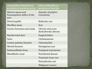
  • 1. ANATOMIC PERIAPICAL RADIOLUCENCIES(FALSE) TRUE PERIAPICAL RADIOLUCENCIES Marrow spaces and hematopoietic defect of the jaws Sequelae of pulpitis Granuloma Dental papilla Radicular cyst Maxillary sinus Scar Incisive foramina and canals Chronic and acute dentoalveolar abscess Nasolacrimal duct Surgical defect Naris Cholesteatoma Greater palatine foramen Osteomyelitis Mental foramen Dentigerous cyst Submandibular fossa Periapical cementoma Mandibular canal Periodontal disease Traumatic bone cyst Nonradicular cyst Malignant tumors
  • 2.  TRUE PERIAPICAL RADIOLUCENCIES represents lesions that are truly located in contact with apex of a tooth  Their shadow cannot be shifted from the periapex by taking additional radiographs at different angles • FALSE PERIAPICAL RADIOLUCENCIES Are produced by anatomic cavities or lytic bony lesions that donot contact the apex of a tooth  The radiolucent shadows may be shifted from the periapex by taking additional radiograph at different angles
  • 3.
  • 7.
  • 8.  Most common type  Result of a successful attempt by the periapical tissues to neutralize and confine the irritating toxic products(prostaglandins,kinins,lysosomes and endotoxins)that are escaping from the root canal  Low grade inflammation  Microstructure consists of proliferating endothelial cells,capillaries,young fibroblasts,a minimal amount of collagen and chronic inflammatory cells  FOUR TYPES ON BASIS OF HISTOPATHOLOGY  Exudative  Granulomatous  Granulofibrous  fibrous
  • 9.  Radiographically,lesion is a rather well circumscribed radiolucency somewhat rounded in shape and surrounding the apex of the tooth  May or may not have a thin radiopaque(hyperostotic)border.  Radiographs reveal presence of deep restorations,extensive caries,fractures or a narrower pulp canal  Periapical radiolucency of diameter less than 1.6cm is considered as periapical granuloma  Electrical and thermal pulp testing will usually indicate that pulp is non-vital
  • 10. Tooth is completely asymptomatic Absence of sensitivity to percussion Crown may have darker colour than of its neighbours because of blood pigments that have diffused into empty dentinal tubules Swelling and expansion of cortical plate is unusual since they rarely reach a size to produce such an effect
  • 11. GRANULOMA Small round or oval radiolucency <1.6cm in diameter surrounding apex of tooth with or without well-circumscribed borders
  • 12.  Second most common  Inflammatory cyst  Origin from cell rest of Malassez  Orginate in pre-existing periapical granuloma  As the masses of proliferating epithelial nests increase in size, the central cells start to degenerate and liquefy  Leads to liquid-filled cavity lined with epithelium  Cysts continues to grow because of a combination of factors 1. The products from the cell lysis are probably irritating and may provide growth stimulus 2. Epilthelial cells discharged into the cyst lumen increase the protein content and hence the osmotic pressure of cyst
  • 13. fluid leading to more water diffusion into lumen 3. The pressure exerted by the enlarging cyst on the alveolar bone induces osteoclastic action and resorption of the periapical bone  FEATURES  The more pronounced the hyperostotic border of the lesion, the more likely is the lesion to be a cyst  Should measure atleast 1.6cm in diameter  An untreated cyst may slowly enlarge and cause expansion of cortical plates,observed clinically as dome like swelling on the alveolus over the periapical region of involved tooth
  • 14. The swelling may develop on either the buccal or lingual side of alveolar process and will be covered with normal-appearing Mucosa Initially,bony hard to palpation,but later it may demonstrate A crackling sound(crepitus) as cortical plates become thinned Usually,the cortical plates remain intact Large cysts may involve a complete quadrant with some of the Teeth occasionally mobile and some of the pulps non-vital Root resorption is also seen the causative tooth and the alveolar swelling are painless in case of sterile cysts,however when infected develops pain
  • 15. RADICULAR CYST Round or oval well- defined radiolucency >1.6cm in diameter
  • 16.  Well defined radiolucency at apex of untreated asymptomatic tooth with nonvital or diseased pulp:90% cases dental granuloma or radicular cyst(1.6cm or more=radicular cyst)  PERIAPICAL SCAR: if well sealed, persistent asymptomatic nonenlarging radiolucency associated with teeth which received non-surgical endodontic treatment for granuloma and cyst  Asymptomatic radiolucency persisting after root resection can be either a scar or surgical defect  PERIAPICAL CEMENTOMA: pulp is vital and mostly involve lower teeth ,mostly incisors  TRAUMATIC BONE CYST :pulp is vital and 90% occurs in mandible(molar,premolar,incisor region)
  • 17.  Extraction  Conservative root canal therapy  Cyst respond well to nonsurgical endodontic treatment:2 theories 1. Instrumentation beyond apex permits the cyst to drain into canal 2. When root canal is sealed the irritating products from gangrenous pulp are no longer present and inflammation subsides  Biopsy via buccal window to determine the nature of lesion  Apical curettage or root resection procedure if 2cm or larger(radicular cyst)  Retrograde filling and periapical curettage if root canal is nonnegotiable
  • 18. For large radicular cysts with considerable bone loss Surgical enucleation Restoration of defect with graft,preferably autogenous bone Marsupialization Decompression Decompression with delayed enucleation Creation of common chamber with maxillary sinus or nasal cavity Sequential post surgical radiographs to ensure regression of defect
  • 19.  Composed of dense fibrous tissue  Situated at periapex of a pulpless tooth in which rootcanals have been filled  Represented by granuloma,cyst or abscess whose healing has stopped in the formation of dense scar tissue(cicatrix) rather than bone  In some instances,granulation tissue during resolution slowly organizes with production of more collagen fibers leading to dense collagenous connective tissue scar :permanent and radiolucent  Microscopically : few spindle shaped fibroblasts scattered throughout dense collagen bundles  Scarring periapical granuloma: relatively young ,less dense scar experiencing intermittent inflammation
  • 20. Well circumscribed radiolucency that is more or less round resembling granuloma and cyst radiographically but usually smaller Asymptomatic Occurs mostly in anterior region of maxilla Majority of involved teeth have been endodontically treated MANAGEMENT No treatment required
  • 21. SCAR Well defined borders Remain constant in size over years asymptomatic
  • 22.  Primary or neoteric abscesses: associated with teeth that have not developed apparent radiolucent lesion • Acute apical periodontitis/acute periapical abscess  Secondary or recrudescent abscesses: develop in a previously existing asymptomatic periapical radiolucent lesion PRIMARY ABSCESS:  Radiographically normal periapical region  Occurs due to rapid spread of virulent bacteria from canal to periapical tissues  Very sensitive tooth and perhaps alveolar swelling  No resorption of bone due to sudden onset and course  Frequent swelling of the periodontal ligament which force the tooth out slightly from its socket leading to an increased periodontal ligament space around entire root
  • 23. Chronic or acute type depending on 1. Number and virulence of invading organism 2. Resistance of host 3. Type and timing of treatment  MICROORGANISMS: Streptococci,Staphylococci, Bacteroides, Peptococcus, Peptostreptococci, Actinomyces, Eubacterium, Fusobacterium
  • 24.  RADIOGRAPHIC  periapical radiolucency is a feature of secondary abscess  Radiolucency may vary in size  Depending on duration ,acuteness and chronicity,margins may be well defined with hyperostotic border to poorly defined in chronic cases  Sometimes present as blurred area of lessened density than surrounding bone  Deep restorations, caries, narrowed pulp chamber or canals : suggestive of non-vital pulp  Roots may show resorption of apex
  • 26. MICROSCOPICALLY Central region of necrosis with dense accumulation of PMN leukocytes surrounded by inflamed connective tissue wall of varying thickness CLINICALLY Very painful to percussion No response to electric pulp tests Ice application relieves pain while heat aggravates the pain Progression leads to penetration of cortical plates, formation of space infection and sinus tracts Warmth of overlying skin or mucosa Elevation of systemic temperature
  • 27. A small proliferation of granulomatous tissue often forms on the surface referred to as parules pain subsides when drainage is established COMPLICATIONS Osteomyelitis Septicemia Septic emboli Ludwig’s angina Cavernous sinus thrombosis
  • 28.  Drainage established by opening pulp chamber and passing file through canal into periapical region  If not possible,TREPHINATION procedure done:opening made through mucosa and bone to abscess at apex  A THROUGH AND THROUGH DRAIN may be placed in abscess in case of vestibular and lingual space infection along with frequent irrigation with H2O2 solution and saline  sample of pus for culture and sensitivity test  Penicillin therapy(not less than 500mg qid for atleast 5 days)/erythromycin  Antibiotics before extraction  In case of draining sinus,exact location of the abscess can be located by inserting a gutta-percha cone to the extent of sinus and making radiograph of that area
  • 29.  Occurs in patients with periapical abscess with underlying systemic disease or who have received large doses of radiation therapy  Defined as an infection of bone that involves all 3 components: periosteum, cortex, and marrow  Osteitis: localized ; Osteomyelitis: diffuse  ACUTE OSTEOMYELITIS: Similar to acute primary alveolar abscess, rapid onset and course, no bone resorption, thus radiolucency may not be present  CHRONIC OSTEOMYELITIS: low –grade infection of bone if untreated leads to extensive bone destruction,produces radiolucent lesion
  • 30. Chronic osteomyelitis demonstrate 4 distinct radiographic picture 1. Completely radiolucent 2. Mixed radiolucent and radiopaque 3. Completely radiopaque 4. Garre’s osteomyelitis FEATURES  Seldom observed in maxilla because of rich blood supply  Tooth associated: non-vital pulp,may be sensitive to percussion and previously associated with periapical abscess  Periapical radiolucency somewhat rounded in shape with poorly defined and ragged borders
  • 31. Sequestrum present as radiopacity within a radiolucency Draining tract may be seen as radiolucency through cortical plate beneath sinus opening on mucosa or skin Fever,malaise Swelling on bone and mucosa around osteomyelitis
  • 33.  If area of bone destruction is large and region is not painful:Eosinophilic granuloma:biopsy  If only alveolar portion of jawbone is infected:Alveolar abscess  If tooth suspected is in fracture line or if an uncontrolled systemic disease is present: Chronic Osteomyelitis  Paget’s disease: several bones with classic wool appearance MANAGEMENT  Best treatment will be to extract the offending tooth rather than conserving it by endodontic procedure
  • 34. Arise from elements in periodontal ligament Other names: 1. Periapical cemental dysplasia 2. Periapical osteofibroma 3. Periapical osteofibrosis  Arise on a reactive basis rather than on neoplastic basis  3 stages in their development which are radiographically apparent
  • 35. 1. Early (osteolytic or fibroblastic)stage: radiolucent 2. Cellular fibroblastic stroma with small foci of calcified material 2. Intermediate stage: radiolucent area containing radiopaque foci 3. Final stage(mature lesion):well-defined solid ,homogeneous radiopacity surrounded in most cases by thin radiolucent border  Calcified material may cementum,bone or both(differentiated using polarising microscopy)  FEATURES  In early stage ,they occur as radiolucencies that are somewhat rounded,have well-defined borders and are associated with teeth having vital pulp
  • 36. Blacks are commonly affected 80% occur in women 90% occur in mandible Asymptomatic Seldom exceeds 1 cm in diameter MANAGEMENT Treatment required only if radiographic changes present If infection or expansion of cortical plate: Surgical enucleation If inflammation of pulp:conservative endodontic treatment
  • 37. 1. Hemorrhagic bone cyst 2. Extravasation cyst 3. Simple bone cyst 4. Progressive bony cyst 5. Blood cyst  FALSE CYST(no epithelial lining)  Localized aberration in normal bone remodeling or metabolism
  • 38. FEATURES A history of trauma may or may not be present Asymptomatic except when it reaches size sufficient to cause expansion of jaws Well-defined cyst-like radiolucency above the mandibular canal either round and positioned somewhat symmetrically about periapex of root OR More elongated and oriented mesiodistally extending superiorly between premolar and molar roots producing scalloped appearance No aspiration results but sometimes serosanguineous fluid or some blood may be obtained Mandible>Maxilla Premolar-molar region most common location
  • 39. TREATMENT 1. open the area surgically 2. Establish a diagnosis of traumatic bone cyst 3. Remove the tissue debris present 4. Curette the walls of bony cavity to induce bleeding 5. Close the soft tissue flap securely  Antibiotics  ALTERNATE METHOD:injecting venous blood into bony defect(good results but objected because sometimes in periodic radiographic follow-up examination, the radiolucency is found to be expanding)
  • 40.
  • 41.  WOOD AND GOAZ  BASIC ORAL RADIOLOGY: ANIL GOVINDRAO GHOM