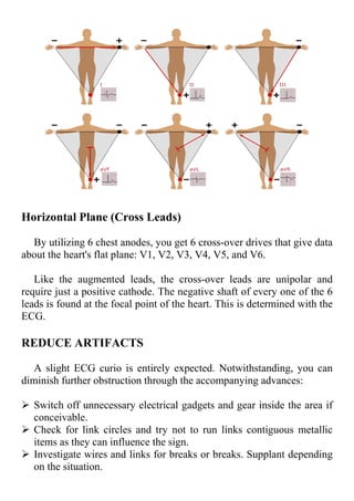
ELECTROCARDIOGRAM -ELECTRICAL ACTIVITY OF THE HEART
AND MEASUREMENT EXPLAINED
What is ECG
An electrocardiogram (ECG) is a realistic record delivered by
an electrocardiograph that gives insights concerning one's pulse and
some other related irregularities; it portrays assuming that the heart has
expanded because of hypertension (hypertension) or proof of a
myocardial dead tissue beforehand (coronary failure if any).
Electrocardiogram (ECG) is one of the most widely recognized and
pragmatic tests for all medications. It is not difficult to perform, painless
yields results in a flash, and is valuable for recognizing many heart
conditions.
Why ECG has to be performed
Two principal types of information are given by an ECG:
Deciding the time taken for an electromagnetic heartbeat to go
through the heart to find assuming that areas of the heart are exhausted
or excessively huge initially, a specialist will decide how long it
requires for the electromagnetic heartbeat to go through the heart by
computing time stretches on the ECG. Whether the electrical action is
regular or lazy, quick or sporadic, sorting out how long a heartbeat
requires to go starts with one piece of the heart and then onto the next.
Second, a cardiologist might have the option to see whether regions of
the heart are excessively enormous or exhausted by estimating how
much electrical action that moves through the heart muscle. Ten
cathodes are mounted on the arms of the patient and on the highest point
of the heart in a customary 12-lead ECG. The typical strength of the
electrical capability of the heart is then determined from 12 unique
points ("leads") and revealed throughout some undefined time frame
(normally 10 seconds). All through the cardiovascular stage, the
absolute power and direction of the electrical depolarization of the heart
are seen at every second.
How is an ECG done?
An ECG is a protected and effortless test that generally requires a
couple of moments.
Utilizing cement patches to tie leads from an electrocardiograph
framework to the skin on your hands, legs, and chest. This prompts your
heart to understand signs and send this data to the electrocardiograph.
On a paper strip or on a screen, the PC then prints the text.
Before the patches are connected, one is normally approached to take
off the upper dress, the chest should be cleaned or shaved. When the
patches are put on, clinic staff offers an outfit to cover themselves. The
ECG test requires about a couple of moments.
ECG Waveform
An ECG has three fundamental parts: the P wave, which signifies
depolarising atria; the QRS complex, which indicates the depolarization
of the ventricles; and the T wave addresses repolarising ventricles.
During each heartbeat, a sound heart has an arranged course of
depolarization that beginnings with pacemaker cells in the sinoatrial hub,
reaches out all through the chamber, and travels through the
atrioventricular hub into its group and into the filaments of Purkinje,
spreading all through the ventricles and to one side.
The electrical action happens in a little fix of pacemaker cells called
the sinus hub during a customary heartbeat. This delivers a little blip
called the P wave when the motivation invigorates the atria. It then
enacts the primary siphoning chambers, and the ventricles, and produces
the huge all over in the center, the QRS complex. The last T wave is a
period of recovery as the drive switches over the ventricles and goes
back.
The Relation between Electrocardiogram (ECG) and
Electrical Movement of Cardiovascular Cycle
An extraordinary bundle of cells, the sino-atrial hub (SA hub),
likewise normally alluded to as a characteristic or essential pacemaker,
is situated on the upper right-hand side of the right chamber. This piece
of the heart produces the electrical drive which then passes down
different offices of the heart. The way and term of this electrical action
of the heart are followed utilizing an exceptional machine and the such
following is called an electrocardiogram. It is an easy technique
ordinarily used to screen heart well-being. An ordinary cardiovascular
cycle has the accompanying advances:
SA hub sends the electrical motivation, it ventures first through atrial
muscles causing their depolarization.
This depolarization prompts atrial withdrawal, siphoning the blood to
the ventricles.
Then the electrical motivation goes to the atrioventricular hub, the
heap of His and Purkinje filaments causing the depolarization of
ventricular muscles.
Depolarization of ventricular muscles happens to make the ventricles
agree and siphon the blood out of the heart.
Simultaneously, atrial repolarization happens and atria unwind.
This is trailed by ventricular unwinding attributable to the repolarization
of ventricular muscles.
MEASUREMENT
A 12-lead ECG portrays the heart's electrical action by recording data
through 12 alternate points of view. Consider it 12 distinct perspectives
of an item woven together to make a durable story - the ECG
translation.
These 12 perspectives are gathered by setting cathodes or little, tacky
patches on the chest (precordial), wrists, and lower legs. These cathodes
are associated with a machine that enlists the heart's electrical action.
Einthoven Triangle
Einthoven's triangle makes sense of why there are 6 front-facing
leads when there are only 4 appendage anodes.
The rule behind Einthoven's triangle depicts how terminals RA, LA,
and LL don't just keep the electrical activity of the heart corresponding
to themselves through the aVR, aVL, and aVF leads. They likewise
relate with one another to frame drives I (RA to LA), II (RA to LL), and
III (LL to LA).
Therefore, they structure a symmetrical triangle. Consequently, it's
known as Einthoven's triangle, named after Willem Einthoven who
created the primary pragmatic ECG.
Remember that RL is nonpartisan (otherwise called point zero where
the electrical flow is estimated). RL doesn't come up in ECG readings
and is considered as an establishing lead that limits ECG curio.
What is LEAD
A lead is a brief look at the electrical movement of the heart from a
specific point.Set forth plainly, a lead resembles a viewpoint. In 12-lead
ECG, there are 10 anodes giving 12 points of view of the heart's action
utilizing various points through two electrical planes - vertical and flat
planes.
Vertical plane (Front facing Leads):
By utilizing 4 appendage cathodes, you get 6 front facing drives that
give data about the heart's upward plane:
 Lead I
 Lead II
 Lead III
 Expanded Vector Right (aVR)
 Expanded Vector Left (aVL)
 Expanded vector foot (aVF)
Drives I, II, and III require a negative and positive cathode
(bipolarity) for checking. Then again, the augmented leads - aVR, aVL,
and aVF-are unipolar and require just a positive cathode for observing.
Horizontal Plane (Cross Leads)
By utilizing 6 chest anodes, you get 6 cross-over drives that give data
about the heart's flat plane: V1, V2, V3, V4, V5, and V6.
Like the augmented leads, the cross-over leads are unipolar and
require just a positive cathode. The negative shaft of every one of the 6
leads is found at the focal point of the heart. This is determined with the
ECG.
REDUCE ARTIFACTS
A slight ECG curio is entirely expected. Notwithstanding, you can
diminish further obstruction through the accompanying advances:
 Switch off unnecessary electrical gadgets and gear inside the area if
conceivable.
 Check for link circles and try not to run links contiguous metallic
items as they can influence the sign.
 Investigate wires and links for breaks or breaks. Supplant depending
on the situation.
 In the event that is conceivable, use flood silencers with the power
supply.
 Guarantee that channels and preamplifiers are properly changed.
 Guarantee safe association between persistent link and the ECG
gadget.
 Twofold checks for holes between connectors.

ECG - Definition and Measurement techniques .pdf

  • 1.
    ELECTROCARDIOGRAM -ELECTRICAL ACTIVITYOF THE HEART AND MEASUREMENT EXPLAINED What is ECG An electrocardiogram (ECG) is a realistic record delivered by an electrocardiograph that gives insights concerning one's pulse and some other related irregularities; it portrays assuming that the heart has expanded because of hypertension (hypertension) or proof of a myocardial dead tissue beforehand (coronary failure if any). Electrocardiogram (ECG) is one of the most widely recognized and pragmatic tests for all medications. It is not difficult to perform, painless yields results in a flash, and is valuable for recognizing many heart conditions. Why ECG has to be performed Two principal types of information are given by an ECG: Deciding the time taken for an electromagnetic heartbeat to go through the heart to find assuming that areas of the heart are exhausted or excessively huge initially, a specialist will decide how long it requires for the electromagnetic heartbeat to go through the heart by computing time stretches on the ECG. Whether the electrical action is regular or lazy, quick or sporadic, sorting out how long a heartbeat requires to go starts with one piece of the heart and then onto the next. Second, a cardiologist might have the option to see whether regions of the heart are excessively enormous or exhausted by estimating how much electrical action that moves through the heart muscle. Ten cathodes are mounted on the arms of the patient and on the highest point of the heart in a customary 12-lead ECG. The typical strength of the electrical capability of the heart is then determined from 12 unique points ("leads") and revealed throughout some undefined time frame (normally 10 seconds). All through the cardiovascular stage, the
  • 2.
    absolute power anddirection of the electrical depolarization of the heart are seen at every second. How is an ECG done? An ECG is a protected and effortless test that generally requires a couple of moments. Utilizing cement patches to tie leads from an electrocardiograph framework to the skin on your hands, legs, and chest. This prompts your heart to understand signs and send this data to the electrocardiograph. On a paper strip or on a screen, the PC then prints the text. Before the patches are connected, one is normally approached to take off the upper dress, the chest should be cleaned or shaved. When the patches are put on, clinic staff offers an outfit to cover themselves. The ECG test requires about a couple of moments. ECG Waveform An ECG has three fundamental parts: the P wave, which signifies depolarising atria; the QRS complex, which indicates the depolarization of the ventricles; and the T wave addresses repolarising ventricles.
  • 3.
    During each heartbeat,a sound heart has an arranged course of depolarization that beginnings with pacemaker cells in the sinoatrial hub, reaches out all through the chamber, and travels through the atrioventricular hub into its group and into the filaments of Purkinje, spreading all through the ventricles and to one side. The electrical action happens in a little fix of pacemaker cells called the sinus hub during a customary heartbeat. This delivers a little blip called the P wave when the motivation invigorates the atria. It then enacts the primary siphoning chambers, and the ventricles, and produces the huge all over in the center, the QRS complex. The last T wave is a period of recovery as the drive switches over the ventricles and goes back. The Relation between Electrocardiogram (ECG) and Electrical Movement of Cardiovascular Cycle An extraordinary bundle of cells, the sino-atrial hub (SA hub), likewise normally alluded to as a characteristic or essential pacemaker, is situated on the upper right-hand side of the right chamber. This piece of the heart produces the electrical drive which then passes down different offices of the heart. The way and term of this electrical action of the heart are followed utilizing an exceptional machine and the such following is called an electrocardiogram. It is an easy technique ordinarily used to screen heart well-being. An ordinary cardiovascular cycle has the accompanying advances: SA hub sends the electrical motivation, it ventures first through atrial muscles causing their depolarization. This depolarization prompts atrial withdrawal, siphoning the blood to the ventricles. Then the electrical motivation goes to the atrioventricular hub, the heap of His and Purkinje filaments causing the depolarization of ventricular muscles.
  • 4.
    Depolarization of ventricularmuscles happens to make the ventricles agree and siphon the blood out of the heart. Simultaneously, atrial repolarization happens and atria unwind. This is trailed by ventricular unwinding attributable to the repolarization of ventricular muscles. MEASUREMENT A 12-lead ECG portrays the heart's electrical action by recording data through 12 alternate points of view. Consider it 12 distinct perspectives of an item woven together to make a durable story - the ECG translation. These 12 perspectives are gathered by setting cathodes or little, tacky patches on the chest (precordial), wrists, and lower legs. These cathodes are associated with a machine that enlists the heart's electrical action. Einthoven Triangle Einthoven's triangle makes sense of why there are 6 front-facing leads when there are only 4 appendage anodes. The rule behind Einthoven's triangle depicts how terminals RA, LA, and LL don't just keep the electrical activity of the heart corresponding to themselves through the aVR, aVL, and aVF leads. They likewise relate with one another to frame drives I (RA to LA), II (RA to LL), and III (LL to LA). Therefore, they structure a symmetrical triangle. Consequently, it's known as Einthoven's triangle, named after Willem Einthoven who created the primary pragmatic ECG. Remember that RL is nonpartisan (otherwise called point zero where the electrical flow is estimated). RL doesn't come up in ECG readings and is considered as an establishing lead that limits ECG curio.
  • 5.
    What is LEAD Alead is a brief look at the electrical movement of the heart from a specific point.Set forth plainly, a lead resembles a viewpoint. In 12-lead ECG, there are 10 anodes giving 12 points of view of the heart's action utilizing various points through two electrical planes - vertical and flat planes. Vertical plane (Front facing Leads): By utilizing 4 appendage cathodes, you get 6 front facing drives that give data about the heart's upward plane:  Lead I  Lead II  Lead III  Expanded Vector Right (aVR)  Expanded Vector Left (aVL)  Expanded vector foot (aVF) Drives I, II, and III require a negative and positive cathode (bipolarity) for checking. Then again, the augmented leads - aVR, aVL, and aVF-are unipolar and require just a positive cathode for observing.
  • 6.
    Horizontal Plane (CrossLeads) By utilizing 6 chest anodes, you get 6 cross-over drives that give data about the heart's flat plane: V1, V2, V3, V4, V5, and V6. Like the augmented leads, the cross-over leads are unipolar and require just a positive cathode. The negative shaft of every one of the 6 leads is found at the focal point of the heart. This is determined with the ECG. REDUCE ARTIFACTS A slight ECG curio is entirely expected. Notwithstanding, you can diminish further obstruction through the accompanying advances:  Switch off unnecessary electrical gadgets and gear inside the area if conceivable.  Check for link circles and try not to run links contiguous metallic items as they can influence the sign.  Investigate wires and links for breaks or breaks. Supplant depending on the situation.
  • 7.
     In theevent that is conceivable, use flood silencers with the power supply.  Guarantee that channels and preamplifiers are properly changed.  Guarantee safe association between persistent link and the ECG gadget.  Twofold checks for holes between connectors.