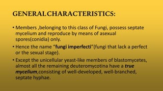
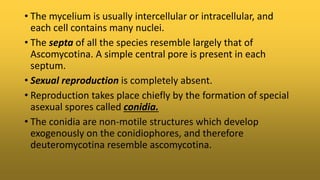
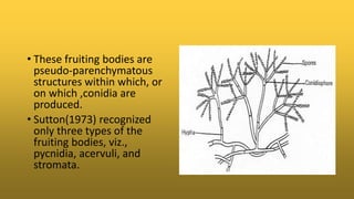
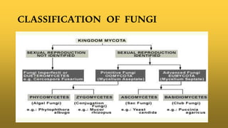
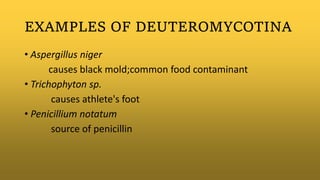
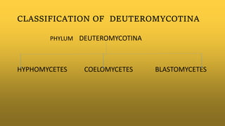
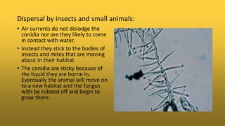
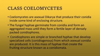
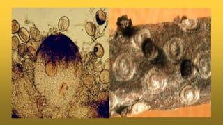
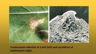
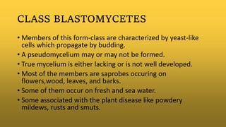
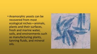
PRESENTED BY:
Vivek kumar
M.Sc MICROBIOLOGY
Bangalore University
GENERALCHARACTERISTICS:
• Members ,belonging to this class of Fungi, possess septate
mycelium and reproduce by means of asexual
spores(conidia) only.
• Hence the name “fungi imperfecti”(fungi that lack a perfect
or the sexual stage).
• Except the unicellular yeast-like members of blastomycetes,
almost all the remaining deuteromycotina have a true
mycelium,consisting of well-developed, well-branched,
septate hyphae.
• The mycelium is usually intercellular or intracellular, and
each cell contains many nuclei.
• The septa of all the species resemble largely that of
Ascomycotina. A simple central pore is present in each
septum.
• Sexual reproduction is completely absent.
• Reproduction takes place chiefly by the formation of special
asexual spores called conidia.
• The conidia are non-motile structures which develop
exogenously on the conidiophores, and therefore
deuteromycotina resemble ascomycotina.
• The conidia are highly variable in shape, size, colour etc.
• They are hyaline or variously coloured, unicellular or
multicellular, and transversely septate or contain both
transverse as well as longitudinal septa.
• They may be oval, elongated, spherical, star-shaped, curved,
threadlike, disc-shaped, coiled, and of other shape.
• The conidia are produced either directly on the
conidiophores or in some special types of fruiting bodies
such as synnemota, acervuli, sporodochia or pycnidia.
• These fruiting bodies are
pseudo-parenchymatous
structures within which, or
on which ,conidia are
produced.
• Sutton(1973) recognized
only three types of the
fruiting bodies, viz.,
pycnidia, acervuli, and
stromata.
CLASSIFICATION OF FUNGI
PARASEXUAL CYCLE
• It is a process in which plasmogamy, karyogamy and
haploidization takes place, but not in particular place in the
thallus nor at any specific period during its life cycle.
• FORMATION OF HETEROKARYOTIC MYCELIUM:
Heterokaryon formation refers to the condition by which
genetically different nuclei are associated in a common
cytoplasm.
• OCCASIONAL KARYOGAMY:
Following initial fusion of hyphal cells, to form a genetically
different cell, mitotic division perpetuates the cell and
mycelium that is made up of genetically, different nuclei is
formed.
• HAPLOIDIZATION:
This haploidization is NOT meiosis.
It is in fact, a series of errors in mitosis.
A sequential loss of chromosomes will eventually give rise
to a haploid nucleus.
EXAMPLES OF DEUTEROMYCOTINA
• Aspergillus niger
causes black mold;common food contaminant
• Trichophyton sp.
causes athlete's foot
• Penicillium notatum
source of penicillin
CLASSIFICATION OF DEUTEROMYCOTINA
PHYLUM DEUTEROMYCOTINA
HYPHOMYCETES COELOMYCETES BLASTOMYCETES
CLASS HYPHOMYCETES
• Hyphomycetes are asexual reproductive structures produced
directly on their substrate without any kind of enclosing tissues.
• The most common function of hyphomycetes is reproduction and
dispersal, although in some species the conidia may act as
gametes or "spermatia" that can fertilize an incipient dikaryon.
• Hyphomycetes come in a staggering variety of forms.
• This immense diversity reflects the role these forms play in the
dispersal of the fungus producing them.
• Each species grows in a particular habitat.
• When the nutrients in this habitat are exhausted the fungus
must ensure that its offspring find their way to a similar
source of nutrition.
• Getting their conidia to this new place, often a very small
target, requires precise dispersal mechanisms.
• Dispersal mechanisms includes:
-Air dispersal
-Water dispersal
-Dispersal by insects and small other animals.
AIR DISPERSAL:
-Epicoccum nigrum and Nigrospora
sphaerica, are examples of fungi that
actually propel their conidia into the
air using mechanisms that force the
conidium away from its subtending
cell.
• The large black conidium is still
attached to its cell awaiting lift-off.
• There are two cells that have
already discharged their conidia.
• The tube that directed water
pressure up against the bottom of
the conidium, shooting it away.
• WATER DISPERSAL:
Many hyphomycetes are dispersed
by water rather than air. Some of
these species actually live out their
lives in water while others only use
water as a means of dispersing their
conidia.
Fungi often grow in habitats that
are periodically flooded but are
usually dry. When water levels rise
the conidia are able to float away.
These conidia are not as likely to
trap air but will become attached to
bubbles of foam.
Conidia of aquatic hyphomycetes are
easy to collect and observe under the
microscope. They often collect and
concentrate in the foam of running
streams.
Dispersal by insects and small animals:
• Air currents do not dislodge the
conidia nor are they likely to come
in contact with water.
• Instead they stick to the bodies of
insects and mites that are moving
about in their habitat.
• The conidia are sticky because of
the liquid they are borne in.
Eventually the animal will move on
to a new habitat and the fungus
with be rubbed off and begin to
grow there.
• The class hphomycetes is classified into 4 orders based on
the presence or absence of conidia and degree of the
aggregation of the conidiophores into specialized structures
called synnetama.
• Orders:
• Hyphomycetales
• Stilbellables
• Tuberculariales
• Agonomycetales
• The disease caused by
hyphomycetes is called as
HYALOHYPHOMYCOSIS. It is
caused by a number of
hyaline(non-dematiaceous)
hyphomycetes where the
tissue morphology of the
causitive organism is
mycelial.
CLASS COELOMYCETES
• Coelomycetes are asexual Dikarya that produce their conidia
inside some kind of enclosing structure.
• The fungal hyphae germinate from conidia and form an
aggregated mass until they form a fertile layer of densely
packed conidiophores.
• Conidiophores are simple or branched hyphae that develop
specialized cells (conidiogenous) from which asexual conidia
are produced. It is this mass of hyphae that create the
fruiting structure known as a conidiomata.
• If this structure completely surrounds the conidia it is called
a pycnidium; if the conidia are borne in a more open
structure with a wall only around the lower part it is an
acervulus.
• The pycnidium is very light coloured where it is sunken in the
host tissues but dark on top.
• This is a common phenomenon among pycnidial fungi.
• Most pycnidia release their spores in a wet mass that is
extruded out through the apical pore (ostiole). These often
collect in large amounts and are either transported away by
insects or will flow along with rain water.
• Pycnidia resemble the fruiting structures of ascomycetes
called ascomata
• The second main type of conidomata are acervuli.
• Like pycnidia, acervuli also produce a mat of closely packed
conidiophores but instead grow in a raised mass.
• Unlike pycnidia, acervuli are typically open and cup-shaped
and the opening by which conidia are released is much larger
than that of a pycnidium.
• More commonly these fungi grow on living and dead plants.
• Some pycnidia and acervuli can be highly complex and be
composed of several cavities.
• Orders:
• Melanconiales (producing spores in acervuli)
• Sphaeropsidales (producing spores in pycnidia)
• Pycnothyriales (produces pycnothyrial conidiomata.)
Coelomycete infection in a leaf (left) and pycnidium of
coelomycete (right)
CLASS BLASTOMYCETES
• Members of this form-class are characterized by yeast-like
cells which propagate by budding.
• A pseudomycelium may or may not be formed.
• True mycelium is either lacking or is not well developed.
• Most of the members are saprobes occuring on
flowers,wood, leaves, and barks.
• Some of them occur on fresh and sea water.
• Some associated with the plant disease like powdery
mildews, rusts and smuts.
• Anamorphic yeasts can be
recovered from most
ecological niches—animals,
plants and their surfaces,
fresh and marine water,
soils, and environments such
as manufacturing plants,
tanning fluids, and mineral
oils.
• They comprises anamorphic (asexual or imperfect) yeast
fungi that lack fruit bodies (conidiomata), have no
dikaryophase, and are usually unicellular rather than
filamentous.
• The thallus consists of individual cells.
• Approximately 80 genera comprising about 600 species are
recognized.
• Blastomycetes are of great economic importance in two
respects: the production of products and the spoilage of raw
materials and products.
• Blastomycetes are also recognized pathogens in medicine.
• Both Candida, causing candidiasis or candidosis, and
Cryptococcus, causing cryptococcosis, are opportunistic
pathogens that cause systemic infections only in individuals
with lowered resistance.
• Blastomyces dermatitidis is the causal agent of
blastomycosis, an invasive and often serious fungal infection
found occasionally in humans and other animals in regions
where the fungus is endemic.
• The causal organism is a fungus living in soil and wet,
decaying wood, often in an area close to a waterway such as
a lake, river or stream.
• Blastomycosis is generally
readily treatable with
systemic antifungal drugs
once it is correctly
diagnosed; however,
delayed diagnosis is very
common except in highly
endemic areas.
BIBILIOGRAPHY
 AN INTRODUCTION TO MYCOLOGY:
• Alexopoulos, Mims,Blackwell
 AN INTRODUCTION TO MYCOLOGY
• R.S Mehrotra,KR Aneja
• Classes of deuteromycotina-website.nbm-mnb.ca › Home › Diversity
and classification
THANK YOU

Deuteromycotina

  • 1.
    PRESENTED BY: Vivek kumar M.ScMICROBIOLOGY Bangalore University
  • 2.
    GENERALCHARACTERISTICS: • Members ,belongingto this class of Fungi, possess septate mycelium and reproduce by means of asexual spores(conidia) only. • Hence the name “fungi imperfecti”(fungi that lack a perfect or the sexual stage). • Except the unicellular yeast-like members of blastomycetes, almost all the remaining deuteromycotina have a true mycelium,consisting of well-developed, well-branched, septate hyphae.
  • 3.
    • The myceliumis usually intercellular or intracellular, and each cell contains many nuclei. • The septa of all the species resemble largely that of Ascomycotina. A simple central pore is present in each septum. • Sexual reproduction is completely absent. • Reproduction takes place chiefly by the formation of special asexual spores called conidia. • The conidia are non-motile structures which develop exogenously on the conidiophores, and therefore deuteromycotina resemble ascomycotina.
  • 4.
    • The conidiaare highly variable in shape, size, colour etc. • They are hyaline or variously coloured, unicellular or multicellular, and transversely septate or contain both transverse as well as longitudinal septa. • They may be oval, elongated, spherical, star-shaped, curved, threadlike, disc-shaped, coiled, and of other shape. • The conidia are produced either directly on the conidiophores or in some special types of fruiting bodies such as synnemota, acervuli, sporodochia or pycnidia.
  • 5.
    • These fruitingbodies are pseudo-parenchymatous structures within which, or on which ,conidia are produced. • Sutton(1973) recognized only three types of the fruiting bodies, viz., pycnidia, acervuli, and stromata.
  • 6.
  • 7.
    PARASEXUAL CYCLE • Itis a process in which plasmogamy, karyogamy and haploidization takes place, but not in particular place in the thallus nor at any specific period during its life cycle. • FORMATION OF HETEROKARYOTIC MYCELIUM: Heterokaryon formation refers to the condition by which genetically different nuclei are associated in a common cytoplasm.
  • 8.
    • OCCASIONAL KARYOGAMY: Followinginitial fusion of hyphal cells, to form a genetically different cell, mitotic division perpetuates the cell and mycelium that is made up of genetically, different nuclei is formed. • HAPLOIDIZATION: This haploidization is NOT meiosis. It is in fact, a series of errors in mitosis. A sequential loss of chromosomes will eventually give rise to a haploid nucleus.
  • 10.
    EXAMPLES OF DEUTEROMYCOTINA •Aspergillus niger causes black mold;common food contaminant • Trichophyton sp. causes athlete's foot • Penicillium notatum source of penicillin
  • 11.
    CLASSIFICATION OF DEUTEROMYCOTINA PHYLUMDEUTEROMYCOTINA HYPHOMYCETES COELOMYCETES BLASTOMYCETES
  • 12.
    CLASS HYPHOMYCETES • Hyphomycetesare asexual reproductive structures produced directly on their substrate without any kind of enclosing tissues. • The most common function of hyphomycetes is reproduction and dispersal, although in some species the conidia may act as gametes or "spermatia" that can fertilize an incipient dikaryon. • Hyphomycetes come in a staggering variety of forms. • This immense diversity reflects the role these forms play in the dispersal of the fungus producing them.
  • 13.
    • Each speciesgrows in a particular habitat. • When the nutrients in this habitat are exhausted the fungus must ensure that its offspring find their way to a similar source of nutrition. • Getting their conidia to this new place, often a very small target, requires precise dispersal mechanisms. • Dispersal mechanisms includes: -Air dispersal -Water dispersal -Dispersal by insects and small other animals.
  • 14.
    AIR DISPERSAL: -Epicoccum nigrumand Nigrospora sphaerica, are examples of fungi that actually propel their conidia into the air using mechanisms that force the conidium away from its subtending cell. • The large black conidium is still attached to its cell awaiting lift-off. • There are two cells that have already discharged their conidia. • The tube that directed water pressure up against the bottom of the conidium, shooting it away.
  • 15.
    • WATER DISPERSAL: Manyhyphomycetes are dispersed by water rather than air. Some of these species actually live out their lives in water while others only use water as a means of dispersing their conidia. Fungi often grow in habitats that are periodically flooded but are usually dry. When water levels rise the conidia are able to float away. These conidia are not as likely to trap air but will become attached to bubbles of foam. Conidia of aquatic hyphomycetes are easy to collect and observe under the microscope. They often collect and concentrate in the foam of running streams.
  • 16.
    Dispersal by insectsand small animals: • Air currents do not dislodge the conidia nor are they likely to come in contact with water. • Instead they stick to the bodies of insects and mites that are moving about in their habitat. • The conidia are sticky because of the liquid they are borne in. Eventually the animal will move on to a new habitat and the fungus with be rubbed off and begin to grow there.
  • 17.
    • The classhphomycetes is classified into 4 orders based on the presence or absence of conidia and degree of the aggregation of the conidiophores into specialized structures called synnetama. • Orders: • Hyphomycetales • Stilbellables • Tuberculariales • Agonomycetales
  • 18.
    • The diseasecaused by hyphomycetes is called as HYALOHYPHOMYCOSIS. It is caused by a number of hyaline(non-dematiaceous) hyphomycetes where the tissue morphology of the causitive organism is mycelial.
  • 19.
    CLASS COELOMYCETES • Coelomycetesare asexual Dikarya that produce their conidia inside some kind of enclosing structure. • The fungal hyphae germinate from conidia and form an aggregated mass until they form a fertile layer of densely packed conidiophores. • Conidiophores are simple or branched hyphae that develop specialized cells (conidiogenous) from which asexual conidia are produced. It is this mass of hyphae that create the fruiting structure known as a conidiomata.
  • 20.
    • If thisstructure completely surrounds the conidia it is called a pycnidium; if the conidia are borne in a more open structure with a wall only around the lower part it is an acervulus. • The pycnidium is very light coloured where it is sunken in the host tissues but dark on top. • This is a common phenomenon among pycnidial fungi. • Most pycnidia release their spores in a wet mass that is extruded out through the apical pore (ostiole). These often collect in large amounts and are either transported away by insects or will flow along with rain water.
  • 21.
    • Pycnidia resemblethe fruiting structures of ascomycetes called ascomata • The second main type of conidomata are acervuli. • Like pycnidia, acervuli also produce a mat of closely packed conidiophores but instead grow in a raised mass. • Unlike pycnidia, acervuli are typically open and cup-shaped and the opening by which conidia are released is much larger than that of a pycnidium.
  • 23.
    • More commonlythese fungi grow on living and dead plants. • Some pycnidia and acervuli can be highly complex and be composed of several cavities. • Orders: • Melanconiales (producing spores in acervuli) • Sphaeropsidales (producing spores in pycnidia) • Pycnothyriales (produces pycnothyrial conidiomata.)
  • 25.
    Coelomycete infection ina leaf (left) and pycnidium of coelomycete (right)
  • 26.
    CLASS BLASTOMYCETES • Membersof this form-class are characterized by yeast-like cells which propagate by budding. • A pseudomycelium may or may not be formed. • True mycelium is either lacking or is not well developed. • Most of the members are saprobes occuring on flowers,wood, leaves, and barks. • Some of them occur on fresh and sea water. • Some associated with the plant disease like powdery mildews, rusts and smuts.
  • 27.
    • Anamorphic yeastscan be recovered from most ecological niches—animals, plants and their surfaces, fresh and marine water, soils, and environments such as manufacturing plants, tanning fluids, and mineral oils.
  • 28.
    • They comprisesanamorphic (asexual or imperfect) yeast fungi that lack fruit bodies (conidiomata), have no dikaryophase, and are usually unicellular rather than filamentous. • The thallus consists of individual cells. • Approximately 80 genera comprising about 600 species are recognized. • Blastomycetes are of great economic importance in two respects: the production of products and the spoilage of raw materials and products.
  • 29.
    • Blastomycetes arealso recognized pathogens in medicine. • Both Candida, causing candidiasis or candidosis, and Cryptococcus, causing cryptococcosis, are opportunistic pathogens that cause systemic infections only in individuals with lowered resistance. • Blastomyces dermatitidis is the causal agent of blastomycosis, an invasive and often serious fungal infection found occasionally in humans and other animals in regions where the fungus is endemic. • The causal organism is a fungus living in soil and wet, decaying wood, often in an area close to a waterway such as a lake, river or stream.
  • 30.
    • Blastomycosis isgenerally readily treatable with systemic antifungal drugs once it is correctly diagnosed; however, delayed diagnosis is very common except in highly endemic areas.
  • 32.
    BIBILIOGRAPHY  AN INTRODUCTIONTO MYCOLOGY: • Alexopoulos, Mims,Blackwell  AN INTRODUCTION TO MYCOLOGY • R.S Mehrotra,KR Aneja • Classes of deuteromycotina-website.nbm-mnb.ca › Home › Diversity and classification
  • 33.