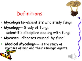
Definitions
• Mycologists--scientists whostudy fungi
• Mycology--Study of fungi.
scientific discipline dealing with fungi
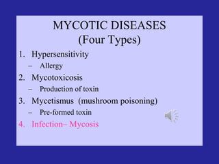
• Mycoses--diseases caused by fungi
• Medical Mycology--- is the study of
mycoses of man and their etiologic agents
4.
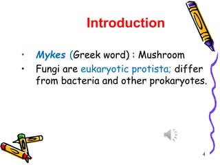
Introduction
• Mykes (Greekword) : Mushroom
• Fungi are eukaryotic protista; differ
from bacteria and other prokaryotes.
4
5.
History
• The abilityof fungi to invade plant and animal
tissue was observed in early 19 th century but
the first documented animal infection by any
fungus was made by Bassi, who in 1835 studied
the muscardine disease of silkworm and proved
the that the infection was caused by a fungus
Beauveria bassiana.
• In 1910 Raymond Sabouraud published his book
Les Teignes, which was a comprehensive study of
dermatophytic fungi. He is also regarded as
father of medical mycology.
6.
Hapitate of fungi:
•Fungi inhabit almost every niche in the
environment and humans are exposed to
these organisms in various fields of life.
7.
Beneficial Effects ofFungi:
1. Decomposition - nutrient and carbon recycling.
2. Biosynthetic factories. The fermentation
property is used for the industrial production of
alcohols, fats, citric, oxalic and gluconic acids.
3. Important sources of antibiotics, such as
Penicillin.
4. Model organisms for biochemical and genetic
studies. Eg: Neurospora crassa
5. Saccharomyces cerviciae is extensively used in
recombinant DNA technology, which includes the
Hepatitis B Vaccine.
8.
6. Some fungiare edible (mushrooms).
7. Yeasts provide nutritional supplements such as
vitamins and cofactors.
8. Penicillium is used to flavour Roquefort and
Camembert cheeses.
9. Ergot produced by Claviceps purpurea contains
medically important alkaloids that help in inducing
uterine contractions, controlling bleeding and
treating migraine.
9.
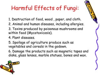
Harmful Effects ofFungi:
1. Destruction of food, wood , paper, and cloth.
2. Animal and human diseases, including allergies.
3. Toxins produced by poisonous mushrooms and
within food (Mycotoxicosis).
4. Plant diseases.
5. Spoilage of agriculture produce such as
vegetables and cereals in the godown.
6. Damage the products such as magnetic tapes and
disks, glass lenses, marble statues, bones and wax.
10.
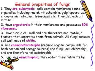
General properties offungi:
1. They are eukaryotic; cells contain membrane bound cell
organelles including nuclei, mitochondria, golgi apparatus,
endoplasmic reticulum, lysosomes etc. They also exhibit
mitosis.
2. Have ergosterols in their membranes and possesses 80S
ribosomes.
3. Have a rigid cell wall and are therefore non-motile, a
feature that separates them from animals. All fungi possess
cell wall made of chitin.
4. Are chemoheterotrophs (require organic compounds for
both carbon and energy sources) and fungi lack chlorophyll
and are therefore not autotrophic.
5. Fungi are osmiotrophic; they obtain their nutrients by
absorption.
11.
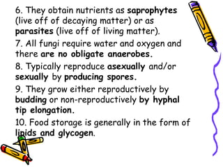
6. They obtainnutrients as saprophytes
(live off of decaying matter) or as
parasites (live off of living matter).
7. All fungi require water and oxygen and
there are no obligate anaerobes.
8. Typically reproduce asexually and/or
sexually by producing spores.
9. They grow either reproductively by
budding or non-reproductively by hyphal
tip elongation.
10. Food storage is generally in the form of
lipids and glycogen.
12.
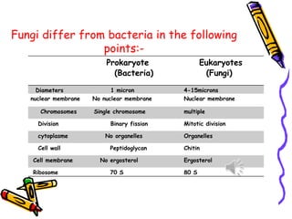
Fungi differ frombacteria in the following
points:-
Prokaryote
(Bacteria)
Eukaryotes
(Fungi)
Diameters 1 micron 4-15microns
nuclear membrane No nuclear membrane Nuclear membrane
Chromosomes Single chromosome multiple
Division Binary fission Mitotic division
cytoplasme No organelles Organelles
Cell wall Peptidoglycan Chitin
Cell membrane No ergosterol Ergosterol
Ribosome 70 S 80 S
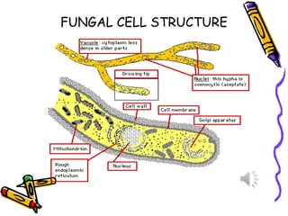
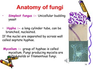
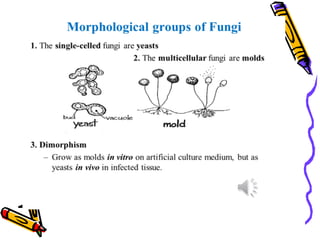
Anatomy of fungi
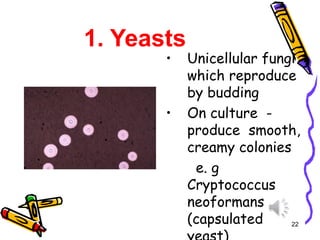
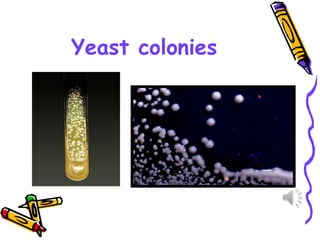
•Simplest fungus :- Unicellular budding
yeast
• Hypha :- a long cylinder tube, can be
branched, nucleated.
If the nuclei are separated by across wall
called septate hyphae.
Mycelium :- group of hyphae is called
mycelium. Fungi producing mycelia are
called molds or filamentous fungi.
14
15.
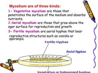
Mycelium are ofthree kinds:
1- Vegetative mycelium are those that
penetrates the surface of the medium and absorbs
nutrients.
2-Aerial mycelium are those that grow above the
agar surface for reproduction and growth
3- Fertile mycelium are aerial hyphae that bear
reproductive structures such as conidia or
sporangia.
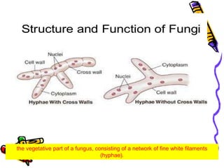
16.
the vegetative partof a fungus, consisting of a network of fine white filaments
(hyphae).
17.
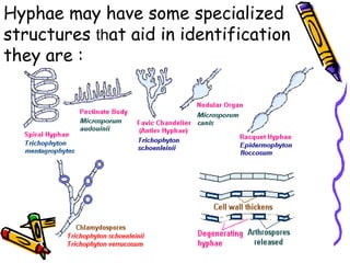
Hyphae may havesome specialized
structures that aid in identification
they are :
18.
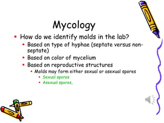
Mycology
▪ How dowe identify molds in the lab?
▪ Based on type of hyphae (septate versus non-
septate)
▪ Based on color of mycelium
▪ Based on reproductive structures
▪ Molds may form either sexual or asexual spores
▪ Sexual spores
▪ Asexual spores,
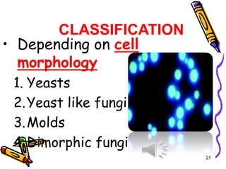
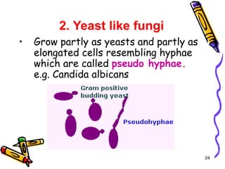
2. Yeast likefungi
• Grow partly as yeasts and partly as
elongated cells resembling hyphae
which are called pseudo hyphae.
e.g. Candida albicans
24
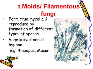
25.
3.Molds/ Filamentous
fungi
• Formtrue mycelia &
reproduce by
formation of different
types of spores.
• Vegetative/ aerial
hyphae
e.g. Rhizopus, Mucor
25
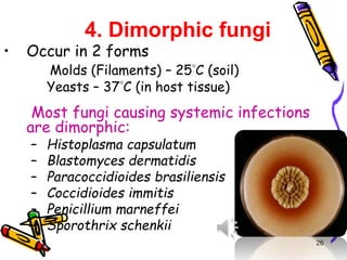
26.
4. Dimorphic fungi
•Occur in 2 forms
Molds (Filaments) – 25C (soil)
Yeasts – 37C (in host tissue)
Most fungi causing systemic infections
are dimorphic:
– Histoplasma capsulatum
– Blastomyces dermatidis
– Paracoccidioides brasiliensis
– Coccidioides immitis
– Penicillium marneffei
– Sporothrix schenkii
26
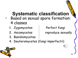
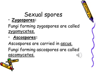
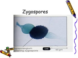
Sexual spores
• Zygospores:
Fungiforming zygospores are called
zygomycetes.
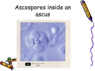
• Ascospores:
Ascospores are carried in ascus.
Fungi forming ascospores are called
ascomycetes.
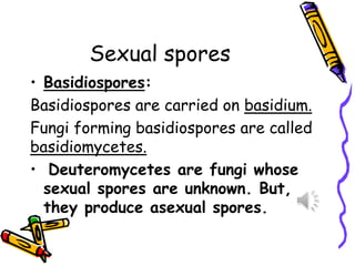
Sexual spores
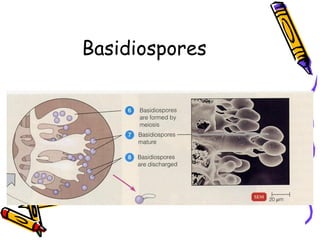
• Basidiospores:
Basidiosporesare carried on basidium.
Fungi forming basidiospores are called
basidiomycetes.
• Deuteromycetes are fungi whose
sexual spores are unknown. But,
they produce asexual spores.
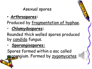
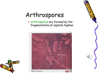
• Arthrospores:
Produced byfragmentation of hyphae.
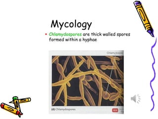
• Chlamydospores:
Rounded thick walled spores produced
by candida fungus.
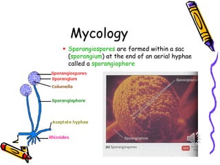
• Sporangiospores:
Spores formed within a sac called
sporangium. Formed by zygomycetes.
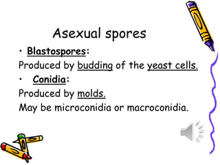
Asexual spores
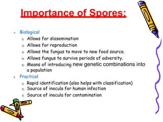
Importance of Spores:
A.Biological
1) Allows for dissemination
2) Allows for reproduction
3) Allows the fungus to move to new food source.
4) Allows fungus to survive periods of adversity.
5) Means of introducing new genetic combinations into
a population
B. Practical
1) Rapid identification (also helps with classification)
2) Source of inocula for human infection
3) Source of inocula for contamination
46.
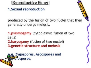
Reproductive Fungi
1.Sexual reproduction
producedby the fusion of two nuclei that then
generally undergo meiosis.
1.plasmogamy (cytoplasmic fusion of two
cells)
2.karyogamy (fusion of two nuclei)
3.genetic structure and meiosis
e.g. Zygospores, Ascospores and
Basidiospores.
47.
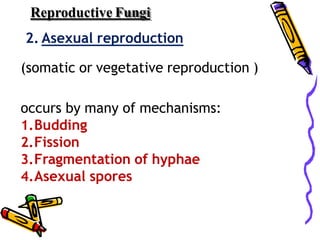
Reproductive Fungi
2. Asexualreproduction
(somatic or vegetative reproduction )
occurs by many of mechanisms:
1.Budding
2.Fission
3.Fragmentation of hyphae
4.Asexual spores
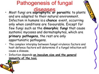
Pathogenesis of fungal
diseases
•Most fungi are saprophytic or parasitic to plants
and are adapted to their natural environment.
Infection in humans is a chance event, occurring
only when conditions are favourable. Except for
few fungi such as the dimorphic fungi that cause
systemic mycoses and dermatophytes, which are
primary pathogens, the rest are only
opportunistic pathogens.
• The complex interplay between fungal virulence factors and
host defence factors will determine if a fungal infection will
cause a disease.
• Infection depends on inoculum size and the general
immunity of the host.
51.
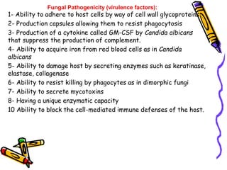
Fungal Pathogenicity (virulencefactors):
1- Ability to adhere to host cells by way of cell wall glycoproteins
2- Production capsules allowing them to resist phagocytosis
3- Production of a cytokine called GM-CSF by Candida albicans
that suppress the production of complement.
4- Ability to acquire iron from red blood cells as in Candida
albicans
5- Ability to damage host by secreting enzymes such as keratinase,
elastase, collagenase
6- Ability to resist killing by phagocytes as in dimorphic fungi
7- Ability to secrete mycotoxins
8- Having a unique enzymatic capacity
10 Ability to block the cell-mediated immune defenses of the host.
52.
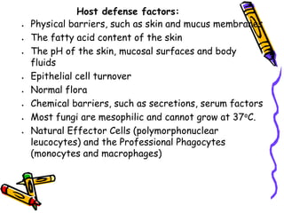
Host defense factors:
•Physical barriers, such as skin and mucus membranes
• The fatty acid content of the skin
• The pH of the skin, mucosal surfaces and body
fluids
• Epithelial cell turnover
• Normal flora
• Chemical barriers, such as secretions, serum factors
• Most fungi are mesophilic and cannot grow at 37oC.
• Natural Effector Cells (polymorphonuclear
leucocytes) and the Professional Phagocytes
(monocytes and macrophages)
Immunity to fungalinfections:
• Mechanism of immunity to fungal infections can
be innate or acquired.
55.
Antifungal drugs
• Mechanismof action of antifungal drugs
• 1- Drugs act on cell membrane:
• e.g. Polyenes and Azoles
• 2-Drugs act on Nucleic acid synthesis
• e.g. 5-flucytosin and griseofulvin
• 3- Drugs act on cell wall
• e.g. Caspofungin
56.
Lab Diagnoses ofMycoses
1- Clinical presentation
–History - Physical Exam
–Mould or Yeast? -Septate hyphae?
2- Culture of organism (days to weeks)
–SDA , SDA with antibiotics ,
BHIA
3 Serology-Antibody or Antigen tests
4 Molecular Biology-PCR
57.
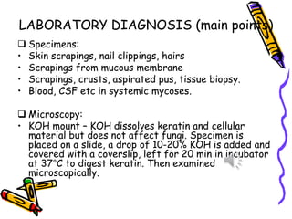
LABORATORY DIAGNOSIS (mainpoints)
❑ Specimens:
• Skin scrapings, nail clippings, hairs
• Scrapings from mucous membrane
• Scrapings, crusts, aspirated pus, tissue biopsy.
• Blood, CSF etc in systemic mycoses.
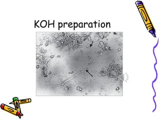
❑ Microscopy:
• KOH mount – KOH dissolves keratin and cellular
material but does not affect fungi. Specimen is
placed on a slide, a drop of 10-20% KOH is added and
covered with a coverslip, left for 20 min in incubator
at 37°C to digest keratin. Then examined
microscopically.
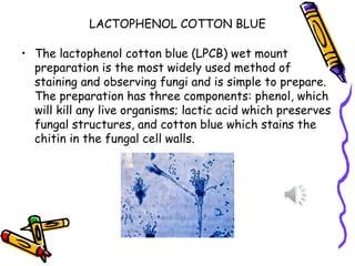
LACTOPHENOL COTTON BLUE
•The lactophenol cotton blue (LPCB) wet mount
preparation is the most widely used method of
staining and observing fungi and is simple to prepare.
The preparation has three components: phenol, which
will kill any live organisms; lactic acid which preserves
fungal structures, and cotton blue which stains the
chitin in the fungal cell walls.
60.
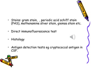
• Stains: gramstain, , periodic acid schiff stain
(PAS), methenamine sliver stain, giemsa stain etc.
• Direct immunofluorescence test
• Histology
• Antigen detection tests eg cryptococcal antigen in
CSF.
61.
❑ Culture:
• Sabouraud’sdextrose agar is commonly used for
fungal culture.
• pH =5.6 does not allow bacterial growth.
• Drugs like chloramphenicol, cyclohexamide and other
antibiotics are added to prevent bacterial or
saprophytic fungal infection.
• Cultures are incubated at two temperatures:
• One tube at 25°C (room temperature)
• One tube at 37°C (incubator).
• This helps reveal fungal dimorphism.
62.
• Cultures areincubated for at least 2-
3 weeks and in some cases upto 6
weeks.
• Cultures are examined
macroscopically for colony
morphology, and microscopically for
fungal morphology.
• Czapek-Dox agar
• Cornmeal agar