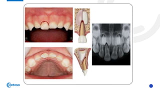
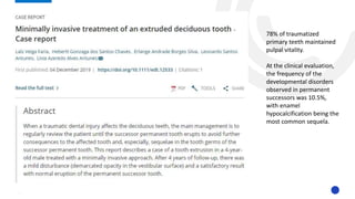
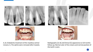
SlideShare a Scribd company logo
DENTAL
EXTRUSION
INTRODUCTION
Trauma to the oral cavity comprises of 5% injuries in people that look for treatment.
Among all facial damages, dental injuries are the most common.
Traumatic dental injuries (TDIs) of permanent teeth occur frequently in children and young adults affecting 10.5–17.3%
of the population. Crown fractures and luxations are the most commonly of all dental injuries.
World Prevalence 22.7% affecting primary teeth
Commonly occur between 2-6 years old
Proper diagnosis, treatment planning, and follow up are important for achieving a favorable outcome. Guidelines
should assist dentists and patients in decision making and in providing the best care possible, both effectively and
efficiently.
DEFINITION OF EXTRUSION:
Displacement of the tooth out of its socket in an incisal/axial direction,
tear of the periodontal ligament along with injury to the apical vessels
2
3
Extrusive luxation injuries make up nearly 2.6% of all traumatic injuries
to the dentition, with subsequent pulp canal obliteration 24% and pulpal
necrosis 64%, as the most common consequence
Humphreys classified extrusive luxation to three categories:
• smaller than or equal to 3mm : MILD
• between 3 and 5mm : MODERATE
• larger than 6mm : SEVERE
He demonstrated that for severely extruded teeth and for those with
closed apices, the risk of pulpal necrosis was greatest
4
• In extrusive luxation, the apical development stage is a key factor in
pulp healing
• In a patient with open apices, the pulp has the potential to heal, usually
following pulp canal obliteration.
• In teeth with closed apices, the probability of pulp revascularization is
low, which leads to pulp necrosis, and can often be detected within the
first eight weeks
• Study investigated pulp necrosis after extrusion in 52 teeth with closed apices
and observed that 51 teeth were necrotized. They concluded that teeth with
fully mature root development were forcefully separated from their blood
supply and were not expected to recover when reinserted in their previous
position
EXTRUSIVE
LUXATION IN
PRIMARY
DENTITION
• Partial displacement of the tooth out
of its socket
• The tooth appears elongated and can
be excessively mobile.
• Occlusal interference may be present
7
CLINICAL FINDINGS
RECOMMENDATIONS
• Periapical film –paralleling technique
• Occlusal
• Radiograph should be taken at the time of initial
presentation for :
-diagnostic purposes
-to establish a baseline
FINDINGS
• Widened periodontal ligament space apically
Please used thyroid collar if needed
8
RADIOGRAPHIC
9
Treatment decisions are based on:
• Degree of displacement
• Excessively displaced : extract
• Mobility
• If excessively mobile or extruded > 3mm : extract under LA
• Interference with the occlusion
• If not interfering, allow spontaneous reposition
• Root formation
• Ability of the child to tolerate the emergency situation
Pulp tests are not reliable in primary dentition
TREATMENT
• Treatment should be
performed by a child-
oriented team with
experience and expertise in
the management of
pediatric dental injuries.
• Extractions have the
potential to cause long-term
dental anxiety
10
PARENT AND PATIENT
EDUCATION
• Extra care when eating not to further
traumatize the injured tooth while
encouraging a return to normal function
as soon as possible
• Soft diet
• Avoid using the tooth in mastication
• To encourage gingival healing and
prevent plaque accumulation, parents
should:
• Clean the affected area with a soft brush or
• Oral toilet using cotton swab combined
with an alcohol-free 0.1%-0.2%
chlorhexidine gluconate mouth rinse twice
a day for 1 wk
11
12
• Clinical examination after
• 1 week
• 6-8 week
• 1 year
• Continue clinical follow up each year until eruption of the permanent
teeth if unfavourable outcome is likely
• Radiographic on follow up: only indicated if clinical findings are
suggestive of pathosis (eg, an unfavourable outcome)
• Parents should be informed to watch for any unfavorable outcomes and
the need to return to the clinic as soon as possible.
• If unfavourable outcomes are identified, treatment is often required
FOLLOW UP
The follow-up treatment,
which frequently requires the
expertise of a child-oriented
team, is outside the scope of
these guidelines
OUTCOMES
FAVORABLE OUTCOME UNFAVORABLE OUTCOME
• Asymptomatic
• Pulp healing with:
- Normal color of the crown or transient
red/ gray or yellow discoloration
- Pulp canal obliteration
- No signs of pulp necrosis and infection
• Continued root development in
immature teeth
• Realignment of the extruded tooth
• No interference with the occlusion
• No disturbance to the development
and/or eruption of the permanent
successor
• Symptomatic
• Signs of pulp necrosis and infection—
such as:
- Sinus tract, gingival swelling, abscess, or
increased mobility
- Persistent dark gray discoloration plus one
or more signs of root canal infection
• Radiographic signs of pulp necrosis and
infection –PA lesion
• No further root development of
immature teeth
• No improvement in the position of the
extruded tooth
• Negative impact on the development
and/or eruption of the permanent
successor 13
EXTRUSIVE
LUXATION IN
PERMANENT
DENTITION
• Partial displacement of the tooth out
of its socket
• The tooth appears elongated
• Has increased mobility
• Occlusal interference may be present
• Likely to have no response to pulp
sensibility test
16
CLINICAL FINDINGS
RECOMMENDATIONS
• Periapical film – 1 x paralleling technique, 2 x vertical/
horizontal angulations
• Occlusal
• Radiograph should be taken at the time of initial
presentation for:
• diagnostic purposes
• to establish a baseline
FINDINGS
• Increased periodontal ligament space apically and laterally
• Tooth not seated in its socket
• Appear elongated incisally
17
RADIOGRAPHIC
18
• Reposition the tooth by gently pushing It back into the tooth socket under local
anaesthesia
• Stabilize the tooth for 2 week using a passive and flexible splint. If alveolar bone
fracture, splint for an additional 4- 8 weeks
• Used up to 0.4mm diameter wire to obtain physiological stabilization
• Maintain repositioned tooth in correct position, favour initial healing, provide comfort and function
• Keep composite and bonding agent away from gingiva to avoid plaque retention and secondary
infection
• Monitor the pulp condition with pulp sensibility tests
• cold test and EPT test
• not reliable in young permanent teeth due to underdeveloped nerve fibres
• not conclusive for pulp necrosis in traumatized teeth because temporary loss of sensibility is
common during post traumatic pulp healing
• If the pulp becomes necrotic and infected, endodontic treatment appropriate to the
tooth's stage of root development is indicated to eliminate any infection, to facilitate
healing and retention of the tooth
TREATMENT
19
• Teeth with incomplete root formation:
• May survive and heal, therefor RCT should be avoided unless there is clinical and radiographical evidence of pulp necrosis or PA
lesion on follow up.
• Pulp exposed – conservative pulp therapy such as pulp capping, partial pulpotomy, cervical pulpotomy. (aim to maintain pulp
and allow continue root dev)
• If the pulp becomes necrotic and there are signs of inflammatory (infection-related) external resorption, root canal treatment
should be started as soon as possible. As such infection is very rapid in children.
• Endodontic procedures suitable for immature teeth should be used (apexification or revascularization)
• Teeth with complete root formation:
• Early endo tx advisable if severely extruded
• Root canal treatment should be started, using intracanal medicaments:
• Corticosteroid/antibiotic paste can be used as anti inflammatory and anti resorptive to prevent external resorption. Left in situ for 6 weeks
• calcium hydroxide is recommended intracanal medicaments and placed at least 2 weeks up to 1 month followed by root canal filling
• teeth with external infection related root resorption
• Start rct immediately
• Used calcium hydroxide as intracanal medicament and placed for 3 weeks and replaced every 3 months until radiolucencies of
resorption lesion disappear.
• Final obturation can be done once bone repair visible radiographically
ENDODONTICTREATMENT
20
• Limited evidence for the use of systemic abx in emergency mx of
luxation injuries
• Use by clinicians if TDI accompanied by soft tissue and other associated
injuries
• Patient medical status may warrant antibiotic coverage
• Used of tetanus booster may be required if injury contaminated –if
doubt refer medical
USE OF ANTIBIOTICS
21
22
23
PARENT AND PATIENT EDUCATION
• Extra care when eating not to further
traumatize the injured tooth while
encouraging a return to normal function as
soon as possible
• Soft diet
• Avoid using the tooth in mastication
• Enforced oral hygiene care
• Avoid participate in contact sports
• To encourage gingival healing and prevent
plaque accumulation, parents should:
• Clean the affected area with a soft brush or
• Oral toilet using cotton swab combined with an
alcohol-free 0.1%-0.2% chlorhexidine gluconate
mouth rinse twice a day for 1 wk
24
25
• Clinical and radiographic examinations both are necessary after:
• 2 weeks after splint removal
• 4 weeks
• 8 weeks
• 12 weeks
• 6 months
• 1 year
• Yearly for at least 5 years
• Parents and patient should be informed to watch for any unfavorable
outcomes and the need to return to the clinic as soon as possible.
• If unfavourable outcomes are identified, treatment is often required
FOLLOW UP
OUTCOMES
FAVORABLE OUTCOME UNFAVORABLE OUTCOME
• Asymptomatic
• Clinical and radiographic signs of
normal or healed periodontium.
• Positive response to pulp sensibility
testing; however, a false negative
response is possible for several months.
• Endodontic treatment should not be
started solely on the basis of no
response to pulp sensibility testing
• No marginal bone loss
• Continued root development in
immature teeth
• Symptomatic
• Pulp necrosis and infection such as:
- Sinus tract, gingival swelling, abscess, or
increased mobility, persistent dark gray
discoloration
• Apical periodontitis
• Breakdown of marginal bone
• External inflammatory (infection-related)
resorption – if this type of resorption
develops, root canal treatment should be
initiated immediately, with the use of calcium
hydroxide as an intracanal medicament.
• Alternatively, corticosteroid/ antibiotic
medicament can be used initially, which is
then followed by calcium hydroxide for at
least 2-3 weeks and change after 3months
26
CASE REPORT
78% of traumatized
primary teeth maintained
pulpal vitality.
At the clinical evaluation,
the frequency of the
developmental disorders
observed in permanent
successors was 10.5%,
with enamel
hypocalcification being the
most common sequela.
29
30
History:
• 9 year old female referred to the trauma center of Shiraz school of dentistry, two days after
an injury to her face and jaws, due to a bicycle accident. No previous trauma was reported.
C/o :
• spontaneous pain over maxilla and
mandibular incisors area
Examination
• Contusion of chin, lips, and nose
• Torn and bleeding gums were observed
• 11, 21- avulsed
• 31, 41, 42- severely extruded – tender to percussion, non-responsive to cold and electric
pulp testing
• Alveolar bone fracture on lower incisors area
31
Radiograph:
• the roots of 11, 21, 31,41, 42 were fully formed, and the apices were
completely closed.
• The periodontal ligament space had widened by more than 5mm
32
Management:
1. LA given, the avulsed 11, 21 were replanted in their appropriate place and splinted using
a flexible 0.5 mm wire
2. 31,41, 42 were first positioned in their correct place, then splinted using a flexible 0.5
mm wire
3. Radiography was taken to ensure correct positioning of the teeth
4. Occlusal adjustment was made to prevent occlusal traumatic interference.
5. Hygiene instructions were provided.
6. RX: Systemic penicillin for seven days and daily mouthwash with 0.12% chlorhexidine
digluconate was prescribed.
7. Endodontic treatment of avulsed 11,21 was started seven days after replantation
1. canals were filled with calcium hydroxide.
2. Final filling of the canals took place 30 days later. S
3. ince it was not possible to obtain a good tugback with gutta-percha, the root canals were sealed
using MTA.
4. The rest of the canals were backfilled using gutta-percha and AH26 sealer
5. Teeth were restored with composite resin
8. the splints were removed after six weeks
33
A, B, Clinical appearance of wire-composite splinting of maxillary and mandibular incisors;
C, D, Radiography was taken to ensure correct positioning of the teeth,
The splint was extended to the primary canines.
34
Follow up:
1. Initially 31,42, 42 did not respond to the sensitivity tests; however, since
there were no signs indicating necrosis, the researchers did not find
endodontic intervention necessary.
2. In clinical and radiographic follow-ups, three, six, nine and twelve months
following the procedure, 31,41,42 showed no signs of necrosis and were
not sensitive to percussion and palpation tests. There were no mobility,
probing defects or discoloration.
3. Radiographically, there was no apical pathosis, root resorption, ankylosis or
marginal bone loss. The teeth still did not show any sign of necrosis after
the 18-month follow-up
4. 31, 41 responded to the cold and electric tests after 12 months follow up
5. 42 showed no response to any of the tests. Due to lack of signs indicating
necrosis, an endodontic intervention was not performed.
6. The patient is still undergoing follow-up.
35
Radiographic (A, B) and Clinical (C) appearance at 18 months
follow-up; Normal color of the crowns and normal position of
the teeth is seen.
A, B, Endodontic treatment of the maxillary central
incisors; C, The splints were removed after 6 weeks.
36
• It is possible that young patients (aged 7 to 15) may have open or partially open apices, even though
the apices appear closed, radiographically
• it may take as long as nine months for normal blood flow to return to the coronal pulp of a
traumatized fully formed tooth. As circulation is restored, the responsiveness to pulp tests might
return
• The 31, 41 responded to the cold and electric tests after the 12-month follow-up
• Some authors suggested endodontic treatment for extruded teeth with complete root formation,
due to high incidence of pulp necrosis in traumatic teeth with complete root formation.
• In contrast, immature teeth should be managed by pulpal monitoring and radiographs, and for cases
where pulpal necrosis occurs, endodontic treatment should be performed.
• Other authors stated that all traumatically extruded teeth with open or closed apices should be
monitored regularly, and in cases where signs of pulp necrosis appear, endodontic treatment should
be initiated.
• In this patient, the sensitivity tests did not provide reliable responses in the recall visits; yet
endodontic intervention was not performed as the teeth showed no signs of necrosis.
• There were also no signs of crown discoloration, periapical lesions or gingival recession.
DISCUSSIONS
37
• Results of this case report showed that in young patients, with closed
apices, the dental pulps might be alive following trauma.
• In such cases, short-interval clinical and radiographic follow-ups are
necessary
• Endodontic treatment must be avoided until the appearance of signs
indicating necrosis
CONCLUSION
38
39
13 teeth with extrusive luxation but no other injury to
dental hard tissue
Result:
1 healed completely
9 showed pulp canal obliterations
3 develop pulp necrosis
No tooth with PCO developed pulp necrosis
Treatment used:
Manual repositioning
Orthodontic repositioning
Stabilization splinting
40
• There are three ways of treating extrusive luxation injuries, namely through manual, orthodontic or surgical repositioning. In
2002, Andreasen et al. argued that the choice of treatment should also depend on its timing in relation to the injury.
• Accordingly, they distinguished between acute (within the first 3 h), subacute (within 24 h) and delayed (after more than 24 h)
treatment.
• Extrusive luxation is normally treated under local anesthesia. After radiographic assessment, the luxated tooth is repositioned
manually and stabilized using a flexible splint. Three teeth were treated using manual repositioning and all these teeth
developed PCO. This approach was used for moderate–mild extrusions with a maximum dislocation of 3 mm.
• When manual repositioning is not possible, surgical and orthodontic repositioning are alternative approaches. This is a slow,
gradual process that can help to safeguard dental pulp vitality, prevent ankylosis and promote periodontal healing, especially
in healthy young subjects
• Surgical repositioning, or intentional replantation: involves the extraction of the extruded tooth followed by irrigation and
gentle cleaning of the socket. The tooth is then reinserted into the socket as quickly as possible. Appropriate endodontic
treatment needs to be performed within 15 days of the dental trauma to prevent the onset of IRR
• orthodontic repositioning technique : was performed using NiTi orthodontic arch wires (0.14/0.16) inserted in preadjusted
edgewise orthodontic brackets. The wire, generating light forces of up to 40gr, produced a gentle and gradual movement of
the extruded tooth in the socket
• In the present study, the treatment was chosen on the basis of the time that had elapsed since the trauma, the severity of the
extrusion and the need to quickly eliminate the dental interference (i.e., occlusal trauma or OT) in order to restore a correct
occlusal relationship.
• Dental interference, if maintained, causes serious hypofunction.
• Accordingly, orthodontic repositioning, which very often consists of a vestibular intrusion movement to reposition palatally
extruded teeth, was chosen for seven luxated teeth.
THANKYOU!
REFERENCES
42
International Association of Dental Traumatology guidelines for
the management of traumatic dental injuries: 1. Fractures and
Luxations (Cecilia Bourguignon et al) may 2020
International Association of Dental Traumatology guidelines for
the management of traumatic dental injuries: 3. Injuries in the
primary dentition (Peter F. Day) may 2020
Pulp Revascularization Following Severe Extrusive Luxation Injury in
Mature Permanent Mandibular Incisors: A Case Report
(Fariborz Moazzami1 and Elham Karami) August 2018

More Related Content

What's hot

Endodontic Root Perforation: Causes, Identification, and Management Presentation
Endodontic Root Perforation: Causes, Identification, and Management PresentationEndodontic Root Perforation: Causes, Identification, and Management Presentation
Endodontic Root Perforation: Causes, Identification, and Management Presentation
Iraqi Dental Academy
 
Hot tooth
Hot toothHot tooth
Surgical removal of Impacted teeth
Surgical removal of Impacted teethSurgical removal of Impacted teeth
Surgical removal of Impacted teeth
Saleh Bakry
 
Dental veneer @
Dental veneer  @Dental veneer  @
Dental veneer @
sheenu vk
 
Periodontal pocket
Periodontal pocketPeriodontal pocket
Periodontal pocket
Enas Elgendy
 
Cavity preparation
Cavity preparationCavity preparation
Cavity preparation
Saeed Bajafar
 
atraumatic restorative treatment
atraumatic restorative treatmentatraumatic restorative treatment
atraumatic restorative treatment
DrAmrita Rastogi
 
Ferrule effect
Ferrule effectFerrule effect
Ferrule effect
Dr. Rutuja Gawarle
 
Wasting diseases of teeth final
Wasting diseases of teeth finalWasting diseases of teeth final
Wasting diseases of teeth final
Prachee Hendre
 
Vital Pulp Therapy
Vital Pulp TherapyVital Pulp Therapy
Vital Pulp Therapy
IAU Dent
 
Cracked tooth syndrome
Cracked tooth syndromeCracked tooth syndrome
Cracked tooth syndrome
fattahaa
 
Endodontics periodontal lesions
Endodontics periodontal lesionsEndodontics periodontal lesions
Endodontics periodontal lesions
Arshe Gs
 
Rationale of endodontics
Rationale of endodonticsRationale of endodontics
Rationale of endodontics
alka shukla
 
The Smear layer in endodontics
The Smear layer in endodonticsThe Smear layer in endodontics
The Smear layer in endodontics
Dr. Arpit Viradiya
 
Vital pulp therapy
Vital pulp therapyVital pulp therapy
Vital pulp therapy
aravindhanarumugam1
 
Standardisation of endodontic instruments
Standardisation of endodontic instrumentsStandardisation of endodontic instruments
Standardisation of endodontic instruments
Krishna Naikwade
 
Laminates Veneers in Dentistry
Laminates Veneers in DentistryLaminates Veneers in Dentistry
Laminates Veneers in Dentistry
Naveed AnJum
 
Management of deep carious
Management of  deep cariousManagement of  deep carious
Management of deep carious
Kainaat Kaur
 
Endodontic flareup
Endodontic flareupEndodontic flareup
Endodontic flareup
Praveena Veena
 
Dental mobility
Dental mobilityDental mobility
Dental mobility
Sannah Jahangir
 

What's hot (20)

Endodontic Root Perforation: Causes, Identification, and Management Presentation
Endodontic Root Perforation: Causes, Identification, and Management PresentationEndodontic Root Perforation: Causes, Identification, and Management Presentation
Endodontic Root Perforation: Causes, Identification, and Management Presentation
 
Hot tooth
Hot toothHot tooth
Hot tooth
 
Surgical removal of Impacted teeth
Surgical removal of Impacted teethSurgical removal of Impacted teeth
Surgical removal of Impacted teeth
 
Dental veneer @
Dental veneer  @Dental veneer  @
Dental veneer @
 
Periodontal pocket
Periodontal pocketPeriodontal pocket
Periodontal pocket
 
Cavity preparation
Cavity preparationCavity preparation
Cavity preparation
 
atraumatic restorative treatment
atraumatic restorative treatmentatraumatic restorative treatment
atraumatic restorative treatment
 
Ferrule effect
Ferrule effectFerrule effect
Ferrule effect
 
Wasting diseases of teeth final
Wasting diseases of teeth finalWasting diseases of teeth final
Wasting diseases of teeth final
 
Vital Pulp Therapy
Vital Pulp TherapyVital Pulp Therapy
Vital Pulp Therapy
 
Cracked tooth syndrome
Cracked tooth syndromeCracked tooth syndrome
Cracked tooth syndrome
 
Endodontics periodontal lesions
Endodontics periodontal lesionsEndodontics periodontal lesions
Endodontics periodontal lesions
 
Rationale of endodontics
Rationale of endodonticsRationale of endodontics
Rationale of endodontics
 
The Smear layer in endodontics
The Smear layer in endodonticsThe Smear layer in endodontics
The Smear layer in endodontics
 
Vital pulp therapy
Vital pulp therapyVital pulp therapy
Vital pulp therapy
 
Standardisation of endodontic instruments
Standardisation of endodontic instrumentsStandardisation of endodontic instruments
Standardisation of endodontic instruments
 
Laminates Veneers in Dentistry
Laminates Veneers in DentistryLaminates Veneers in Dentistry
Laminates Veneers in Dentistry
 
Management of deep carious
Management of  deep cariousManagement of  deep carious
Management of deep carious
 
Endodontic flareup
Endodontic flareupEndodontic flareup
Endodontic flareup
 
Dental mobility
Dental mobilityDental mobility
Dental mobility
 

Similar to Dental Extrusion.pptx

GI System Lecture 2
GI System Lecture 2GI System Lecture 2
GI System Lecture 2
Jofred Martinez
 
Case selection In endodontic cases
Case selection In endodontic casesCase selection In endodontic cases
Case selection In endodontic cases
Partha Sarathi Adhya
 
Exodontia
ExodontiaExodontia
Guidelines for the management of avulsion
Guidelines for the management of avulsionGuidelines for the management of avulsion
Guidelines for the management of avulsion
Rahaf Sn
 
Abused tissue management 2
Abused tissue management 2Abused tissue management 2
Abused tissue management 2
DR PAAVANA
 
Diagnosis and treatment planning in rpd
Diagnosis and treatment planning in rpdDiagnosis and treatment planning in rpd
Diagnosis and treatment planning in rpd
v c
 
Complication of extraction
Complication of extractionComplication of extraction
Complication of extraction
Pushp Shah
 
Endodontic Treatment For Children by professor hasham khan
Endodontic Treatment For Children by professor hasham khanEndodontic Treatment For Children by professor hasham khan
Endodontic Treatment For Children by professor hasham khan
Jamil Kifayatullah
 
Primary teeth trauma managment
Primary teeth trauma managmentPrimary teeth trauma managment
Primary teeth trauma managment
Rahaf Sn
 
Management of displaced tooth
Management of displaced toothManagement of displaced tooth
Management of displaced tooth
Krishnameghana Reddy
 
Traumatic injuries
Traumatic injuriesTraumatic injuries
Traumatic injuries
Dr. Prathamesh Fulsundar
 
Endodontic Emergencies Summary for Students
Endodontic Emergencies Summary for StudentsEndodontic Emergencies Summary for Students
Endodontic Emergencies Summary for Students
Iraqi Dental Academy
 
ABNORMALITIES OF THE GUMS.pptx
ABNORMALITIES OF THE GUMS.pptxABNORMALITIES OF THE GUMS.pptx
ABNORMALITIES OF THE GUMS.pptx
Ramya569989
 
Pedodontics I lecture 10
Pedodontics I lecture 10Pedodontics I lecture 10
Pedodontics I lecture 10
Lama K Banna
 
Mouth preparation for Fixed Prosthodontic treatment.pdf
Mouth preparation for Fixed Prosthodontic treatment.pdfMouth preparation for Fixed Prosthodontic treatment.pdf
Mouth preparation for Fixed Prosthodontic treatment.pdf
Radwa Ibrahim El-tahawi
 
Pulp therapy
Pulp therapyPulp therapy
Pulp therapy
dentpress
 
caseselectionfinal-170514163212.pptx
caseselectionfinal-170514163212.pptxcaseselectionfinal-170514163212.pptx
caseselectionfinal-170514163212.pptx
DentalYoutube
 
Management of displaced tooth
Management of displaced toothManagement of displaced tooth
Management of displaced tooth
Imran Shaikh
 
complications of tooth extraction
complications of tooth extraction complications of tooth extraction
complications of tooth extraction
Dr. Haydar Muneer Salih
 
MANAGEMENT OF AVULSED TEETH-converted.pptx
MANAGEMENT OF AVULSED TEETH-converted.pptxMANAGEMENT OF AVULSED TEETH-converted.pptx
MANAGEMENT OF AVULSED TEETH-converted.pptx
WanNurfazliyana2
 

Similar to Dental Extrusion.pptx (20)

GI System Lecture 2
GI System Lecture 2GI System Lecture 2
GI System Lecture 2
 
Case selection In endodontic cases
Case selection In endodontic casesCase selection In endodontic cases
Case selection In endodontic cases
 
Exodontia
ExodontiaExodontia
Exodontia
 
Guidelines for the management of avulsion
Guidelines for the management of avulsionGuidelines for the management of avulsion
Guidelines for the management of avulsion
 
Abused tissue management 2
Abused tissue management 2Abused tissue management 2
Abused tissue management 2
 
Diagnosis and treatment planning in rpd
Diagnosis and treatment planning in rpdDiagnosis and treatment planning in rpd
Diagnosis and treatment planning in rpd
 
Complication of extraction
Complication of extractionComplication of extraction
Complication of extraction
 
Endodontic Treatment For Children by professor hasham khan
Endodontic Treatment For Children by professor hasham khanEndodontic Treatment For Children by professor hasham khan
Endodontic Treatment For Children by professor hasham khan
 
Primary teeth trauma managment
Primary teeth trauma managmentPrimary teeth trauma managment
Primary teeth trauma managment
 
Management of displaced tooth
Management of displaced toothManagement of displaced tooth
Management of displaced tooth
 
Traumatic injuries
Traumatic injuriesTraumatic injuries
Traumatic injuries
 
Endodontic Emergencies Summary for Students
Endodontic Emergencies Summary for StudentsEndodontic Emergencies Summary for Students
Endodontic Emergencies Summary for Students
 
ABNORMALITIES OF THE GUMS.pptx
ABNORMALITIES OF THE GUMS.pptxABNORMALITIES OF THE GUMS.pptx
ABNORMALITIES OF THE GUMS.pptx
 
Pedodontics I lecture 10
Pedodontics I lecture 10Pedodontics I lecture 10
Pedodontics I lecture 10
 
Mouth preparation for Fixed Prosthodontic treatment.pdf
Mouth preparation for Fixed Prosthodontic treatment.pdfMouth preparation for Fixed Prosthodontic treatment.pdf
Mouth preparation for Fixed Prosthodontic treatment.pdf
 
Pulp therapy
Pulp therapyPulp therapy
Pulp therapy
 
caseselectionfinal-170514163212.pptx
caseselectionfinal-170514163212.pptxcaseselectionfinal-170514163212.pptx
caseselectionfinal-170514163212.pptx
 
Management of displaced tooth
Management of displaced toothManagement of displaced tooth
Management of displaced tooth
 
complications of tooth extraction
complications of tooth extraction complications of tooth extraction
complications of tooth extraction
 
MANAGEMENT OF AVULSED TEETH-converted.pptx
MANAGEMENT OF AVULSED TEETH-converted.pptxMANAGEMENT OF AVULSED TEETH-converted.pptx
MANAGEMENT OF AVULSED TEETH-converted.pptx
 

Recently uploaded

Innovative Minds France's Most Impactful Healthcare Leaders.pdf
Innovative Minds France's Most Impactful Healthcare Leaders.pdfInnovative Minds France's Most Impactful Healthcare Leaders.pdf
Innovative Minds France's Most Impactful Healthcare Leaders.pdf
eurohealthleaders
 
FACIAL NERVE
FACIAL NERVEFACIAL NERVE
FACIAL NERVE
aditigupta1117
 
TEST BANK For Accounting Information Systems, 3rd Edition by Vernon Richardso...
TEST BANK For Accounting Information Systems, 3rd Edition by Vernon Richardso...TEST BANK For Accounting Information Systems, 3rd Edition by Vernon Richardso...
TEST BANK For Accounting Information Systems, 3rd Edition by Vernon Richardso...
rightmanforbloodline
 
CANSA support - Caring for Cancer Patients' Caregivers
CANSA support - Caring for Cancer Patients' CaregiversCANSA support - Caring for Cancer Patients' Caregivers
CANSA support - Caring for Cancer Patients' Caregivers
CANSA The Cancer Association of South Africa
 
Professional Secrecy: Forensic Medicine Lecture
Professional Secrecy: Forensic Medicine LectureProfessional Secrecy: Forensic Medicine Lecture
Professional Secrecy: Forensic Medicine Lecture
DIVYANSHU740006
 
Comprehensive Rainy Season Advisory: Safety and Preparedness Tips.pdf
Comprehensive Rainy Season Advisory: Safety and Preparedness Tips.pdfComprehensive Rainy Season Advisory: Safety and Preparedness Tips.pdf
Comprehensive Rainy Season Advisory: Safety and Preparedness Tips.pdf
Dr Rachana Gujar
 
DECODING THE RISKS - ALCOHOL, TOBACCO & DRUGS.pdf
DECODING THE RISKS - ALCOHOL, TOBACCO & DRUGS.pdfDECODING THE RISKS - ALCOHOL, TOBACCO & DRUGS.pdf
DECODING THE RISKS - ALCOHOL, TOBACCO & DRUGS.pdf
Dr Rachana Gujar
 
Luxurious Spa In Ajman Chandrima Massage Center
Luxurious Spa In Ajman Chandrima Massage CenterLuxurious Spa In Ajman Chandrima Massage Center
Luxurious Spa In Ajman Chandrima Massage Center
Chandrima Spa Ajman
 
Gemma Wean- Nutritional solution for Artemia
Gemma Wean- Nutritional solution for ArtemiaGemma Wean- Nutritional solution for Artemia
Gemma Wean- Nutritional solution for Artemia
smuskaan0008
 
Champions of Health Spotlight On Leaders Shaping Germany's Healthcare.pdf
Champions of Health Spotlight On Leaders Shaping Germany's Healthcare.pdfChampions of Health Spotlight On Leaders Shaping Germany's Healthcare.pdf
Champions of Health Spotlight On Leaders Shaping Germany's Healthcare.pdf
eurohealthleaders
 
Vicarious movements or trick movements_AB.pdf
Vicarious movements or trick movements_AB.pdfVicarious movements or trick movements_AB.pdf
Vicarious movements or trick movements_AB.pdf
Arunima620542
 
Can Allopathy and Homeopathy Be Used Together in India.pdf
Can Allopathy and Homeopathy Be Used Together in India.pdfCan Allopathy and Homeopathy Be Used Together in India.pdf
Can Allopathy and Homeopathy Be Used Together in India.pdf
Dharma Homoeopathy
 
2024 HIPAA Compliance Training Guide to the Compliance Officers
2024 HIPAA Compliance Training Guide to the Compliance Officers2024 HIPAA Compliance Training Guide to the Compliance Officers
2024 HIPAA Compliance Training Guide to the Compliance Officers
Conference Panel
 
R3 Stem Cell Therapy: A New Hope for Women with Ovarian Failure
R3 Stem Cell Therapy: A New Hope for Women with Ovarian FailureR3 Stem Cell Therapy: A New Hope for Women with Ovarian Failure
R3 Stem Cell Therapy: A New Hope for Women with Ovarian Failure
R3 Stem Cell
 
Michigan HealthTech Market Map 2024 with Policy Makers, Academic Innovation C...
Michigan HealthTech Market Map 2024 with Policy Makers, Academic Innovation C...Michigan HealthTech Market Map 2024 with Policy Makers, Academic Innovation C...
Michigan HealthTech Market Map 2024 with Policy Makers, Academic Innovation C...
Levi Shapiro
 
Letter to MREC - application to conduct study
Letter to MREC - application to conduct studyLetter to MREC - application to conduct study
Letter to MREC - application to conduct study
Azreen Aj
 
Tips for Pet Care in winters How to take care of pets.
Tips for Pet Care in winters How to take care of pets.Tips for Pet Care in winters How to take care of pets.
Tips for Pet Care in winters How to take care of pets.
Dinesh Chauhan
 
TEST BANK FOR Health Assessment in Nursing 7th Edition by Weber Chapters 1 - ...
TEST BANK FOR Health Assessment in Nursing 7th Edition by Weber Chapters 1 - ...TEST BANK FOR Health Assessment in Nursing 7th Edition by Weber Chapters 1 - ...
TEST BANK FOR Health Assessment in Nursing 7th Edition by Weber Chapters 1 - ...
rightmanforbloodline
 
Hypertension and it's role of physiotherapy in it.
Hypertension and it's role of physiotherapy in it.Hypertension and it's role of physiotherapy in it.
Hypertension and it's role of physiotherapy in it.
Vishal kr Thakur
 
一比一原版(EUR毕业证)鹿特丹伊拉斯姆斯大学毕业证如何办理
一比一原版(EUR毕业证)鹿特丹伊拉斯姆斯大学毕业证如何办理一比一原版(EUR毕业证)鹿特丹伊拉斯姆斯大学毕业证如何办理
一比一原版(EUR毕业证)鹿特丹伊拉斯姆斯大学毕业证如何办理
gjsma0ep
 

Recently uploaded (20)

Innovative Minds France's Most Impactful Healthcare Leaders.pdf
Innovative Minds France's Most Impactful Healthcare Leaders.pdfInnovative Minds France's Most Impactful Healthcare Leaders.pdf
Innovative Minds France's Most Impactful Healthcare Leaders.pdf
 
FACIAL NERVE
FACIAL NERVEFACIAL NERVE
FACIAL NERVE
 
TEST BANK For Accounting Information Systems, 3rd Edition by Vernon Richardso...
TEST BANK For Accounting Information Systems, 3rd Edition by Vernon Richardso...TEST BANK For Accounting Information Systems, 3rd Edition by Vernon Richardso...
TEST BANK For Accounting Information Systems, 3rd Edition by Vernon Richardso...
 
CANSA support - Caring for Cancer Patients' Caregivers
CANSA support - Caring for Cancer Patients' CaregiversCANSA support - Caring for Cancer Patients' Caregivers
CANSA support - Caring for Cancer Patients' Caregivers
 
Professional Secrecy: Forensic Medicine Lecture
Professional Secrecy: Forensic Medicine LectureProfessional Secrecy: Forensic Medicine Lecture
Professional Secrecy: Forensic Medicine Lecture
 
Comprehensive Rainy Season Advisory: Safety and Preparedness Tips.pdf
Comprehensive Rainy Season Advisory: Safety and Preparedness Tips.pdfComprehensive Rainy Season Advisory: Safety and Preparedness Tips.pdf
Comprehensive Rainy Season Advisory: Safety and Preparedness Tips.pdf
 
DECODING THE RISKS - ALCOHOL, TOBACCO & DRUGS.pdf
DECODING THE RISKS - ALCOHOL, TOBACCO & DRUGS.pdfDECODING THE RISKS - ALCOHOL, TOBACCO & DRUGS.pdf
DECODING THE RISKS - ALCOHOL, TOBACCO & DRUGS.pdf
 
Luxurious Spa In Ajman Chandrima Massage Center
Luxurious Spa In Ajman Chandrima Massage CenterLuxurious Spa In Ajman Chandrima Massage Center
Luxurious Spa In Ajman Chandrima Massage Center
 
Gemma Wean- Nutritional solution for Artemia
Gemma Wean- Nutritional solution for ArtemiaGemma Wean- Nutritional solution for Artemia
Gemma Wean- Nutritional solution for Artemia
 
Champions of Health Spotlight On Leaders Shaping Germany's Healthcare.pdf
Champions of Health Spotlight On Leaders Shaping Germany's Healthcare.pdfChampions of Health Spotlight On Leaders Shaping Germany's Healthcare.pdf
Champions of Health Spotlight On Leaders Shaping Germany's Healthcare.pdf
 
Vicarious movements or trick movements_AB.pdf
Vicarious movements or trick movements_AB.pdfVicarious movements or trick movements_AB.pdf
Vicarious movements or trick movements_AB.pdf
 
Can Allopathy and Homeopathy Be Used Together in India.pdf
Can Allopathy and Homeopathy Be Used Together in India.pdfCan Allopathy and Homeopathy Be Used Together in India.pdf
Can Allopathy and Homeopathy Be Used Together in India.pdf
 
2024 HIPAA Compliance Training Guide to the Compliance Officers
2024 HIPAA Compliance Training Guide to the Compliance Officers2024 HIPAA Compliance Training Guide to the Compliance Officers
2024 HIPAA Compliance Training Guide to the Compliance Officers
 
R3 Stem Cell Therapy: A New Hope for Women with Ovarian Failure
R3 Stem Cell Therapy: A New Hope for Women with Ovarian FailureR3 Stem Cell Therapy: A New Hope for Women with Ovarian Failure
R3 Stem Cell Therapy: A New Hope for Women with Ovarian Failure
 
Michigan HealthTech Market Map 2024 with Policy Makers, Academic Innovation C...
Michigan HealthTech Market Map 2024 with Policy Makers, Academic Innovation C...Michigan HealthTech Market Map 2024 with Policy Makers, Academic Innovation C...
Michigan HealthTech Market Map 2024 with Policy Makers, Academic Innovation C...
 
Letter to MREC - application to conduct study
Letter to MREC - application to conduct studyLetter to MREC - application to conduct study
Letter to MREC - application to conduct study
 
Tips for Pet Care in winters How to take care of pets.
Tips for Pet Care in winters How to take care of pets.Tips for Pet Care in winters How to take care of pets.
Tips for Pet Care in winters How to take care of pets.
 
TEST BANK FOR Health Assessment in Nursing 7th Edition by Weber Chapters 1 - ...
TEST BANK FOR Health Assessment in Nursing 7th Edition by Weber Chapters 1 - ...TEST BANK FOR Health Assessment in Nursing 7th Edition by Weber Chapters 1 - ...
TEST BANK FOR Health Assessment in Nursing 7th Edition by Weber Chapters 1 - ...
 
Hypertension and it's role of physiotherapy in it.
Hypertension and it's role of physiotherapy in it.Hypertension and it's role of physiotherapy in it.
Hypertension and it's role of physiotherapy in it.
 
一比一原版(EUR毕业证)鹿特丹伊拉斯姆斯大学毕业证如何办理
一比一原版(EUR毕业证)鹿特丹伊拉斯姆斯大学毕业证如何办理一比一原版(EUR毕业证)鹿特丹伊拉斯姆斯大学毕业证如何办理
一比一原版(EUR毕业证)鹿特丹伊拉斯姆斯大学毕业证如何办理
 

Dental Extrusion.pptx

  • 2. INTRODUCTION Trauma to the oral cavity comprises of 5% injuries in people that look for treatment. Among all facial damages, dental injuries are the most common. Traumatic dental injuries (TDIs) of permanent teeth occur frequently in children and young adults affecting 10.5–17.3% of the population. Crown fractures and luxations are the most commonly of all dental injuries. World Prevalence 22.7% affecting primary teeth Commonly occur between 2-6 years old Proper diagnosis, treatment planning, and follow up are important for achieving a favorable outcome. Guidelines should assist dentists and patients in decision making and in providing the best care possible, both effectively and efficiently. DEFINITION OF EXTRUSION: Displacement of the tooth out of its socket in an incisal/axial direction, tear of the periodontal ligament along with injury to the apical vessels 2
  • 3. 3 Extrusive luxation injuries make up nearly 2.6% of all traumatic injuries to the dentition, with subsequent pulp canal obliteration 24% and pulpal necrosis 64%, as the most common consequence Humphreys classified extrusive luxation to three categories: • smaller than or equal to 3mm : MILD • between 3 and 5mm : MODERATE • larger than 6mm : SEVERE He demonstrated that for severely extruded teeth and for those with closed apices, the risk of pulpal necrosis was greatest
  • 4. 4 • In extrusive luxation, the apical development stage is a key factor in pulp healing • In a patient with open apices, the pulp has the potential to heal, usually following pulp canal obliteration. • In teeth with closed apices, the probability of pulp revascularization is low, which leads to pulp necrosis, and can often be detected within the first eight weeks • Study investigated pulp necrosis after extrusion in 52 teeth with closed apices and observed that 51 teeth were necrotized. They concluded that teeth with fully mature root development were forcefully separated from their blood supply and were not expected to recover when reinserted in their previous position
  • 6.
  • 7. • Partial displacement of the tooth out of its socket • The tooth appears elongated and can be excessively mobile. • Occlusal interference may be present 7 CLINICAL FINDINGS
  • 8. RECOMMENDATIONS • Periapical film –paralleling technique • Occlusal • Radiograph should be taken at the time of initial presentation for : -diagnostic purposes -to establish a baseline FINDINGS • Widened periodontal ligament space apically Please used thyroid collar if needed 8 RADIOGRAPHIC
  • 9. 9 Treatment decisions are based on: • Degree of displacement • Excessively displaced : extract • Mobility • If excessively mobile or extruded > 3mm : extract under LA • Interference with the occlusion • If not interfering, allow spontaneous reposition • Root formation • Ability of the child to tolerate the emergency situation Pulp tests are not reliable in primary dentition TREATMENT • Treatment should be performed by a child- oriented team with experience and expertise in the management of pediatric dental injuries. • Extractions have the potential to cause long-term dental anxiety
  • 10. 10
  • 11. PARENT AND PATIENT EDUCATION • Extra care when eating not to further traumatize the injured tooth while encouraging a return to normal function as soon as possible • Soft diet • Avoid using the tooth in mastication • To encourage gingival healing and prevent plaque accumulation, parents should: • Clean the affected area with a soft brush or • Oral toilet using cotton swab combined with an alcohol-free 0.1%-0.2% chlorhexidine gluconate mouth rinse twice a day for 1 wk 11
  • 12. 12 • Clinical examination after • 1 week • 6-8 week • 1 year • Continue clinical follow up each year until eruption of the permanent teeth if unfavourable outcome is likely • Radiographic on follow up: only indicated if clinical findings are suggestive of pathosis (eg, an unfavourable outcome) • Parents should be informed to watch for any unfavorable outcomes and the need to return to the clinic as soon as possible. • If unfavourable outcomes are identified, treatment is often required FOLLOW UP The follow-up treatment, which frequently requires the expertise of a child-oriented team, is outside the scope of these guidelines
  • 13. OUTCOMES FAVORABLE OUTCOME UNFAVORABLE OUTCOME • Asymptomatic • Pulp healing with: - Normal color of the crown or transient red/ gray or yellow discoloration - Pulp canal obliteration - No signs of pulp necrosis and infection • Continued root development in immature teeth • Realignment of the extruded tooth • No interference with the occlusion • No disturbance to the development and/or eruption of the permanent successor • Symptomatic • Signs of pulp necrosis and infection— such as: - Sinus tract, gingival swelling, abscess, or increased mobility - Persistent dark gray discoloration plus one or more signs of root canal infection • Radiographic signs of pulp necrosis and infection –PA lesion • No further root development of immature teeth • No improvement in the position of the extruded tooth • Negative impact on the development and/or eruption of the permanent successor 13
  • 15.
  • 16. • Partial displacement of the tooth out of its socket • The tooth appears elongated • Has increased mobility • Occlusal interference may be present • Likely to have no response to pulp sensibility test 16 CLINICAL FINDINGS
  • 17. RECOMMENDATIONS • Periapical film – 1 x paralleling technique, 2 x vertical/ horizontal angulations • Occlusal • Radiograph should be taken at the time of initial presentation for: • diagnostic purposes • to establish a baseline FINDINGS • Increased periodontal ligament space apically and laterally • Tooth not seated in its socket • Appear elongated incisally 17 RADIOGRAPHIC
  • 18. 18 • Reposition the tooth by gently pushing It back into the tooth socket under local anaesthesia • Stabilize the tooth for 2 week using a passive and flexible splint. If alveolar bone fracture, splint for an additional 4- 8 weeks • Used up to 0.4mm diameter wire to obtain physiological stabilization • Maintain repositioned tooth in correct position, favour initial healing, provide comfort and function • Keep composite and bonding agent away from gingiva to avoid plaque retention and secondary infection • Monitor the pulp condition with pulp sensibility tests • cold test and EPT test • not reliable in young permanent teeth due to underdeveloped nerve fibres • not conclusive for pulp necrosis in traumatized teeth because temporary loss of sensibility is common during post traumatic pulp healing • If the pulp becomes necrotic and infected, endodontic treatment appropriate to the tooth's stage of root development is indicated to eliminate any infection, to facilitate healing and retention of the tooth TREATMENT
  • 19. 19 • Teeth with incomplete root formation: • May survive and heal, therefor RCT should be avoided unless there is clinical and radiographical evidence of pulp necrosis or PA lesion on follow up. • Pulp exposed – conservative pulp therapy such as pulp capping, partial pulpotomy, cervical pulpotomy. (aim to maintain pulp and allow continue root dev) • If the pulp becomes necrotic and there are signs of inflammatory (infection-related) external resorption, root canal treatment should be started as soon as possible. As such infection is very rapid in children. • Endodontic procedures suitable for immature teeth should be used (apexification or revascularization) • Teeth with complete root formation: • Early endo tx advisable if severely extruded • Root canal treatment should be started, using intracanal medicaments: • Corticosteroid/antibiotic paste can be used as anti inflammatory and anti resorptive to prevent external resorption. Left in situ for 6 weeks • calcium hydroxide is recommended intracanal medicaments and placed at least 2 weeks up to 1 month followed by root canal filling • teeth with external infection related root resorption • Start rct immediately • Used calcium hydroxide as intracanal medicament and placed for 3 weeks and replaced every 3 months until radiolucencies of resorption lesion disappear. • Final obturation can be done once bone repair visible radiographically ENDODONTICTREATMENT
  • 20. 20 • Limited evidence for the use of systemic abx in emergency mx of luxation injuries • Use by clinicians if TDI accompanied by soft tissue and other associated injuries • Patient medical status may warrant antibiotic coverage • Used of tetanus booster may be required if injury contaminated –if doubt refer medical USE OF ANTIBIOTICS
  • 21. 21
  • 22. 22
  • 23. 23
  • 24. PARENT AND PATIENT EDUCATION • Extra care when eating not to further traumatize the injured tooth while encouraging a return to normal function as soon as possible • Soft diet • Avoid using the tooth in mastication • Enforced oral hygiene care • Avoid participate in contact sports • To encourage gingival healing and prevent plaque accumulation, parents should: • Clean the affected area with a soft brush or • Oral toilet using cotton swab combined with an alcohol-free 0.1%-0.2% chlorhexidine gluconate mouth rinse twice a day for 1 wk 24
  • 25. 25 • Clinical and radiographic examinations both are necessary after: • 2 weeks after splint removal • 4 weeks • 8 weeks • 12 weeks • 6 months • 1 year • Yearly for at least 5 years • Parents and patient should be informed to watch for any unfavorable outcomes and the need to return to the clinic as soon as possible. • If unfavourable outcomes are identified, treatment is often required FOLLOW UP
  • 26. OUTCOMES FAVORABLE OUTCOME UNFAVORABLE OUTCOME • Asymptomatic • Clinical and radiographic signs of normal or healed periodontium. • Positive response to pulp sensibility testing; however, a false negative response is possible for several months. • Endodontic treatment should not be started solely on the basis of no response to pulp sensibility testing • No marginal bone loss • Continued root development in immature teeth • Symptomatic • Pulp necrosis and infection such as: - Sinus tract, gingival swelling, abscess, or increased mobility, persistent dark gray discoloration • Apical periodontitis • Breakdown of marginal bone • External inflammatory (infection-related) resorption – if this type of resorption develops, root canal treatment should be initiated immediately, with the use of calcium hydroxide as an intracanal medicament. • Alternatively, corticosteroid/ antibiotic medicament can be used initially, which is then followed by calcium hydroxide for at least 2-3 weeks and change after 3months 26
  • 28. 78% of traumatized primary teeth maintained pulpal vitality. At the clinical evaluation, the frequency of the developmental disorders observed in permanent successors was 10.5%, with enamel hypocalcification being the most common sequela.
  • 29. 29
  • 30. 30 History: • 9 year old female referred to the trauma center of Shiraz school of dentistry, two days after an injury to her face and jaws, due to a bicycle accident. No previous trauma was reported. C/o : • spontaneous pain over maxilla and mandibular incisors area Examination • Contusion of chin, lips, and nose • Torn and bleeding gums were observed • 11, 21- avulsed • 31, 41, 42- severely extruded – tender to percussion, non-responsive to cold and electric pulp testing • Alveolar bone fracture on lower incisors area
  • 31. 31 Radiograph: • the roots of 11, 21, 31,41, 42 were fully formed, and the apices were completely closed. • The periodontal ligament space had widened by more than 5mm
  • 32. 32 Management: 1. LA given, the avulsed 11, 21 were replanted in their appropriate place and splinted using a flexible 0.5 mm wire 2. 31,41, 42 were first positioned in their correct place, then splinted using a flexible 0.5 mm wire 3. Radiography was taken to ensure correct positioning of the teeth 4. Occlusal adjustment was made to prevent occlusal traumatic interference. 5. Hygiene instructions were provided. 6. RX: Systemic penicillin for seven days and daily mouthwash with 0.12% chlorhexidine digluconate was prescribed. 7. Endodontic treatment of avulsed 11,21 was started seven days after replantation 1. canals were filled with calcium hydroxide. 2. Final filling of the canals took place 30 days later. S 3. ince it was not possible to obtain a good tugback with gutta-percha, the root canals were sealed using MTA. 4. The rest of the canals were backfilled using gutta-percha and AH26 sealer 5. Teeth were restored with composite resin 8. the splints were removed after six weeks
  • 33. 33 A, B, Clinical appearance of wire-composite splinting of maxillary and mandibular incisors; C, D, Radiography was taken to ensure correct positioning of the teeth, The splint was extended to the primary canines.
  • 34. 34 Follow up: 1. Initially 31,42, 42 did not respond to the sensitivity tests; however, since there were no signs indicating necrosis, the researchers did not find endodontic intervention necessary. 2. In clinical and radiographic follow-ups, three, six, nine and twelve months following the procedure, 31,41,42 showed no signs of necrosis and were not sensitive to percussion and palpation tests. There were no mobility, probing defects or discoloration. 3. Radiographically, there was no apical pathosis, root resorption, ankylosis or marginal bone loss. The teeth still did not show any sign of necrosis after the 18-month follow-up 4. 31, 41 responded to the cold and electric tests after 12 months follow up 5. 42 showed no response to any of the tests. Due to lack of signs indicating necrosis, an endodontic intervention was not performed. 6. The patient is still undergoing follow-up.
  • 35. 35 Radiographic (A, B) and Clinical (C) appearance at 18 months follow-up; Normal color of the crowns and normal position of the teeth is seen. A, B, Endodontic treatment of the maxillary central incisors; C, The splints were removed after 6 weeks.
  • 36. 36 • It is possible that young patients (aged 7 to 15) may have open or partially open apices, even though the apices appear closed, radiographically • it may take as long as nine months for normal blood flow to return to the coronal pulp of a traumatized fully formed tooth. As circulation is restored, the responsiveness to pulp tests might return • The 31, 41 responded to the cold and electric tests after the 12-month follow-up • Some authors suggested endodontic treatment for extruded teeth with complete root formation, due to high incidence of pulp necrosis in traumatic teeth with complete root formation. • In contrast, immature teeth should be managed by pulpal monitoring and radiographs, and for cases where pulpal necrosis occurs, endodontic treatment should be performed. • Other authors stated that all traumatically extruded teeth with open or closed apices should be monitored regularly, and in cases where signs of pulp necrosis appear, endodontic treatment should be initiated. • In this patient, the sensitivity tests did not provide reliable responses in the recall visits; yet endodontic intervention was not performed as the teeth showed no signs of necrosis. • There were also no signs of crown discoloration, periapical lesions or gingival recession. DISCUSSIONS
  • 37. 37 • Results of this case report showed that in young patients, with closed apices, the dental pulps might be alive following trauma. • In such cases, short-interval clinical and radiographic follow-ups are necessary • Endodontic treatment must be avoided until the appearance of signs indicating necrosis CONCLUSION
  • 38. 38
  • 39. 39 13 teeth with extrusive luxation but no other injury to dental hard tissue Result: 1 healed completely 9 showed pulp canal obliterations 3 develop pulp necrosis No tooth with PCO developed pulp necrosis Treatment used: Manual repositioning Orthodontic repositioning Stabilization splinting
  • 40. 40 • There are three ways of treating extrusive luxation injuries, namely through manual, orthodontic or surgical repositioning. In 2002, Andreasen et al. argued that the choice of treatment should also depend on its timing in relation to the injury. • Accordingly, they distinguished between acute (within the first 3 h), subacute (within 24 h) and delayed (after more than 24 h) treatment. • Extrusive luxation is normally treated under local anesthesia. After radiographic assessment, the luxated tooth is repositioned manually and stabilized using a flexible splint. Three teeth were treated using manual repositioning and all these teeth developed PCO. This approach was used for moderate–mild extrusions with a maximum dislocation of 3 mm. • When manual repositioning is not possible, surgical and orthodontic repositioning are alternative approaches. This is a slow, gradual process that can help to safeguard dental pulp vitality, prevent ankylosis and promote periodontal healing, especially in healthy young subjects • Surgical repositioning, or intentional replantation: involves the extraction of the extruded tooth followed by irrigation and gentle cleaning of the socket. The tooth is then reinserted into the socket as quickly as possible. Appropriate endodontic treatment needs to be performed within 15 days of the dental trauma to prevent the onset of IRR • orthodontic repositioning technique : was performed using NiTi orthodontic arch wires (0.14/0.16) inserted in preadjusted edgewise orthodontic brackets. The wire, generating light forces of up to 40gr, produced a gentle and gradual movement of the extruded tooth in the socket • In the present study, the treatment was chosen on the basis of the time that had elapsed since the trauma, the severity of the extrusion and the need to quickly eliminate the dental interference (i.e., occlusal trauma or OT) in order to restore a correct occlusal relationship. • Dental interference, if maintained, causes serious hypofunction. • Accordingly, orthodontic repositioning, which very often consists of a vestibular intrusion movement to reposition palatally extruded teeth, was chosen for seven luxated teeth.
  • 42. REFERENCES 42 International Association of Dental Traumatology guidelines for the management of traumatic dental injuries: 1. Fractures and Luxations (Cecilia Bourguignon et al) may 2020 International Association of Dental Traumatology guidelines for the management of traumatic dental injuries: 3. Injuries in the primary dentition (Peter F. Day) may 2020 Pulp Revascularization Following Severe Extrusive Luxation Injury in Mature Permanent Mandibular Incisors: A Case Report (Fariborz Moazzami1 and Elham Karami) August 2018