Downloaded 156 times

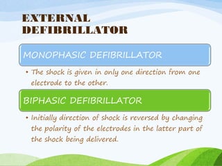


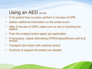

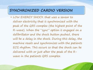
![INDICATIONS
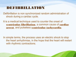
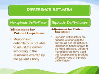
• Supraventricular tachycardia (atrioventricular nodal
reentrant tachycardia [AVNRT] and atrioventricular
reentrant tachycardia [AVRT])
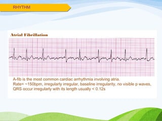
• Atrial fibrillation
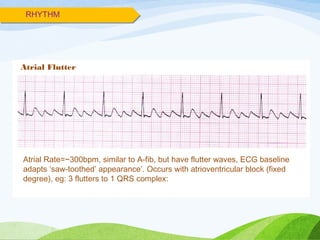
• Atrial flutter (types I and II)
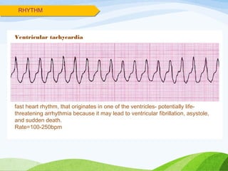
• Ventricular tachycardia with pulse.](https://image.slidesharecdn.com/defibrillation-170604161922/85/Defibrillation-94-320.jpg)
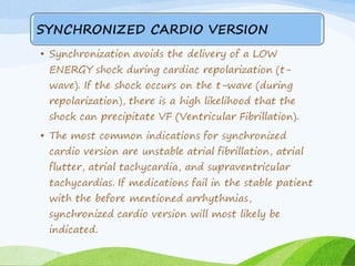
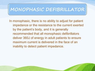
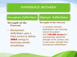
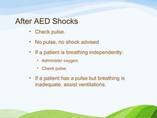
![SPECIAL POPULATION
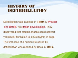
Cardioversion in patients with permanent
pacemakers/ICDs
• Cardioversion in patients with permanent
pacemaker/ICD should be performed with extra care.
Improper technique may damage the device, lead
system, or myocardial tissue, resulting in device
malfunction. The electrode paddle or patch should be at
least 12 cm from the pulse generator and
anteroposterior paddle position.[15, 16]
The lowest amount of
energy should be used during cardioversion, based on
the patient’s clinical condition. After cardioversion, the
pacemaker/ICD should be interrogated to ensure normal
function of the device.](https://image.slidesharecdn.com/defibrillation-170604161922/85/Defibrillation-113-320.jpg)


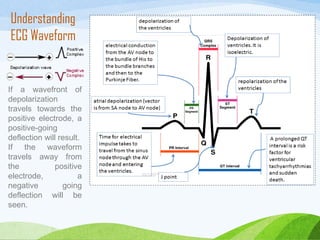
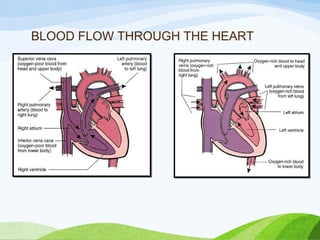
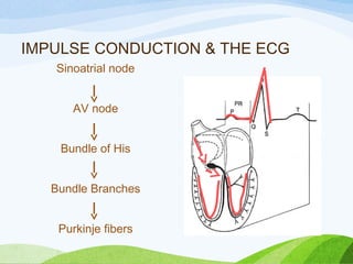
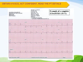
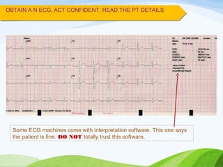
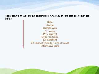
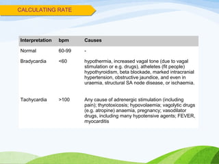
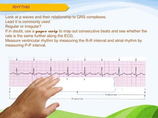
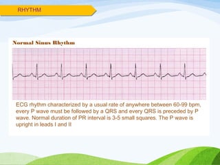
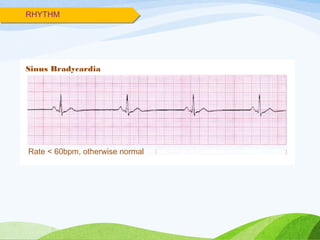
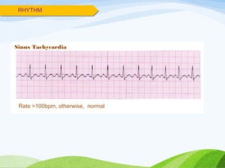
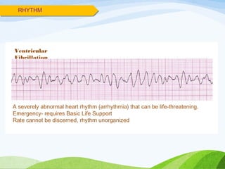
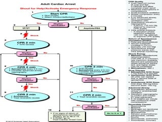
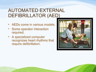
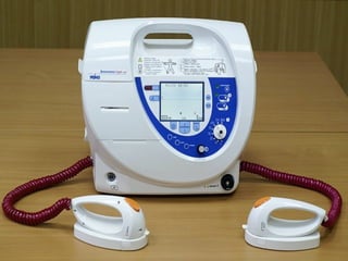
Defibrillation is a medical procedure used to treat life-threatening heart rhythm disturbances such as ventricular fibrillation and pulseless ventricular tachycardia by administering electric shocks to the heart. Developed in the late 19th century, modern variations include implantable cardioverter-defibrillators and automated external defibrillators, with biphasic defibrillators being more effective and safer than monophasic types. Proper ECG interpretation is crucial for diagnosing various cardiac conditions, using techniques to assess heart rate, rhythm, and potential arrhythmias.


