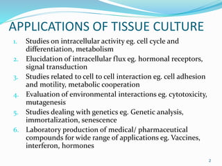
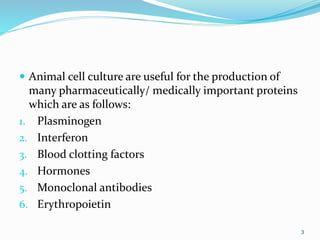
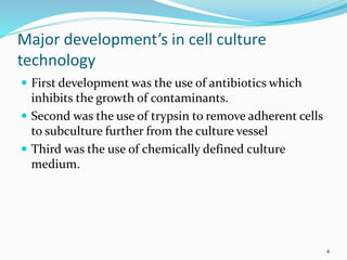
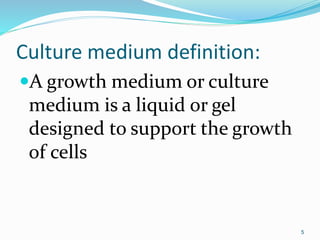
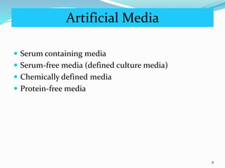
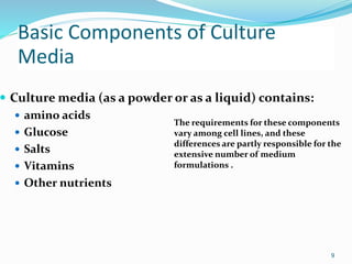
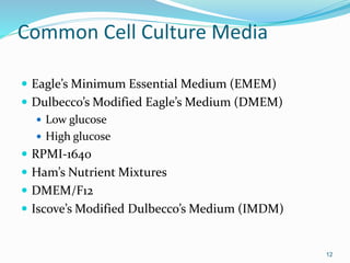
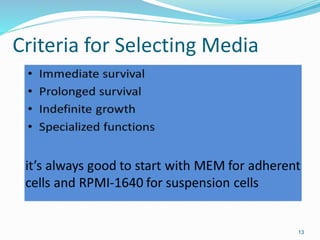
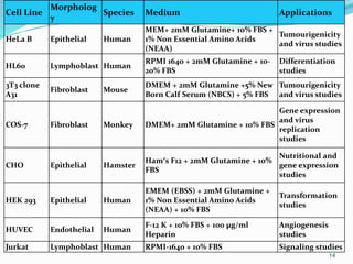
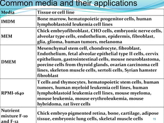
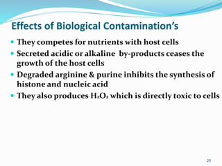
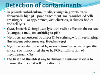
This document discusses various applications of tissue culture, including intracellular studies, elucidation of signaling pathways, studies of cell interactions, and evaluation of environmental interactions. It notes that animal cell culture is useful for producing pharmaceutical proteins like interferon, blood clotting factors, and monoclonal antibodies. Major developments in cell culture technology included the use of antibiotics, trypsin for subculturing, and chemically defined media. Common cell culture media include MEM, DMEM, RPMI-1640, and F-12, and selection depends on factors like the cell line, species, and intended application.
Role of serum and supplements in culture medium k.skailash saini
ROLE OF SERUM AND SUPPLEMENTS IN CULTURE MEDIA
Serum is a complex mix of albumins, growth factors and growth inhibitors.
Serum is one of the most important components of cell culture media and serves as a source for amino acids, proteins, vitamins (particularly fat-soluble vitamins such as A, D, E, and K), carbohydrates, lipids, hormones, growth factors, minerals, and trace elements.
Serum from fetal and calf bovine sources are commonly used to support the growth of cells in culture.
Fetal serum is a rich source of growth factors and is appropriate for cell cloning and for the growth of fastidious cells.
Calf serum is used in contact-inhibition studies because of its lower growth-promoting properties.
Normal growth media often contain 2-10% of serum.
Supplementation of media with serum serves the following functions :
Serum provides the basic nutrients (both in the solution as well as bound to the proteins) for cells.
Serum provides several growth factors and hormones involved in growth promotion and specialized cell function.
It provides several binding proteins like albumin, transferrin, which can carry other molecules into the cell. For example: albumin carries lipids, vitamins, hormones, etc. into cells.
It also supplies proteins, like fibronectin, which promote the attachment of cells to the substrate. It also provides spreading factors that help the cells to spread out before they begin to divide.
It provides protease inhibitors which protect cells from proteolysis.
It also provides minerals, like Na+, K+, Zn2+, Fe2+, etc.
It increases the viscosity of the medium and thus, protects cells from mechanical damages during agitation of suspension cultures.
It also acts a buffer.
Due to the presence of both growth factors and inhibitors, the role of serum in cell culture is very complex.
Unfortunately, in addition to serving various functions, the use of serum in tissue culture applications has several drawbacks .
This presentation presents an overview of definition, equipment, various cell culturing methods,
characterization, and applications of animal cell culture. This presentation also contains MCQs to acquaint reader about types of question asked in various competitive examinations.
This presentation gives you the detailed description of various cells & organs of immune systems that participates (particularly, in combination), make communication between themselves to regulate the whole immune system very precisely.
Animal cell culture, application by kk sahuKAUSHAL SAHU
INTRODUCTION
HISTORY
CELL CULTURE IN TWO DIMENSION
CELL CULTURE IN THREE DIMENSION
APPLICATION:-
VACCINES
PRODUCTION OF HIGH VALUE THERAPEUTICS
TRANSGENIC ANIMAL
GENE THERAPY
TISSUE ENGINEERING
CONCLUSION
REFRENCES
BIOMIMESYS® Liver represents a new generation of mimetic HydroscaffoldS™ for 3D culture of hepatocyte-like cells. Available in a ready-to-use format, it enables the culture of hepatocytes and hepatocyte-like cells under physiological conditions that are representative of the microenvironment found in the liver. The highly porous nature of the Hydroscaffold™ allows the rapid uptake of nutrients, oxygen, etc. into the cells to create a reproducible study model for all downstream analyses used with 3D hepatocyte-like cell culture (metabolism, toxicity…).
IOMIMESYS® Oncology is a new generation of mimetic Hydroscaffolds™ for 3D cell culture. Available in a ready-to-use format, it enables the culture of cells under physiological conditions that are representative of the microenvironment found in whole tissues. The highly porous structure of BIOMIMESYS® allows cells to diffuse into the 3D matrix, where they can fix and begin to develop. The structure and conformation of cells cultured in BIOMIMESYS®, such as cancer cells that spontaneously form spheroids, strongly resemble the structures formed in vivo. Also, the functionality of the BIOMIMESYS® matrix is proved by the expression of genes and proteins at levels that are again more similar to those found in vivo. Cells cultured in BIOMIMESYS® give results that are more physiological than those grown in 2D culture and are closer to those obtained in vivo.
A growth medium or culture medium is a liquid or gel designated to support the growth of microorganisms,cells,or small plants.
Culture media generally comprise an appropriate sourcde of energy and compounds which regulate the cell cycle.
A typical culture media is composed of a complement of amino acids,vitamins,inorganic salts,glucose, and serum as a source of growth factors, hormones, and attachment factors.
In addition to nutrients, the medium also helps maintain pH and osmolarity.
Imporatant amino acids, trace elements, growth factor,hormone,transport protein and adhesion factor are added.
Adhesion factor added are main components of intercellular substance such as fibronectin,collagen and laminin.
Primary purpose of introducing SFM is to promote the specific growth of a particular type of cell.
Exapmple of one serum free media: DCMM-1 SFM. High protein serum free medium, designed for hybridoma cell growth and monoclonal antibody production.
Equipments used , types of culture and media, subculturing, secondary culture, finite & continuous cell lines, cryopreservation and applications of cell culture
The chemical compounds produced by plants are collectively referred to as phytochemicals. Biotechnologists have special interest in plant tissue culture for the large scale production of commercially important compounds. These include pharmaceuticals, flavours, fragrances, cosmetics, food additives, feed stocks and antimicrobials.
Most of these products are secondary metabolites— chemical compounds that do not participate in metabolism of plants. Thus, secondary metabolites are not directly needed by plants as they do not perform any physiological function (as is the case with primary metabolites such as amino acids, nucleic acids etc.). Although the native plants are capable of producing the secondary metabolites of commercial interest, tissue culture systems are preferred.
A cell line is a product of immortal cells that are used for biological research.
Cells used for cell lines are immortal, that happens if a cell is cancerous.
The cells can perpetuate division indefinitely which is unlike regular cells which can only divide approximately 50 times.
Human cell lines
MCF-7 breast cancer
HL 60 Leukemia
HEK-293 Human embryonic kidney
HeLa Henrietta lacks
Primate cell lines
Vero African green monkey kidney epithelial cells
Cos-7 African green monkey kidney cells
And others such as CHO from hamster, sf9 & sf21 from insect cells.
In this modern era, many companies are focusing on the research of monoclonal antibodies to treat cancer and other autoimmune diseases. Hence, cell culture plays an important role to produce therapeutic proteins. To make process efficient we should know alpha and gamma about cell line. The growth of cells in the medium is the most important area or checkpoint. The type of cell line is also discussed here.
Cell Applications, Inc. is a leading authority and global provider of primary human and animal cell types, complimented by optimized media and cell biology products to serve your research & development goals. Primary Cells, derived directly from tissue, maintain physiological relevance and find increasing use in life science research and pharmaceutical drug discovery. In business since 1994, we have perfected the isolation, purification, subculture and growth of human and animal primary cells. Available options include proliferating or cryopreserved cells, 3D models, 96 well plated versions, disease models, or cells pre-screened for metabolic regulation markers and signal pathways.
The term ‘Tissue culture ’ refers to the culture of whole organism, tissue fragments as well as dispersed cell on a suitable nutrient medium. Tissue culture is divided into the two broad groups, i) cultures that facilitates cell to cell interactions and signaling among cell and allow their study and ii) those in which cell to cell interactions and signaling are missing. The first group consist of three distinct types of culture system, viz., (i) Organ culture( in this, whole embryonic organs or small tissue fragments are cultured in vitro in such a manner that they retain their tissue architecture, i.e., the characteristic distribution of various cell types in the given organ) (ii) Histotypic cultures ( in this, individual cell lineages are first isolated from organ, purified and multiplied; they are grown separately to high density in three-dimensional matrix to study interactions and signaling among homologous cells. (iii) Organotypic cultures ( in this, cells of different lineages are mixed together in specific ratios and spatial relationships in order to recreate a component of concerned organ). The second group consists of cell culture either as monolayer (cells are obtained either by enzymatic or mechanical dispersal of tissues into individual cells or by spontaneous migration of cells from an explants or as suspension culture.
The first step in sample preparation is isolating proteins from their source. Usually, proteins are isolated from cells or tissues via lysis. Lysis breaks down the cell membrane to separate proteins from the non-soluble parts of the cell. A number of lysis buffers can be used to prepare samples for western blotting.
micro teaching on communication m.sc nursing.pdfAnurag Sharma
Microteaching is a unique model of practice teaching. It is a viable instrument for the. desired change in the teaching behavior or the behavior potential which, in specified types of real. classroom situations, tends to facilitate the achievement of specified types of objectives.
Couples presenting to the infertility clinic- Do they really have infertility...Sujoy Dasgupta
Dr Sujoy Dasgupta presented the study on "Couples presenting to the infertility clinic- Do they really have infertility? – The unexplored stories of non-consummation" in the 13th Congress of the Asia Pacific Initiative on Reproduction (ASPIRE 2024) at Manila on 24 May, 2024.
These simplified slides by Dr. Sidra Arshad present an overview of the non-respiratory functions of the respiratory tract.
Learning objectives:
1. Enlist the non-respiratory functions of the respiratory tract
2. Briefly explain how these functions are carried out
3. Discuss the significance of dead space
4. Differentiate between minute ventilation and alveolar ventilation
5. Describe the cough and sneeze reflexes
Study Resources:
1. Chapter 39, Guyton and Hall Textbook of Medical Physiology, 14th edition
2. Chapter 34, Ganong’s Review of Medical Physiology, 26th edition
3. Chapter 17, Human Physiology by Lauralee Sherwood, 9th edition
4. Non-respiratory functions of the lungs https://academic.oup.com/bjaed/article/13/3/98/278874
Title: Sense of Smell
Presenter: Dr. Faiza, Assistant Professor of Physiology
Qualifications:
MBBS (Best Graduate, AIMC Lahore)
FCPS Physiology
ICMT, CHPE, DHPE (STMU)
MPH (GC University, Faisalabad)
MBA (Virtual University of Pakistan)
Learning Objectives:
Describe the primary categories of smells and the concept of odor blindness.
Explain the structure and location of the olfactory membrane and mucosa, including the types and roles of cells involved in olfaction.
Describe the pathway and mechanisms of olfactory signal transmission from the olfactory receptors to the brain.
Illustrate the biochemical cascade triggered by odorant binding to olfactory receptors, including the role of G-proteins and second messengers in generating an action potential.
Identify different types of olfactory disorders such as anosmia, hyposmia, hyperosmia, and dysosmia, including their potential causes.
Key Topics:
Olfactory Genes:
3% of the human genome accounts for olfactory genes.
400 genes for odorant receptors.
Olfactory Membrane:
Located in the superior part of the nasal cavity.
Medially: Folds downward along the superior septum.
Laterally: Folds over the superior turbinate and upper surface of the middle turbinate.
Total surface area: 5-10 square centimeters.
Olfactory Mucosa:
Olfactory Cells: Bipolar nerve cells derived from the CNS (100 million), with 4-25 olfactory cilia per cell.
Sustentacular Cells: Produce mucus and maintain ionic and molecular environment.
Basal Cells: Replace worn-out olfactory cells with an average lifespan of 1-2 months.
Bowman’s Gland: Secretes mucus.
Stimulation of Olfactory Cells:
Odorant dissolves in mucus and attaches to receptors on olfactory cilia.
Involves a cascade effect through G-proteins and second messengers, leading to depolarization and action potential generation in the olfactory nerve.
Quality of a Good Odorant:
Small (3-20 Carbon atoms), volatile, water-soluble, and lipid-soluble.
Facilitated by odorant-binding proteins in mucus.
Membrane Potential and Action Potential:
Resting membrane potential: -55mV.
Action potential frequency in the olfactory nerve increases with odorant strength.
Adaptation Towards the Sense of Smell:
Rapid adaptation within the first second, with further slow adaptation.
Psychological adaptation greater than receptor adaptation, involving feedback inhibition from the central nervous system.
Primary Sensations of Smell:
Camphoraceous, Musky, Floral, Pepperminty, Ethereal, Pungent, Putrid.
Odor Detection Threshold:
Examples: Hydrogen sulfide (0.0005 ppm), Methyl-mercaptan (0.002 ppm).
Some toxic substances are odorless at lethal concentrations.
Characteristics of Smell:
Odor blindness for single substances due to lack of appropriate receptor protein.
Behavioral and emotional influences of smell.
Transmission of Olfactory Signals:
From olfactory cells to glomeruli in the olfactory bulb, involving lateral inhibition.
Primitive, less old, and new olfactory systems with different path
Prix Galien International 2024 Forum ProgramLevi Shapiro
June 20, 2024, Prix Galien International and Jerusalem Ethics Forum in ROME. Detailed agenda including panels:
- ADVANCES IN CARDIOLOGY: A NEW PARADIGM IS COMING
- WOMEN’S HEALTH: FERTILITY PRESERVATION
- WHAT’S NEW IN THE TREATMENT OF INFECTIOUS,
ONCOLOGICAL AND INFLAMMATORY SKIN DISEASES?
- ARTIFICIAL INTELLIGENCE AND ETHICS
- GENE THERAPY
- BEYOND BORDERS: GLOBAL INITIATIVES FOR DEMOCRATIZING LIFE SCIENCE TECHNOLOGIES AND PROMOTING ACCESS TO HEALTHCARE
- ETHICAL CHALLENGES IN LIFE SCIENCES
- Prix Galien International Awards Ceremony
Knee anatomy and clinical tests 2024.pdfvimalpl1234
This includes all relevant anatomy and clinical tests compiled from standard textbooks, Campbell,netter etc..It is comprehensive and best suited for orthopaedicians and orthopaedic residents.
Report Back from SGO 2024: What’s the Latest in Cervical Cancer?bkling
Are you curious about what’s new in cervical cancer research or unsure what the findings mean? Join Dr. Emily Ko, a gynecologic oncologist at Penn Medicine, to learn about the latest updates from the Society of Gynecologic Oncology (SGO) 2024 Annual Meeting on Women’s Cancer. Dr. Ko will discuss what the research presented at the conference means for you and answer your questions about the new developments.
Acute scrotum is a general term referring to an emergency condition affecting the contents or the wall of the scrotum.
There are a number of conditions that present acutely, predominantly with pain and/or swelling
A careful and detailed history and examination, and in some cases, investigations allow differentiation between these diagnoses. A prompt diagnosis is essential as the patient may require urgent surgical intervention
Testicular torsion refers to twisting of the spermatic cord, causing ischaemia of the testicle.
Testicular torsion results from inadequate fixation of the testis to the tunica vaginalis producing ischemia from reduced arterial inflow and venous outflow obstruction.
The prevalence of testicular torsion in adult patients hospitalized with acute scrotal pain is approximately 25 to 50 percent
These lecture slides, by Dr Sidra Arshad, offer a quick overview of physiological basis of a normal electrocardiogram.
Learning objectives:
1. Define an electrocardiogram (ECG) and electrocardiography
2. Describe how dipoles generated by the heart produce the waveforms of the ECG
3. Describe the components of a normal electrocardiogram of a typical bipolar leads (limb II)
4. Differentiate between intervals and segments
5. Enlist some common indications for obtaining an ECG
Study Resources:
1. Chapter 11, Guyton and Hall Textbook of Medical Physiology, 14th edition
2. Chapter 9, Human Physiology - From Cells to Systems, Lauralee Sherwood, 9th edition
3. Chapter 29, Ganong’s Review of Medical Physiology, 26th edition
4. Electrocardiogram, StatPearls - https://www.ncbi.nlm.nih.gov/books/NBK549803/
5. ECG in Medical Practice by ABM Abdullah, 4th edition
6. ECG Basics, http://www.nataliescasebook.com/tag/e-c-g-basics
TEST BANK for Operations Management, 14th Edition by William J. Stevenson, Ve...kevinkariuki227
TEST BANK for Operations Management, 14th Edition by William J. Stevenson, Verified Chapters 1 - 19, Complete Newest Version.pdf
Recomendações da OMS sobre cuidados maternos e neonatais para uma experiência pós-natal positiva.
Em consonância com os ODS – Objetivos do Desenvolvimento Sustentável e a Estratégia Global para a Saúde das Mulheres, Crianças e Adolescentes, e aplicando uma abordagem baseada nos direitos humanos, os esforços de cuidados pós-natais devem expandir-se para além da cobertura e da simples sobrevivência, de modo a incluir cuidados de qualidade.
Estas diretrizes visam melhorar a qualidade dos cuidados pós-natais essenciais e de rotina prestados às mulheres e aos recém-nascidos, com o objetivo final de melhorar a saúde e o bem-estar materno e neonatal.
Uma “experiência pós-natal positiva” é um resultado importante para todas as mulheres que dão à luz e para os seus recém-nascidos, estabelecendo as bases para a melhoria da saúde e do bem-estar a curto e longo prazo. Uma experiência pós-natal positiva é definida como aquela em que as mulheres, pessoas que gestam, os recém-nascidos, os casais, os pais, os cuidadores e as famílias recebem informação consistente, garantia e apoio de profissionais de saúde motivados; e onde um sistema de saúde flexível e com recursos reconheça as necessidades das mulheres e dos bebês e respeite o seu contexto cultural.
Estas diretrizes consolidadas apresentam algumas recomendações novas e já bem fundamentadas sobre cuidados pós-natais de rotina para mulheres e neonatos que recebem cuidados no pós-parto em unidades de saúde ou na comunidade, independentemente dos recursos disponíveis.
É fornecido um conjunto abrangente de recomendações para cuidados durante o período puerperal, com ênfase nos cuidados essenciais que todas as mulheres e recém-nascidos devem receber, e com a devida atenção à qualidade dos cuidados; isto é, a entrega e a experiência do cuidado recebido. Estas diretrizes atualizam e ampliam as recomendações da OMS de 2014 sobre cuidados pós-natais da mãe e do recém-nascido e complementam as atuais diretrizes da OMS sobre a gestão de complicações pós-natais.
O estabelecimento da amamentação e o manejo das principais intercorrências é contemplada.
Recomendamos muito.
Vamos discutir essas recomendações no nosso curso de pós-graduação em Aleitamento no Instituto Ciclos.
Esta publicação só está disponível em inglês até o momento.
Prof. Marcus Renato de Carvalho
www.agostodourado.com
Pulmonary Thromboembolism - etilogy, types, medical- Surgical and nursing man...VarunMahajani
Disruption of blood supply to lung alveoli due to blockage of one or more pulmonary blood vessels is called as Pulmonary thromboembolism. In this presentation we will discuss its causes, types and its management in depth.
Ozempic: Preoperative Management of Patients on GLP-1 Receptor Agonists Saeid Safari
Preoperative Management of Patients on GLP-1 Receptor Agonists like Ozempic and Semiglutide
ASA GUIDELINE
NYSORA Guideline
2 Case Reports of Gastric Ultrasound
Ozempic: Preoperative Management of Patients on GLP-1 Receptor Agonists
Day2 tissue culture
1.
2. APPLICATIONS OF TISSUE CULTURE
1. Studies on intracellular activity eg. cell cycle and
differentiation, metabolism
2. Elucidation of intracellular flux eg. hormonal receptors,
signal transduction
3. Studies related to cell to cell interaction eg. cell adhesion
and motility, metabolic cooperation
4. Evaluation of environmental interactions eg. cytotoxicity,
mutagenesis
5. Studies dealing with genetics eg. Genetic analysis,
immortalization, senescence
6. Laboratory production of medical/ pharmaceutical
compounds for wide range of applications eg. Vaccines,
interferon, hormones
2
3. Animal cell culture are useful for the production of
many pharmaceutically/ medically important proteins
which are as follows:
1. Plasminogen
2. Interferon
3. Blood clotting factors
4. Hormones
5. Monoclonal antibodies
6. Erythropoietin
3
4. Major development’s in cell culture
technology
First development was the use of antibiotics which
inhibits the growth of contaminants.
Second was the use of trypsin to remove adherent cells
to subculture further from the culture vessel
Third was the use of chemically defined culture
medium.
4
5. Culture medium definition:
A growth medium or culture
medium is a liquid or gel
designed to support the growth
of cells
5
6. Media Type Examples
Natural media
Biological Fluids
plasma, serum, lymph,
human placental cord serum,
amniotic fluid
Tissue Extracts
Extract of liver, spleen,
tumors, leucocytes and bone
marrow, extract of bovine
embryo and chick embryo
Clots coagulants or plasma clots
Artificial media
Balanced salt
solutions
Eagle’s BS, Hank’s BS,
Basal media MEM DMEM
Complex media RPMI-1640, IMDM
6
Types of Cell culture media
7. Natural Media
Very useful
Lack of knowledge of the exact composition of these
natural media
7
8. Artificial Media
Serum containing media
Serum-free media (defined culture media)
Chemically defined media
Protein-free media
8
9. Basic Components of Culture
Media
Culture media (as a powder or as a liquid) contains:
amino acids
Glucose
Salts
Vitamins
Other nutrients
The requirements for these components
vary among cell lines, and these
differences are partly responsible for the
extensive number of medium
formulations .
9
10. Natural buffering system
HEPES
Phenol red as a pH indicator (yellow or purple)
Inorganic salt
Amino Acids (L-glutamine)
Carbohydrates
Proteins and Peptides (important in serum-free media.
Serum is a rich source of proteins and includes albumin,
transferrin, aprotinin, fetuin, and fibronectin
Fatty Acids and Lipids
Vitamins
Trace Elements
10
14. Cell Line
Morpholog
y
Species Medium Applications
HeLa B Epithelial Human
MEM+ 2mM Glutamine+ 10% FBS +
1% Non Essential Amino Acids
(NEAA)
Tumourigenicity
and virus studies
HL60 Lymphoblast Human
RPMI 1640 + 2mM Glutamine + 10-
20% FBS
Differentiation
studies
3T3 clone
A31
Fibroblast Mouse
DMEM + 2mM Glutamine +5% New
Born Calf Serum (NBCS) + 5% FBS
Tumourigenicity
and virus studies
COS-7 Fibroblast Monkey DMEM+ 2mM Glutamine + 10% FBS
Gene expression
and virus
replication
studies
CHO Epithelial Hamster
Ham′s F12 + 2mM Glutamine + 10%
FBS
Nutritional and
gene expression
studies
HEK 293 Epithelial Human
EMEM (EBSS) + 2mM Glutamine +
1% Non Essential Amino Acids
(NEAA) + 10% FBS
Transformation
studies
HUVEC Endothelial Human
F-12 K + 10% FBS + 100 µg/ml
Heparin
Angiogenesis
studies
Jurkat Lymphoblast Human RPMI-1640 + 10% FBS Signaling studies
14
15. Common media and their applications
Media Tissue or cell line
IMDM
Bone marrow, hematopoietic progenitor cells, human
lymphoblastoid leukemia cell lines
MEM
Chick embryofibroblast, CHO cells, embryonic nerve cells,
alveolar type cells, endothelium, epidermis, fibroblast,
glia, glioma, human tumors, melanoma
DMEM
Mesenchymal stem cell, chondrocyte, fibroblast,
Endothelium, fetal alveolar epithelial type II cells, cervix
epithelium, gastrointestinal cells, mouse neuroblastoma,
porcine cells from thyroid glands, ovarian carcinoma cell
lines, skeleton muscle cells, sertoli cells, Syrian hamster
fibroblast
RPMI-1640
T cells and thymocytes, hematopoietic stem cells, human
tumors, human myeloid leukemia cell lines, human
lymphoblastoid leukemia cell lines, mouse myeloma,
mouse leukemia, mouse erythroleukemia, mouse
hybridoma, rat liver cells
Nutrient
mixture F-10
and F-12
Chick embryo pigmented retina, bone, cartilage, adipose
tissue, embryonic lung cells, skeletal muscle cells 15
16. Media Supplements
Serum in Media
Basic nutrients
Growth factors and hormones
Binding proteins
Promote attachment of cells to the substrate
Protease inhibitors
Provides minerals, like Na+, K+, Zn2+, Fe2+, etc
Protects cells from mechanical damages during agitation of
suspension cultures
Acts a buffer
Antibiotics
16
17. Aseptic conditions
1. Switch on the laminar flow cabinet 20 mts prior to start
working
2. Swab all bottle tops & necks with 70% ethanol
3. If working on the bench use a Bunsen flame
4. Flame all bottle necks & pipette by passing very quickly
through the hottest part of the flame
5. Avoiding placing caps & pipettes down on the bench; practice
holding bottle tops with the little finger
6. Work either left to right or vice versa, so that all material goe
to one side, once finished
17
18. 7. Clean up spills immediately & always leave
the work place neat & tidy
8. Never use the same media bottle for different
cell lines.
9. If caps are dropped or bottles touched
unconditionally touched, replace them with
new ones
10. Necks of glass bottles prefer heat at least for
60 secs at a temperature of 200 C
11. Never use stock of materials during handling
of cells.
18
19. Contaminant’s of cell culture
Cell culture contaminants of two types
Chemical-difficult to detect caused by
endotoxins, plasticizers, metal ions or traces
of disinfectants that are invisible
Biological-cause visible effects on the culture
they are mycoplasma, yeast, bacteria or fungus
or also from cross-contamination of cells from
other cell lines
19
20. Effects of Biological Contamination’s
They competes for nutrients with host cells
Secreted acidic or alkaline by-products ceases the
growth of the host cells
Degraded arginine & purine inhibits the synthesis of
histone and nucleic acid
They also produces H2O2 which is directly toxic to cells
20
21. Detection of contaminants
In general: turbid culture media, change in growth rates,
abnormally high pH, poor attachment, multi-nucleated cells,
graining cellular appearance, vacuolization, inclusion bodies
and cell lysis
Yeast, bacteria & fungi usually shows visible effect on the culture
(changes in medium turbidity or pH)
Mycoplasma detected by direct DNA staining with intercalating
fluorescent substances e.g. Hoechst 33258
Mycoplasma also detected by enzyme immunoassay by specific
antisera or monoclonal abs or by PCR amplification of
mycoplasmal RNA
The best and the oldest way to eliminate contamination is to
discard the infected cell lines directly
21