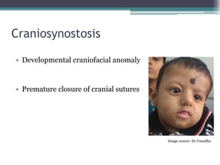
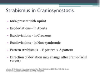
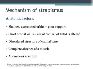
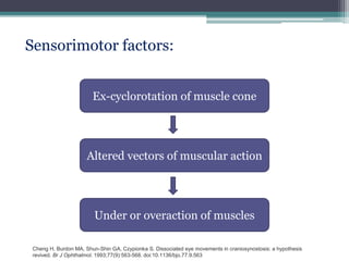
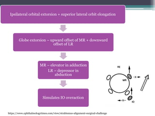
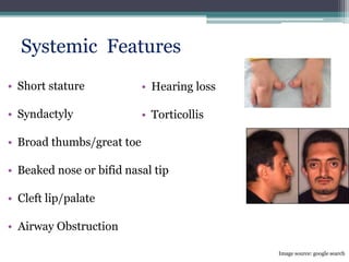
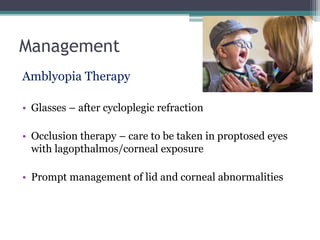
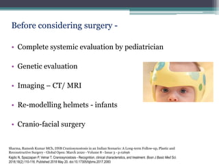
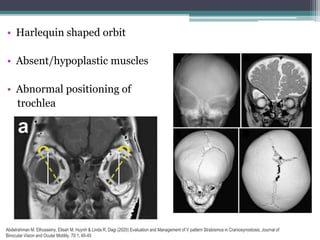
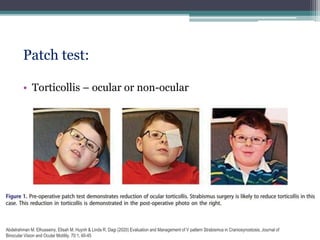
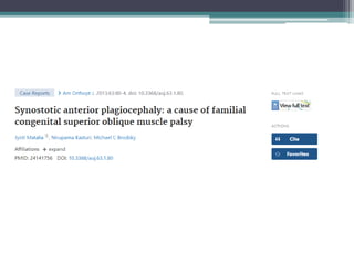
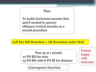
Strabismus is common in patients with craniosynostosis. The document discusses craniosynostosis, the various types and causes. Strabismus in craniosynostosis can be due to anatomical abnormalities of the orbits and extraocular muscles as well as sensorimotor factors. Evaluation involves a thorough eye examination and imaging. Management includes amblyopia treatment, craniofacial surgery, and strabismus surgery which can be challenging due to anatomical variations. Two case examples are presented of patients with plagiocephaly who presented with superior oblique muscle abnormalities but responded well to inferior oblique weakening procedures. Proper evaluation and customized surgical planning is important for achieving good outcomes in strabism
Η Άσηπτη Νέκρωση του Μηνοειδούς: Τι δεν μάθαμε σε 100 χρόνια- Kienbock's dise...Nikos Darlis
State fo the art on Kienbock's disease (avascular necrosis of the lunate). One handred years after its first description, it remains a source for controversies. What is new and what is outdated? Where do we go next?
Digitized manual palpation: a new method of evaluating posture and its defor...ROBERT ELBAUM
The aim of this study was to be able to apply this method for adolescents with idiopathic scoliosis and to try to draw the following elements: postural equilibrium parameters, evaluate the asymmetries present at the spine deformations, make an assessment of the curvature profile both in the frontal and sagittal plane and also a count of the position of the shoulders
Case Review #46: 56 year old female with Adult Idiopathic Scoliosis and Burst...Robert Pashman
56 year old female with Idiopathic Scoliosis, status post burst fracture, presented with junctional kyphosis. Dr. Pashman treated the patient with a posterior spinal fusion from T2-pelvis. Spinal curvature was a KIM/SRP Classification 3.
A Radiological Approach to CraniosynostosisFelice D'Arco
Presentation Summary: Normal Cranial Development (Anatomy and Genetic), Imaging Technique (how to do 3D CT, when to do MRI, why to do not do Plain Film), Imaging Patterns of Craniosynostosis, Associated Complications, Pitfalls.
Title: Sense of Taste
Presenter: Dr. Faiza, Assistant Professor of Physiology
Qualifications:
MBBS (Best Graduate, AIMC Lahore)
FCPS Physiology
ICMT, CHPE, DHPE (STMU)
MPH (GC University, Faisalabad)
MBA (Virtual University of Pakistan)
Learning Objectives:
Describe the structure and function of taste buds.
Describe the relationship between the taste threshold and taste index of common substances.
Explain the chemical basis and signal transduction of taste perception for each type of primary taste sensation.
Recognize different abnormalities of taste perception and their causes.
Key Topics:
Significance of Taste Sensation:
Differentiation between pleasant and harmful food
Influence on behavior
Selection of food based on metabolic needs
Receptors of Taste:
Taste buds on the tongue
Influence of sense of smell, texture of food, and pain stimulation (e.g., by pepper)
Primary and Secondary Taste Sensations:
Primary taste sensations: Sweet, Sour, Salty, Bitter, Umami
Chemical basis and signal transduction mechanisms for each taste
Taste Threshold and Index:
Taste threshold values for Sweet (sucrose), Salty (NaCl), Sour (HCl), and Bitter (Quinine)
Taste index relationship: Inversely proportional to taste threshold
Taste Blindness:
Inability to taste certain substances, particularly thiourea compounds
Example: Phenylthiocarbamide
Structure and Function of Taste Buds:
Composition: Epithelial cells, Sustentacular/Supporting cells, Taste cells, Basal cells
Features: Taste pores, Taste hairs/microvilli, and Taste nerve fibers
Location of Taste Buds:
Found in papillae of the tongue (Fungiform, Circumvallate, Foliate)
Also present on the palate, tonsillar pillars, epiglottis, and proximal esophagus
Mechanism of Taste Stimulation:
Interaction of taste substances with receptors on microvilli
Signal transduction pathways for Umami, Sweet, Bitter, Sour, and Salty tastes
Taste Sensitivity and Adaptation:
Decrease in sensitivity with age
Rapid adaptation of taste sensation
Role of Saliva in Taste:
Dissolution of tastants to reach receptors
Washing away the stimulus
Taste Preferences and Aversions:
Mechanisms behind taste preference and aversion
Influence of receptors and neural pathways
Impact of Sensory Nerve Damage:
Degeneration of taste buds if the sensory nerve fiber is cut
Abnormalities of Taste Detection:
Conditions: Ageusia, Hypogeusia, Dysgeusia (parageusia)
Causes: Nerve damage, neurological disorders, infections, poor oral hygiene, adverse drug effects, deficiencies, aging, tobacco use, altered neurotransmitter levels
Neurotransmitters and Taste Threshold:
Effects of serotonin (5-HT) and norepinephrine (NE) on taste sensitivity
Supertasters:
25% of the population with heightened sensitivity to taste, especially bitterness
Increased number of fungiform papillae
Report Back from SGO 2024: What’s the Latest in Cervical Cancer?bkling
Are you curious about what’s new in cervical cancer research or unsure what the findings mean? Join Dr. Emily Ko, a gynecologic oncologist at Penn Medicine, to learn about the latest updates from the Society of Gynecologic Oncology (SGO) 2024 Annual Meeting on Women’s Cancer. Dr. Ko will discuss what the research presented at the conference means for you and answer your questions about the new developments.
TEST BANK for Operations Management, 14th Edition by William J. Stevenson, Ve...kevinkariuki227
TEST BANK for Operations Management, 14th Edition by William J. Stevenson, Verified Chapters 1 - 19, Complete Newest Version.pdf
ARTIFICIAL INTELLIGENCE IN HEALTHCARE.pdfAnujkumaranit
Artificial intelligence (AI) refers to the simulation of human intelligence processes by machines, especially computer systems. It encompasses tasks such as learning, reasoning, problem-solving, perception, and language understanding. AI technologies are revolutionizing various fields, from healthcare to finance, by enabling machines to perform tasks that typically require human intelligence.
MANAGEMENT OF ATRIOVENTRICULAR CONDUCTION BLOCK.pdfJim Jacob Roy
Cardiac conduction defects can occur due to various causes.
Atrioventricular conduction blocks ( AV blocks ) are classified into 3 types.
This document describes the acute management of AV block.
New Drug Discovery and Development .....NEHA GUPTA
The "New Drug Discovery and Development" process involves the identification, design, testing, and manufacturing of novel pharmaceutical compounds with the aim of introducing new and improved treatments for various medical conditions. This comprehensive endeavor encompasses various stages, including target identification, preclinical studies, clinical trials, regulatory approval, and post-market surveillance. It involves multidisciplinary collaboration among scientists, researchers, clinicians, regulatory experts, and pharmaceutical companies to bring innovative therapies to market and address unmet medical needs.
These simplified slides by Dr. Sidra Arshad present an overview of the non-respiratory functions of the respiratory tract.
Learning objectives:
1. Enlist the non-respiratory functions of the respiratory tract
2. Briefly explain how these functions are carried out
3. Discuss the significance of dead space
4. Differentiate between minute ventilation and alveolar ventilation
5. Describe the cough and sneeze reflexes
Study Resources:
1. Chapter 39, Guyton and Hall Textbook of Medical Physiology, 14th edition
2. Chapter 34, Ganong’s Review of Medical Physiology, 26th edition
3. Chapter 17, Human Physiology by Lauralee Sherwood, 9th edition
4. Non-respiratory functions of the lungs https://academic.oup.com/bjaed/article/13/3/98/278874
Ozempic: Preoperative Management of Patients on GLP-1 Receptor Agonists Saeid Safari
Preoperative Management of Patients on GLP-1 Receptor Agonists like Ozempic and Semiglutide
ASA GUIDELINE
NYSORA Guideline
2 Case Reports of Gastric Ultrasound
5. CALVARIAL
SUTURES
Deposition
of osteoid at
the sutural
margins
Surface
apposition and
resorption
(remodeling) of
the bone
Centrifugal
displacement
by the
expanding
brain
Kajdic N, Spazzapan P, Velnar T. Craniosynostosis - Recognition, clinical characteristics, and treatment. Bosn J Basic Med
Sci. 2018;18(2):110-116. Published 2018 May 20. doi:10.17305/bjbms.2017.2083
6. Cells undergo transformation
Migrate to the sutures
Growth regulators
FGF, TGF-β Mechanical signals
Fusion of sutures
Duramater
Genetic
Mutations/
Environmental
causes
Premature
Closure of
Sutures
Kajdic N, Spazzapan P, Velnar T. Craniosynostosis - Recognition, clinical characteristics, and treatment. Bosn J Basic Med
Sci. 2018;18(2):110-116. Published 2018 May 20. doi:10.17305/bjbms.2017.2083
9. Kajdic N, Spazzapan P, Velnar T. Craniosynostosis - Recognition, clinical characteristics, and treatment. Bosn J Basic Med
Sci. 2018;18(2):110-116. Published 2018 May 20. doi:10.17305/bjbms.2017.2083
Types
Kimonis V, Gold JA, Hoffman TL, Panchal J, Boyadjiev SA. Genetics of craniosynostosis. Semin Pediatr Neurol. 2007;14:150–161
10. Ko JM. Genetic Syndromes Associated with Craniosynostosis. J Korean Neurosurg Soc. 2016;59(3):187-191.
doi:10.3340/jkns.2016.59.3.187
11. In India
• Most common – Coronal synostosis
• Syndromic – Apert’s
Sharma, Ramesh Kumar MCh, DNB Craniosynostosis in an Indian Scenario: A Long-term Follow-up, Plastic and
Reconstructive Surgery - Global Open: March 2020 - Volume 8 - Issue 3 - p e2696
13. Strabismus in Craniosynostosis
• 60% present with squint
• Esodeviations - in Aperts
• Exodeviations - in Crouzons
• Exodeviations - in Non-syndromic
• Pattern strabismus – V pattern > A pattern
• Direction of deviation may change after cranio-facial
surgery
Lehman S. Strabismus in craniosynostosis. Curr Opin Ophthalmol. 2006 Oct;17(5):432-4. doi:
10.1097/01.icu.0000243017.90004.5b. PMID: 16932059.
14. Mechanism of strabismus
Anatomic factors:
• Shallow, exorotated orbits – poor support
• Short orbital walls – arc of contact of EOM is altered
• Disordered structure of cranial base
• Complete absence of a muscle
• Anomalous insertion
Cheng H, Burdon MA, Shun-Shin GA, Czypionka S. Dissociated eye movements in craniosynostosis: a hypothesis
revived. Br J Ophthalmol. 1993;77(9):563-568. doi:10.1136/bjo.77.9.563
15. Sensorimotor factors:
Ex-cyclorotation of muscle cone
Altered vectors of muscular action
Under or overaction of muscles
Cheng H, Burdon MA, Shun-Shin GA, Czypionka S. Dissociated eye movements in craniosynostosis: a hypothesis
revived. Br J Ophthalmol. 1993;77(9):563-568. doi:10.1136/bjo.77.9.563
16. Ipsilateral orbital extorsion + superior lateral orbit elongation
Globe extorsion – upward offset of MR + downward
offset of LR
MR – elevator in adduction
LR – depressor in
abduction
Simulates IO overaction
https://www.ophthalmologytimes.com/view/strabismus-alignment-surgical-challenge
18. Evaluation
• History – Birth, Developmental, Family
• Vision
• Cycloplegic refraction
• Examination of adnexa and slit lamp examination
19. Squint assessment:
• Fusion – to be tested in preferred gaze before
performing cover test
• To be assessed and quantified in all gazes and head-tilts
• Extra-ocular movements – both on ductions and
versions
• Torsion demonstrated on fundus examination
20. Other Ophthalmic features
• Dystopia
• Hypertelorism
• Amblyopia
• Nystagmus
• Proptosis
• Ptosis
• Lagophthalmos
• Corneal exposure
Image source: google search
21. • Short stature
• Syndactyly
• Broad thumbs/great toe
• Beaked nose or bifid nasal tip
• Cleft lip/palate
• Airway Obstruction
• Hearing loss
• Torticollis
Image source: google search
Systemic Features
22. Management
Amblyopia Therapy
• Glasses – after cycloplegic refraction
• Occlusion therapy – care to be taken in proptosed eyes
with lagopthalmos/corneal exposure
• Prompt management of lid and corneal abnormalities
23. Before considering surgery -
• Complete systemic evaluation by pediatrician
• Genetic evaluation
• Imaging – CT/ MRI
• Re-modelling helmets - infants
• Cranio-facial surgery
Kajdic N, Spazzapan P, Velnar T. Craniosynostosis - Recognition, clinical characteristics, and treatment. Bosn J Basic Med Sci.
2018;18(2):110-116. Published 2018 May 20. doi:10.17305/bjbms.2017.2083
Sharma, Ramesh Kumar MCh, DNB Craniosynostosis in an Indian Scenario: A Long-term Follow-up, Plastic and
Reconstructive Surgery - Global Open: March 2020 - Volume 8 - Issue 3 - p e2696
24. Imaging of brain and orbit -
• Orbital anatomy
• Extraocular muscle position
• Excyclorotation of EOM
• Dysgenesis
25. • Harlequin shaped orbit
• Absent/hypoplastic muscles
• Abnormal positioning of
trochlea
Abdelrahman M. Elhusseiny, Elisah M. Huynh & Linda R. Dagi (2020) Evaluation and Management of V pattern Strabismus in Craniosynostosis, Journal of
Binocular Vision and Ocular Motility, 70:1, 40-45
26. Patch test:
• Torticollis – ocular or non-ocular
Abdelrahman M. Elhusseiny, Elisah M. Huynh & Linda R. Dagi (2020) Evaluation and Management of V pattern Strabismus in Craniosynostosis, Journal of
Binocular Vision and Ocular Motility, 70:1, 40-45
27. Timing of surgery
Before or after Craniofacial surgery?
Diamond et al
• Change in squint seen in only 9/120 patients after
cranio-facial surgery.
• Advocates early squint correction
Morax et al
• Change in squint seen in 9/10 patients after cranio-facial
surgery
• Advocates squint surgery after primary surgery
Diamond GR, Katowitz JA, Whitaker LH, et al. Ocular alignment after craniofacial reconstruction. Am J Ophthalmol. 1980;90(2):248–250.
Morax S. Change in eye position after cranio-facial surgery. J Maxillofac Surg. 1984;12(2):47–55
28. Pre-operative counselling
• Realistic expectations
• Pattern may not be fully corrected
• Surgery will not change shape of globe/face
• Need for multiple surgeries
• Surgery may improve binocularity (in very young
children) but not vision
29. Surgical Approach
• FDT and exaggerated traction test – SO and IO function
• SO tuck or IO weakening
• Recession/Resection of Horizontal Recti with vertical
displacement
• Recession/Resection of Horizontal recti with slanting
30. • Conjunctival incision – Limbal incision for recti
Fornix incision for obliques
• Radial relaxing incisions recommended
• Identify muscles correctly before hooking – For IO,
identify/hook LR and IR first – direct viewing better than
MRI/CT
Jean D A Carruthers, Strabismus in Craniosynostosis syndromes. Clinical Strabismus Management – Rosenbaum
Santiago, pages 393-403
31. • If LR and MR at approximate normal horizontal
meridian – true IO over action – requires IO weakening
procedures
• Severe IO overaction – Anterior transposition of IO (
Bell’s phenomenon to be noted pre-op)
• No IO over-action and no displacement of LR – SO
underaction – SO tuck
Jean D A Carruthers, Strabismus in Craniosynostosis syndromes. Clinical Strabismus Management – Rosenbaum
Santiago, pages 393-403
32. For V pattern EXotropia :
MR resection with down-shift
LR recession with up-shift
MR
LR LE
For V pattern ESotropia:
B/L MR Recession – Lower margin
recessed more than upper margin
Vertical Displacement:
Slanting:
33. IO Weakening Procedures
• IO Recession –
upto 14 mm
• Anterior transposition –
Alters path of muscle
Stager D Jr, Dao LM, Felius J. Uses of the Inferior Oblique Muscle in Strabismus Surgery. Middle East Afr J Ophthalmol.
2015;22(3):292-297. doi:10.4103/0974-9233.159723
34. • Anterior Nasal transposition –
Transposed to a location over the nasal half of the inferior
rectus muscle
• Myectomy –
Removal of a segment of IO without detaching from
insertion
35.
36.
37. Case 1
• 6 year old
• Uneventful perinatal and
developmental history
• Tilting of head to watch
TV
• Using glasses and had
undergone amblyopia
therapy
• Facial asymmetry with right-
sided forehead depression
• Left head tilt
• VA: 20/20 and 20/25 with -
1.50D cyl X 150˚
• Stereopsis : 400 secs of arc
• 12 PD right hypertropia
increasing in LG and RHT
• Right IOOA
• Extorsion in fundus in RE
Matalia J, Kasturi N, Brodsky MC. Synostotic anterior plagiocephaly: a cause of familial congenital superior oblique muscle
palsy. Am Orthopt J. 2013;63:80-4. doi: 10.3368/aoj.63.1.80. PMID: 24141756.
38. CT scan:
• Right- sided frontal
plagiocephaly
• Lateral displacement of
the superior rectus
muscle
• Medial displacement of
the inferior rectus muscle
on the affected side
Right inferior oblique anterior transposition under GA
39. CASE 2
• 2 year old
• Uneventful perinatal and
developmental history
• Head tilt to right since 1
year
• Facial asymmetry with left
sided flattening of
forehead
• Right head tilt
• TAC: 20/84 at 84 cm
• left hypertropia -
increasing on RG and LHT
• Left IOOA
• Extorsion in fundus in LE
Left inferior oblique myectomy under GA
40. 1 year later :
• Intermittent outward deviation of eyes
• Visual acuity: OU: 20/25
• IDS of 15 PD with B/L DVD
• Extra-ocular movements: Full
• Treated with glasses
After 1 more year:
• IDS – 30 PD with poor control with B/L DVD
B/L Lateral Rectus recession with half tendon width
infra-displacement
41. Discussion
• Synostotic plagiocephaly - indistinguishable from
neurogenic congenital superior oblique palsy
• Older patients - facial asymmetry simulate longstanding
superior oblique palsy. However, it involves the forehead
and lower face.
• Though unable to evaluate the presence or absence of
trochlear retrodisplacement on orbital images,
heterotopias of muscles was confirmed.
Matalia J, Kasturi N, Brodsky MC. Synostotic anterior plagiocephaly: a cause of familial congenital superior oblique muscle
palsy. Am Orthopt J. 2013;63:80-4. doi: 10.3368/aoj.63.1.80. PMID: 24141756.
42. • Anatomical aberrations could necessitate a
different surgical approach
• Excellent responses to weakening of the inferior
oblique muscle in these and other patients with
unilateral coronal synostosis
Matalia J, Kasturi N, Brodsky MC. Synostotic anterior plagiocephaly: a cause of familial congenital superior oblique muscle
palsy. Am Orthopt J. 2013;63:80-4. doi: 10.3368/aoj.63.1.80. PMID: 24141756.
43. Our Case
35 year old male
Squint since birth
Using glasses since 20 years
Facial asymmetry
Ptosis
44. Image credits: Dr.Bhanumathi
Hirschberg: 15 ° LXT
Cover test: LXT – AXT with R/L with DVD
PBCT : 35 PD BI + 25 PD BD (in front of RE) in all gazes and headtilts
EOMs: B/L SO and IO over action
MRI Brain and Orbit: Fusion of coronal sutures on right side
Frontal Plagiocephaly
45. Plan:
To tackle horizontal muscles first
and if needed to operate
obliques/vertical muscles as a
second procedure
Left Eye MR Resection + LR Recession under MAC
Post op at 1 month:
10 PD BD for near
25 PD BD with 8 PD BI for distance
Convergence Exercises
Patient
happy
with
Outcome
46. Take Home Message
• Strabismus in craniosynostosis is difficult to treat
• Differentiate from true muscle palsies and true muscle
over-action
• CT/MRI before considering surgery
• Surgical approach can be complicated and can achieve
varied results
Take a look at these pictures. As soon as you see this baby, you notice that he isnt a normal looking child. But what are you actually seeing? Keep these pictures in mind and we will get back to them in a while.
A craniosynostosis is a developmental anomaly which occurs as a consequence of abnormal and non-physiological sutural fusion. When one or more sutures are prematurely closed, the compensatory growth starts perpendicular to the patent sutures since the brain still grows and expands in the direction of lower resistance. The result is an abnormally shaped skull and also, in more severe cases, increased intracranial pressure (ICP), as well as sensory, respiratory and neurological dysfunction
The fusion of the sutures is mainly regulated by the dura mater, which interacts with the overlying tissues of the cranial vault. The dura mater provides many important regulators of growth, such as intercellular signals (for example, signaling mediated by fibroblast growth factor [FGF] and transforming growth factor beta [TGF-β] have been suggested to be vital in this process), mechanical signals, and cells which undergo transformation and migrate to the sutures. This complex signaling cascade can be disrupted by a large number of genetic mutations, leading to an abnormal development of the cranial sutures [9-14]. Finally, this may result in a premature fusion. Genes most commonly mutated in craniosynostosis are
Craniosynostoses are classified according to the sutures and the frequencies of these different types of craniosynostoses are as follows: Sagittal (≈60%), coronal (≈25%), metopic (≈15%), and lambdoid. The sagittal suture (40–55%) is the most commonly affected, followed by the coronal (20–25%), metopic (5–15%), and lambdoid (<5%) sutures
Non-syndromic – 70%
doi: 10.1097/GOX.0000000000002696
Pictures of B/o Fouziya – 9 days old, 3rd deg CM, FTND, no NICU admission, frontal bossing – acrocephaly, depressed nasal bridge, syndactyly of hands and feet, proptosis, lagophthalomos - ?? Aperts syndrome. Does this baby have strabismus? We will have to wait a while to know since fixation is yet to develop.
Refractive errors are a major cause for visual impairment after strabismus. Hypermetropia more common in syndromic and astigmatism more in nonsyndromic. 20 –35 % amblyopic. Optic atrophy seen in 7 %.
Excyclorotation, defined as the rotation of the / j upper pole of the globe away from the midline, would explain the pattern of dissociated movements simply by invoking Hering's law without the need of assuming over or underaction of - certain muscles. (counterclockwise rotation of the rectus muscles of the right eye and/or clockwise rotation of the rectus muscles of the left eye
Coronal synostosis
While requesting for MRI/CT – mention orbital cuts and to look for EOM specifically. MRI not possible if metal implants have been used for craniofacial surgery
1. Harlequin orbit – retrusion of upper lateral margin, coronal suture on one side fused,
(Kushner) Nischal has indicated that many of these patients have an abnormally tilted union between the base of the skull and the first cervical vertebra. This results in a persistent non-ocular head tilt even after strabismus has been corrected
Diamond et al - evaluated the ocular misalignment after craniofacial reconstruction. They reported that out of 120 patients who underwent complete ocular examination pre-and postoperatively, only nine patients developed a change in their primary position ocular alignment and this was persistently more than 10 prism diopters in only 3 patients. the current trend toward earlier craniofacial intervention (by 4 months of age for endoscopic procedures and close to year of age for cranial vault expansion) makes awaiting completion of the primary craniofacial repair reasonable. In our scenario, such cases are rare and infantc with CS brought to us for squint surgery is rarer still. Amblyopia therapy takes priority and getting consistent measurement of deviation in infants is almost impossible – hence the question of whether to operate before before cranio-facial surgery or not, does not arise.
Lax SO – tuck, IOOA - weaken
Some muscles may be inserted closer to limbus than normal
If Bells is strong/good pre-op, lesser chance of corneal exposure post-op as antr transposition of IO can lead to lagophthalmos
Vertical transposition of the horizontal recti involves either recessing or resecting the muscle and then re-inserting it to the sclera with a vertical offset. In slanting procedures, In V pattern esotropia, bilateral medial rectus muscle recessions are performed, with the upper margin of the muscle being recessed to correct the deviation in upward position and the lower margin being recessed a greater amount according to the downward deviation. In A pattern esotropia the deviation is greater in upward gaze and so more recession is performed on the upper margin than on the lower margin of the muscle. The difference in recession was between 2 and 3 mm in accordance with the differences in measurements.
IO recession: 1.b anterior border is placed 5 mm posterior to the temporal insertion of the inferior rectus muscle
Antr transposition : postoperative limitation of elevation during abduction – IOOA of opp eye
Myectomy: faster than a recession. In addition, there is less risk of surgical intraoperative complication because there is no scleral pass required to reattach the muscle. The disadvantage is the possibility that the muscle may reattach and lead to recurrent overaction.
Antr Nasal trans: 2 mm nasal to the nasal border of the inferior rectus muscle and 2 mm posterior to the inferior rectus insertion. Makes the inferior oblique muscle into a tonic depressor
Myectomy: faster than a recession. In addition, there is less risk of surgical intraoperative complication because there is no scleral pass required to reattach the muscle. The disadvantage is the possibility that the muscle may reattach and lead to recurrent overaction.
IO recession: 1.b anterior border is placed 5 mm posterior to the temporal insertion of the inferior rectus muscle
Antr transposition : postoperative limitation of elevation during abduction – IOOA of opp eye
Antr Nasal trans: 2 mm nasal to the nasal border of the inferior rectus muscle and 2 mm posterior to the inferior rectus insertion. Makes the inferior oblique muscle into a tonic depressor
Dr.Jyoti matalia has published an article in the American orthoptic journal in 2013 written along with Prof Brodsky. They examined two siblings with familial synostotic plagiocephaly and simulated congenital superior oblique palsy who were successfully managed with weakening of the ipsilateral inferior oblique muscles. Let’s take a look at these cases.
Postoperatively, her inferior oblique overaction, hypertropia, and head tilt resolved.
Synostotic plagiocephaly - secondary orbital aberration involving the extraocular muscles, leading to a clinical picture that is otherwise. SO palsy involves deformation of lower 2/3rds of face