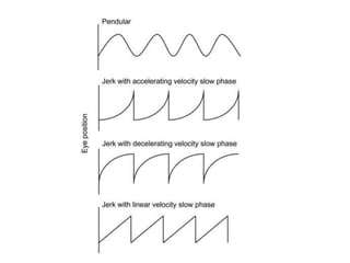
Congenital Nystagmus
Dr Sarah Khan
PATHOPHYSIOLOGY
• Three main mechanisms are involved in
maintenance of the image of the target of
interest on the fovea
• Ocular fixation
• Vestibulo ocular reflex
• Central nervous system as the neural
integrator
• Ocular fixation helps to keep the drifting
image of the object on the fovea and also
suppresses any unwanted saccades
• Vestibulo ocular fixation helps to maintain a
good visual acuity by keeping the eye
movements compensated for changes in the
head posture during various routine activities
• The neural integrator helps to maintain the
eye position in an eccentric position during
different gazes against the pull of suspensory
ligaments and the EOM that tend to bring the
eye back to the central position
• NULL POSITION :
• Position of gaze in which the nystagmus is
minimally ellicited. To keep the eye in this
position, the child may adopt a face turn or a
chin elevation or depression depending on the
null position. This enables the child to keep
the image of the target at the fovea for a
longer time to enable a clear visual acuity (
Foveation period)
• PENDULAR NYSTAGMUS has a sinusoidal
pattern without differentiation into a slow or
fast component
• NEUTRAL ZONE : This is observed in Periodic
alternating nystgamus in which the fast
component changes direction after an interval
of time
• CONJUGATE NYSTAGMUS : Nystagmus in the
two eyes is similar in amplitude, frequency
and direction
• DISCONJUGATE NYSTAGMUS : The direction
of nystagmus in the two eyes is different
• DISSOCIATED NYSTAGMUS : The amplitude of
the nystagmus is different but the direction
remains the same
• JERK NYSTAGMUS has a slow and a fast component and
is named based on the fast component
• Direction of the nystagmus is the direction of its fast
component
• Plane could be horizontal, vertical or rotator or a
combination of these
• Amplitude is the excursion of the nystagmus which
maybe small(<5deg), moderate (5-15deg) and large(
>15deg)
• Frequency Number of beats per second, slow (1-2Hz) ,
medium (3-4Hz), Fast (>5Hz)
• Intensity = Amplitude * Frequency
Classification of Nystagmus
• CEMAS : Classification of Eye Movement
Abnormalities and Strabismus
• By the National Eye Institute
A CLASSIFICATION OF EYE MOVEMENT ABNORMALITIES AND STRABISMUS (CEMAS) Report of a National Eye Institute Sponsored
Workshop , Richard W. Hertle, NEI
NYSTAGMUS AND OCULAR MOTOR
OSCILLATIONS
• A. PHYSIOLOGICAL FIXATIONAL MOVEMENTS
1. MICROTREMOR
2. SLOW DRIFTS
3. MICROSACCADES
A CLASSIFICATION OF EYE MOVEMENT ABNORMALITIES AND STRABISMUS (CEMAS) Report of a National Eye Institute Sponsored
Workshop , Richard W. Hertle, NEI
• B. PHYSIOLOGICAL NYSTAGMUS
1. VESTIBULAR
2. OPTOKINETIC
3. ECCENTRIC GAZE NYSTAGMUS
A CLASSIFICATION OF EYE MOVEMENT ABNORMALITIES AND STRABISMUS (CEMAS) Report of a National Eye Institute Sponsored
Workshop , Richard W. Hertle, NEI
• C. PATHOLOGICAL NYSTAGMUS
1. INFANTILE NYSTAGMUS SYNDROME (INS)
2. FUSION MAL DEVELOPMENT NYSTAGMU
SYNDROME (FMNS)
3. SPASMUS NUTANS SYNDROME
4. VESTIBULAR NYSTAGMUS
a. PERIPHERAL VESTIBULAR IMBALANCE
b. CENTRAL VESTIBULAR IMBALANCE
c. CENTRAL VESTIBULAR INSTABILITY (PAN)
A CLASSIFICATION OF EYE MOVEMENT ABNORMALITIES AND STRABISMUS (CEMAS) Report of a National Eye Institute Sponsored
Workshop , Richard W. Hertle, NEI
5. GAZE HOLDING DEFICIENCY NYSTAGMUS
a. ECCENTRIC GAZE NYSTAGMUS ( AND ASSOCIATED
REBOUND NYSTAGMUS)
b. GAZE INSTABILITY NYSTAGMUS
6. VISION LOSS NYSTAGMUS
a. PRECHIASMAL
b. CHIASMAL
c. POSTCHIASMAL
A CLASSIFICATION OF EYE MOVEMENT ABNORMALITIES AND STRABISMUS (CEMAS) Report of a National Eye Institute Sponsored
Workshop , Richard W. Hertle, NEI
7. OTHER PENDULAR NYSTAGMUS AND
NYSTAGMUS ASSOCIATED WITH DISEASE OF
CENTRAL MYELIN
a. MS, PELIAZAEUS MERZBACHER DISEASE,
COCKAYNE’S PEROXISOMAL DISORDERS,
TOLUENE ABUSE
b. PENDULAR NYSTAGMUS ASSOCIATED WITH
TREMOR OF THE PALATE
c. PENDULAR VERGENCE NYSTAGMUS ASSOCIATED
WITH WHIPLE’S DISEASE
A CLASSIFICATION OF EYE MOVEMENT ABNORMALITIES AND STRABISMUS (CEMAS) Report of a National Eye Institute Sponsored
Workshop , Richard W. Hertle, NEI
8. OCULAR BOBBING (TYPICAL AND ATYPICAL)
9. LID NYSTAGMUS
A CLASSIFICATION OF EYE MOVEMENT ABNORMALITIES AND STRABISMUS (CEMAS) Report of a National Eye Institute Sponsored
Workshop , Richard W. Hertle, NEI
D. Saccadic intrusions and oscillations
• Square wave jerks and oscillations
• Square wave pulses
• Saccadic pulses
• Induced convergence retraction
• Dissociated ocular oscillations
• Hypermetric saccades
• Macrosaccadic oscillations
• Ocular flutter
• Flutter dysmetria
• Opsoclonus
• Psychogenic flutter
• Superior oblique myokymia
E. Generalised disturbance of saccades
F. Generalised disturbance of smooth pursuit
G. Generalised disturbance of vestibular eye
movements
H. Generalised disturbance of optokinetic eye
movements
INFANTILE NYSTAGMUS SYNDROME
• Appears by 0-6 months of age
• Progresses from pendular to jerk type
• Conjugate, uniplanar, horizontal
• Stabilises at 5-6 years of age
• Accelerating slow phase
• Increases on attempt to fixate
• Decreases with convergence
• Marked head posture
• Strong family predisposition
• Abolished during sleep
• May have a latent component
• May show reversal pattern with OKN
Fusion maldevelopment nystagmus
syndrome
• FMNS has an infantile onset
• Conjugate, horizontal and uniplanar
• May appear or increase in mono ocular
fixation
• Linear decelerating slow phase with fast phase
in the direction of the uncovered fixing eye
• Reverses direction with change in fixation
• Intensity decreases with age
Spasmus nutans syndrome
• Constellation of ocular oscillation , head
bobbing and torticollis
• Appears at 4-18 months of age and disappears
by 3 years of age
• Nystagmus increases with convergence
• MRI/CT are normal
Periodic alternating Nystagmus
• This is a central vestibular instability
nystagmus.
• It exhibits a neutral point with a jerk
nystagmus in one direction for 60-90 seconds
which then changes to the other direction for
another 60-90 second
• It, therefore needs to be observed for atleast 3
minutes. It is not affected by visual fixation
• It can be acquired or it maybe a part of the
infantile nystagmus syndrome exhibiting an
accelerating slow phase in the ocular motor
recordings
video
Evaluation
• A good clinical history
• Ante natal history
• Birth history
• Age of onset, associated decreased vision
• Acute onset, diplopia, oscillopsia point
towards acquired causes
• Family history
• Maternal drug intake ante natally
• Or perinatal infections
Examination
• Visual acuity
• Uniocular and binocular
• Near and distance
• With and without head posture
• A complete ocular examination of the anterior
and posterior segments
• Measurement of any face turn
• Goniometer or a protractor
• Helps to decide the amount of surgical
correction required
• Video recording of the ocular movements
• And if available ocular movement tracing
• Neuro imaging is required in cases of
 All acquired nystagmus
 Periodic alternating nystagmus
 Seesaw nytagmus
 Spasms nutans syndrome
 INS with poor vision and disc pallor
Electrooculography
Infrared oculography
• Binocular infrared oculography BIRO
• Video oculography which traces ocular
movements using infra red techniques
• Electromagnetic search coil method
• Scleral contact lenses are worn
Scleral search coils
ICS ChartR VNG
Drug treatments
• Anticholinergics, anti histaminics
• Acetazoleamide effective in familial periodic
ataxia with nystagmus
• Baclofen decreases symptoms in PAN (
increases the inhibitory effect of GABA on the
vestibular nuclei)
• Botulinum toxin injections
Optical treatment
• In congenital nystagmus, convergence and
eccentric gaze decreases nystagmus
• To induce convergence , 7D base out prism
can be placed in each spectacle lens
• In young patients -1.00 D can be added to the
spectacle correction
• If null zone is in a horizontal eccentric gaze,
prism apices pointing towards the null zone
may help
• Contact lenses correct refractive errors more
effectively , and the tactile sensory feedback
might decrease the intensity of nystagmus
Surgery
• To shift the null position from eccentric
position to primary position
• Increase foveation
• To reduce a an abnormal head position
NYSTAGMUS +++
INTENSITY OF NYSTAGMUS REDUCED IN LEVOVERSION
NYSTAGMUS +
HENCE PATIENT HAS A FACE
TURN TO THE RIGHT
It may be good to wait till 4-5 yrs to allow for
maturation of binocular visual system
To correct the head posture
• Anderson’s surgery : Weakening the yoke
muscles responsible for the slow phase of
nystagmus
• Kestenbaum’s procedure : 5mm resection
recession of all four horizontal recti to move
the null position to the primary position
• Goto’s surgery : Resection of the recti that
move the eye away from the null position
• Modified Park’s Kestenbaum procedure :
Recession of one MR 5mm, and LR 7mm
And resection of other MR 6mm and LR 8mm
• Augmented modified Kestenbaum’s :
If the head turn is up to 30 degree,
The values of recessions and resections can be
increased by 40%
• Spielmann’s Procedure : in head turn more
than 20 degree, Kestenbaum’s procedure with
posterior fixation sutures on the recessed
muscle
• Pratt Johnson’s surgrery : Equal recession and
resection of all horizontal recti
• Chin elevation : Recession of inferior rectus
which can be combined with resection of
superior rectus
• And vice versa for chin depression
• Head tilt : Vertical shift of horizontal recti or
horizontal shift of vertical recti
• If there is no null position, or null position is in
the primary position
• Aim of surgery in nystagmus is to dampen the
intensity of nystagmus
• Surgically induced exotropia
• Large recession of all horizontal recti
THANK YOU

Childhood Nystagmus

  • 1.
  • 2.
    PATHOPHYSIOLOGY • Three mainmechanisms are involved in maintenance of the image of the target of interest on the fovea • Ocular fixation • Vestibulo ocular reflex • Central nervous system as the neural integrator
  • 3.
    • Ocular fixationhelps to keep the drifting image of the object on the fovea and also suppresses any unwanted saccades
  • 4.
    • Vestibulo ocularfixation helps to maintain a good visual acuity by keeping the eye movements compensated for changes in the head posture during various routine activities
  • 5.
    • The neuralintegrator helps to maintain the eye position in an eccentric position during different gazes against the pull of suspensory ligaments and the EOM that tend to bring the eye back to the central position
  • 6.
    • NULL POSITION: • Position of gaze in which the nystagmus is minimally ellicited. To keep the eye in this position, the child may adopt a face turn or a chin elevation or depression depending on the null position. This enables the child to keep the image of the target at the fovea for a longer time to enable a clear visual acuity ( Foveation period)
  • 7.
    • PENDULAR NYSTAGMUShas a sinusoidal pattern without differentiation into a slow or fast component
  • 8.
    • NEUTRAL ZONE: This is observed in Periodic alternating nystgamus in which the fast component changes direction after an interval of time
  • 9.
    • CONJUGATE NYSTAGMUS: Nystagmus in the two eyes is similar in amplitude, frequency and direction • DISCONJUGATE NYSTAGMUS : The direction of nystagmus in the two eyes is different • DISSOCIATED NYSTAGMUS : The amplitude of the nystagmus is different but the direction remains the same
  • 10.
    • JERK NYSTAGMUShas a slow and a fast component and is named based on the fast component • Direction of the nystagmus is the direction of its fast component • Plane could be horizontal, vertical or rotator or a combination of these • Amplitude is the excursion of the nystagmus which maybe small(<5deg), moderate (5-15deg) and large( >15deg) • Frequency Number of beats per second, slow (1-2Hz) , medium (3-4Hz), Fast (>5Hz) • Intensity = Amplitude * Frequency
  • 11.
    Classification of Nystagmus •CEMAS : Classification of Eye Movement Abnormalities and Strabismus • By the National Eye Institute A CLASSIFICATION OF EYE MOVEMENT ABNORMALITIES AND STRABISMUS (CEMAS) Report of a National Eye Institute Sponsored Workshop , Richard W. Hertle, NEI
  • 12.
    NYSTAGMUS AND OCULARMOTOR OSCILLATIONS • A. PHYSIOLOGICAL FIXATIONAL MOVEMENTS 1. MICROTREMOR 2. SLOW DRIFTS 3. MICROSACCADES A CLASSIFICATION OF EYE MOVEMENT ABNORMALITIES AND STRABISMUS (CEMAS) Report of a National Eye Institute Sponsored Workshop , Richard W. Hertle, NEI
  • 13.
    • B. PHYSIOLOGICALNYSTAGMUS 1. VESTIBULAR 2. OPTOKINETIC 3. ECCENTRIC GAZE NYSTAGMUS A CLASSIFICATION OF EYE MOVEMENT ABNORMALITIES AND STRABISMUS (CEMAS) Report of a National Eye Institute Sponsored Workshop , Richard W. Hertle, NEI
  • 14.
    • C. PATHOLOGICALNYSTAGMUS 1. INFANTILE NYSTAGMUS SYNDROME (INS) 2. FUSION MAL DEVELOPMENT NYSTAGMU SYNDROME (FMNS) 3. SPASMUS NUTANS SYNDROME 4. VESTIBULAR NYSTAGMUS a. PERIPHERAL VESTIBULAR IMBALANCE b. CENTRAL VESTIBULAR IMBALANCE c. CENTRAL VESTIBULAR INSTABILITY (PAN) A CLASSIFICATION OF EYE MOVEMENT ABNORMALITIES AND STRABISMUS (CEMAS) Report of a National Eye Institute Sponsored Workshop , Richard W. Hertle, NEI
  • 15.
    5. GAZE HOLDINGDEFICIENCY NYSTAGMUS a. ECCENTRIC GAZE NYSTAGMUS ( AND ASSOCIATED REBOUND NYSTAGMUS) b. GAZE INSTABILITY NYSTAGMUS 6. VISION LOSS NYSTAGMUS a. PRECHIASMAL b. CHIASMAL c. POSTCHIASMAL A CLASSIFICATION OF EYE MOVEMENT ABNORMALITIES AND STRABISMUS (CEMAS) Report of a National Eye Institute Sponsored Workshop , Richard W. Hertle, NEI
  • 16.
    7. OTHER PENDULARNYSTAGMUS AND NYSTAGMUS ASSOCIATED WITH DISEASE OF CENTRAL MYELIN a. MS, PELIAZAEUS MERZBACHER DISEASE, COCKAYNE’S PEROXISOMAL DISORDERS, TOLUENE ABUSE b. PENDULAR NYSTAGMUS ASSOCIATED WITH TREMOR OF THE PALATE c. PENDULAR VERGENCE NYSTAGMUS ASSOCIATED WITH WHIPLE’S DISEASE A CLASSIFICATION OF EYE MOVEMENT ABNORMALITIES AND STRABISMUS (CEMAS) Report of a National Eye Institute Sponsored Workshop , Richard W. Hertle, NEI
  • 17.
    8. OCULAR BOBBING(TYPICAL AND ATYPICAL) 9. LID NYSTAGMUS A CLASSIFICATION OF EYE MOVEMENT ABNORMALITIES AND STRABISMUS (CEMAS) Report of a National Eye Institute Sponsored Workshop , Richard W. Hertle, NEI
  • 18.
    D. Saccadic intrusionsand oscillations • Square wave jerks and oscillations • Square wave pulses • Saccadic pulses • Induced convergence retraction • Dissociated ocular oscillations • Hypermetric saccades • Macrosaccadic oscillations • Ocular flutter • Flutter dysmetria • Opsoclonus • Psychogenic flutter • Superior oblique myokymia
  • 19.
    E. Generalised disturbanceof saccades F. Generalised disturbance of smooth pursuit G. Generalised disturbance of vestibular eye movements H. Generalised disturbance of optokinetic eye movements
  • 20.
    INFANTILE NYSTAGMUS SYNDROME •Appears by 0-6 months of age • Progresses from pendular to jerk type • Conjugate, uniplanar, horizontal • Stabilises at 5-6 years of age • Accelerating slow phase • Increases on attempt to fixate • Decreases with convergence • Marked head posture
  • 21.
    • Strong familypredisposition • Abolished during sleep • May have a latent component • May show reversal pattern with OKN
  • 23.
    Fusion maldevelopment nystagmus syndrome •FMNS has an infantile onset • Conjugate, horizontal and uniplanar • May appear or increase in mono ocular fixation • Linear decelerating slow phase with fast phase in the direction of the uncovered fixing eye • Reverses direction with change in fixation • Intensity decreases with age
  • 24.
    Spasmus nutans syndrome •Constellation of ocular oscillation , head bobbing and torticollis • Appears at 4-18 months of age and disappears by 3 years of age • Nystagmus increases with convergence • MRI/CT are normal
  • 26.
    Periodic alternating Nystagmus •This is a central vestibular instability nystagmus. • It exhibits a neutral point with a jerk nystagmus in one direction for 60-90 seconds which then changes to the other direction for another 60-90 second
  • 27.
    • It, thereforeneeds to be observed for atleast 3 minutes. It is not affected by visual fixation • It can be acquired or it maybe a part of the infantile nystagmus syndrome exhibiting an accelerating slow phase in the ocular motor recordings
  • 28.
  • 29.
    Evaluation • A goodclinical history • Ante natal history • Birth history • Age of onset, associated decreased vision • Acute onset, diplopia, oscillopsia point towards acquired causes
  • 30.
    • Family history •Maternal drug intake ante natally • Or perinatal infections
  • 31.
    Examination • Visual acuity •Uniocular and binocular • Near and distance • With and without head posture
  • 32.
    • A completeocular examination of the anterior and posterior segments
  • 34.
    • Measurement ofany face turn • Goniometer or a protractor • Helps to decide the amount of surgical correction required
  • 36.
    • Video recordingof the ocular movements • And if available ocular movement tracing
  • 37.
    • Neuro imagingis required in cases of  All acquired nystagmus  Periodic alternating nystagmus  Seesaw nytagmus  Spasms nutans syndrome  INS with poor vision and disc pallor
  • 38.
  • 41.
  • 42.
    • Binocular infraredoculography BIRO • Video oculography which traces ocular movements using infra red techniques
  • 44.
    • Electromagnetic searchcoil method • Scleral contact lenses are worn
  • 45.
  • 46.
  • 47.
    Drug treatments • Anticholinergics,anti histaminics • Acetazoleamide effective in familial periodic ataxia with nystagmus • Baclofen decreases symptoms in PAN ( increases the inhibitory effect of GABA on the vestibular nuclei) • Botulinum toxin injections
  • 48.
    Optical treatment • Incongenital nystagmus, convergence and eccentric gaze decreases nystagmus • To induce convergence , 7D base out prism can be placed in each spectacle lens • In young patients -1.00 D can be added to the spectacle correction
  • 49.
    • If nullzone is in a horizontal eccentric gaze, prism apices pointing towards the null zone may help • Contact lenses correct refractive errors more effectively , and the tactile sensory feedback might decrease the intensity of nystagmus
  • 50.
    Surgery • To shiftthe null position from eccentric position to primary position • Increase foveation • To reduce a an abnormal head position
  • 51.
    NYSTAGMUS +++ INTENSITY OFNYSTAGMUS REDUCED IN LEVOVERSION NYSTAGMUS + HENCE PATIENT HAS A FACE TURN TO THE RIGHT
  • 52.
    It may begood to wait till 4-5 yrs to allow for maturation of binocular visual system
  • 53.
    To correct thehead posture • Anderson’s surgery : Weakening the yoke muscles responsible for the slow phase of nystagmus
  • 54.
    • Kestenbaum’s procedure: 5mm resection recession of all four horizontal recti to move the null position to the primary position
  • 55.
    • Goto’s surgery: Resection of the recti that move the eye away from the null position
  • 56.
    • Modified Park’sKestenbaum procedure : Recession of one MR 5mm, and LR 7mm And resection of other MR 6mm and LR 8mm
  • 57.
    • Augmented modifiedKestenbaum’s : If the head turn is up to 30 degree, The values of recessions and resections can be increased by 40%
  • 58.
    • Spielmann’s Procedure: in head turn more than 20 degree, Kestenbaum’s procedure with posterior fixation sutures on the recessed muscle
  • 59.
    • Pratt Johnson’ssurgrery : Equal recession and resection of all horizontal recti
  • 60.
    • Chin elevation: Recession of inferior rectus which can be combined with resection of superior rectus • And vice versa for chin depression
  • 61.
    • Head tilt: Vertical shift of horizontal recti or horizontal shift of vertical recti
  • 62.
    • If thereis no null position, or null position is in the primary position • Aim of surgery in nystagmus is to dampen the intensity of nystagmus
  • 63.
    • Surgically inducedexotropia • Large recession of all horizontal recti
  • 64.

Editor's Notes

  • #46 Not more than 30 min
  • #47 With interpretation assistant