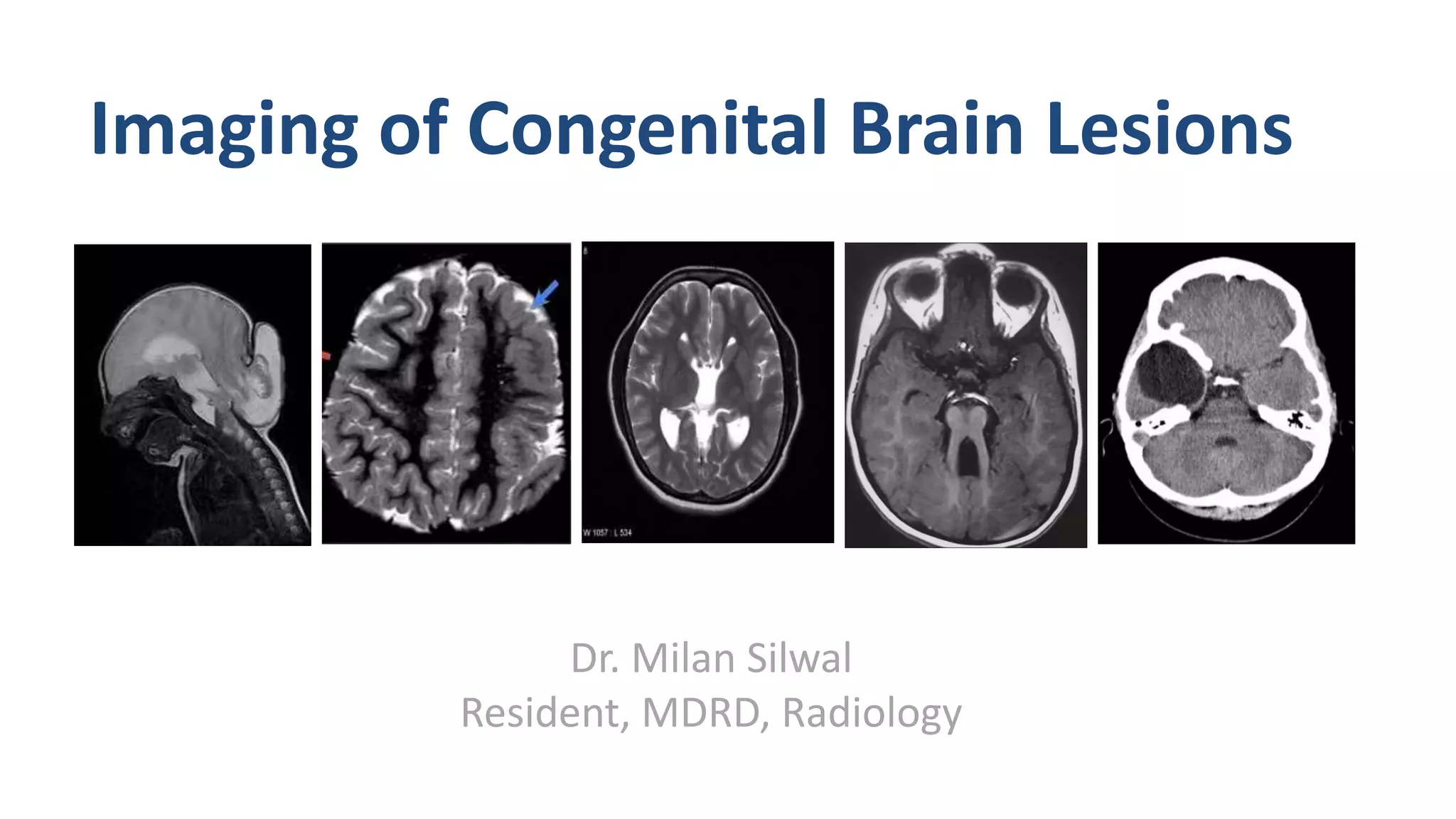
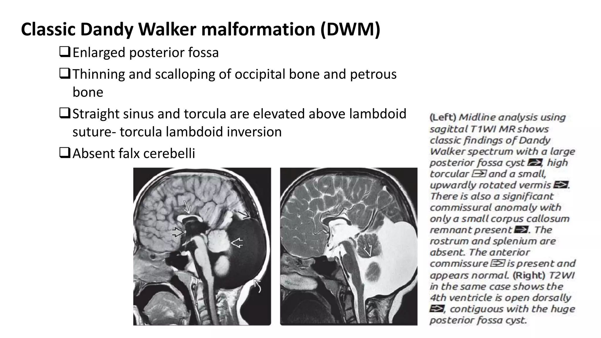
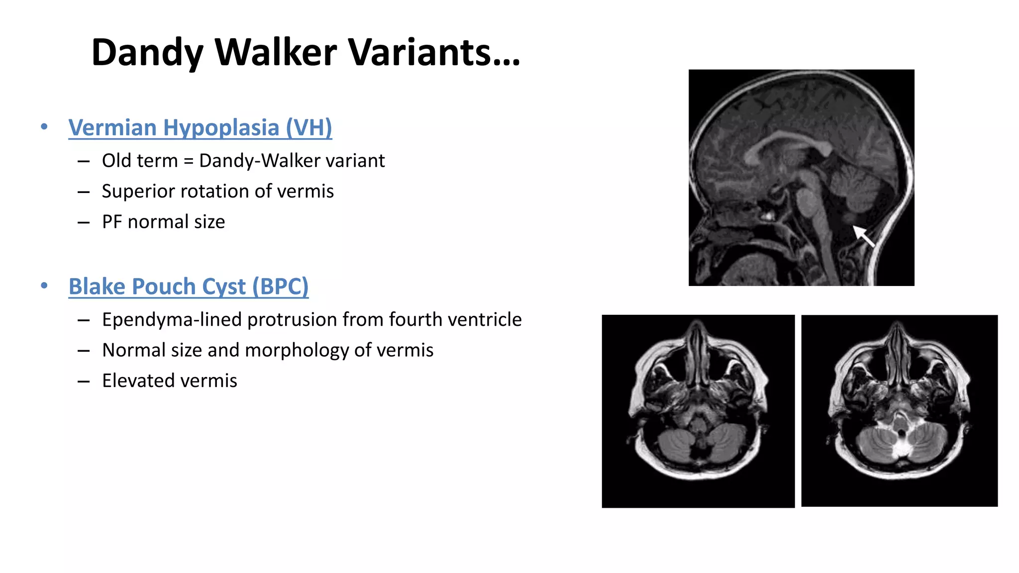
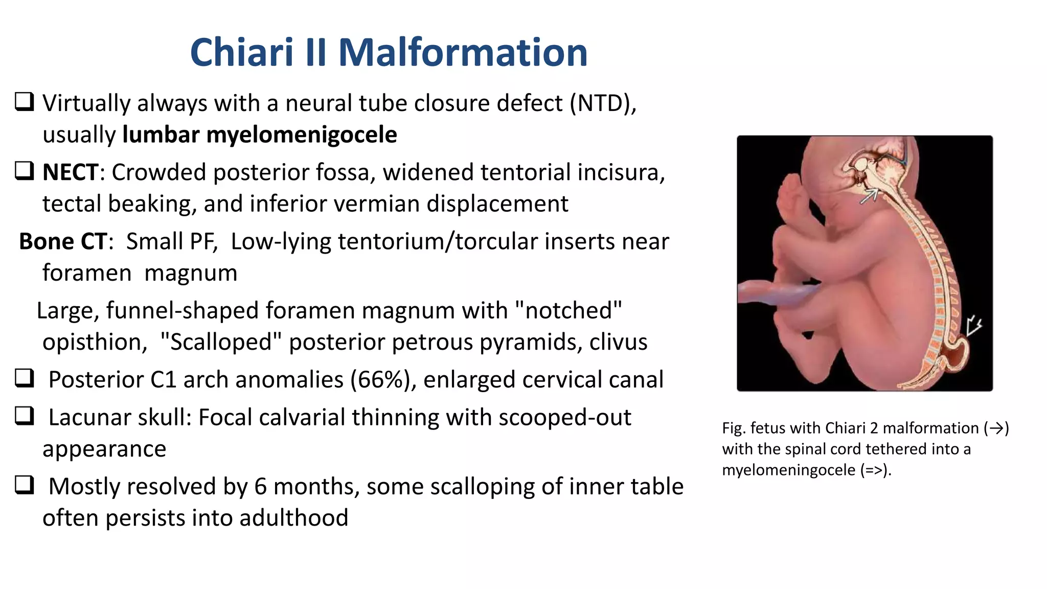
Imaging plays a key role in the diagnosis of diseases of the trachea, bronchi and small airways. The technical advances relating to CT, and in particular the ability to rapidly acquire a volume of data with multidetector CT, has revolutionised the investigation of patients with suspected airway disease.
Tracheal abnormalities can be due to intrinsic or extrinsic causes and may be focal, multifocal or diffuse. CT is now the investigation of choice for suspected bronchiectasis. Asthma remains a clinical diagnosis, but advances in CT technology now allow quantitative assessment of the bronchial wall and this is providing insights into the nature of airway remodelling that occurs in asthma.
Small airways (for practical purposes the bronchioles) are numerous and thus clinical tests are insensitive in detecting disease. This has increased the role and importance of CT in identifying either of the two main categories of small airway disease—constrictive bronchiolitis and exudative bronchiolitis.
In this presentation we will discuss role of high resolution in characterizing normal variant and pathologies of spinal pathologies.
This is a pictoral review.
In this presentation we will discuss role of high resolution in characterizing normal variant and pathologies of spinal pathologies.
This is a pictoral review.
FETAL CENTRAL NERVOUS SYSTEM ANAOMALIES PRESENTATIONkumarramalakshmi
various fetal cns anamolies described with ante nantal and postnatal imaging features of ultrasound, barium study CT and MRI in each entity with representative iamges
It's about HYDROCEPHALUS
TO EXPLAIN ANATOMY OF HUMAN BRAIN
TO INTRODUCE HYDROCEPHALUS
TO DEFINE HYDROCEPHALUS
TO EXPLAIN INCIDENCE OF HYDROCEPHALUS
TO EXPLAIN ETIOLOGY OF HYDROCEPHALUS
TO EXPLAIN PATHOPHYSIOLOGY OF HYDROCEPHALUS
TO EXPLAIN CLINICAL MANIFESTATION OF HYDROCEPHALUS
TO ENLIST DIAGNOSIS & DIAGNOSTIC EVALUATION FOR HYDROCEPHALUS
TO DESCRIBE MANAGEMENT OF HYDROCEPHALUS
TO EXPLAIN COMPLICATION & PROGNOSIS OF HYDROCEPHALUS
These simplified slides by Dr. Sidra Arshad present an overview of the non-respiratory functions of the respiratory tract.
Learning objectives:
1. Enlist the non-respiratory functions of the respiratory tract
2. Briefly explain how these functions are carried out
3. Discuss the significance of dead space
4. Differentiate between minute ventilation and alveolar ventilation
5. Describe the cough and sneeze reflexes
Study Resources:
1. Chapter 39, Guyton and Hall Textbook of Medical Physiology, 14th edition
2. Chapter 34, Ganong’s Review of Medical Physiology, 26th edition
3. Chapter 17, Human Physiology by Lauralee Sherwood, 9th edition
4. Non-respiratory functions of the lungs https://academic.oup.com/bjaed/article/13/3/98/278874
Pulmonary Thromboembolism - etilogy, types, medical- Surgical and nursing man...VarunMahajani
Disruption of blood supply to lung alveoli due to blockage of one or more pulmonary blood vessels is called as Pulmonary thromboembolism. In this presentation we will discuss its causes, types and its management in depth.
Flu Vaccine Alert in Bangalore Karnatakaaddon Scans
As flu season approaches, health officials in Bangalore, Karnataka, are urging residents to get their flu vaccinations. The seasonal flu, while common, can lead to severe health complications, particularly for vulnerable populations such as young children, the elderly, and those with underlying health conditions.
Dr. Vidisha Kumari, a leading epidemiologist in Bangalore, emphasizes the importance of getting vaccinated. "The flu vaccine is our best defense against the influenza virus. It not only protects individuals but also helps prevent the spread of the virus in our communities," he says.
This year, the flu season is expected to coincide with a potential increase in other respiratory illnesses. The Karnataka Health Department has launched an awareness campaign highlighting the significance of flu vaccinations. They have set up multiple vaccination centers across Bangalore, making it convenient for residents to receive their shots.
To encourage widespread vaccination, the government is also collaborating with local schools, workplaces, and community centers to facilitate vaccination drives. Special attention is being given to ensuring that the vaccine is accessible to all, including marginalized communities who may have limited access to healthcare.
Residents are reminded that the flu vaccine is safe and effective. Common side effects are mild and may include soreness at the injection site, mild fever, or muscle aches. These side effects are generally short-lived and far less severe than the flu itself.
Healthcare providers are also stressing the importance of continuing COVID-19 precautions. Wearing masks, practicing good hand hygiene, and maintaining social distancing are still crucial, especially in crowded places.
Protect yourself and your loved ones by getting vaccinated. Together, we can help keep Bangalore healthy and safe this flu season. For more information on vaccination centers and schedules, residents can visit the Karnataka Health Department’s official website or follow their social media pages.
Stay informed, stay safe, and get your flu shot today!
Factory Supply Best Quality Pmk Oil CAS 28578–16–7 PMK Powder in Stockrebeccabio
Factory Supply Best Quality Pmk Oil CAS 28578–16–7 PMK Powder in Stock
Telegram: bmksupplier
signal: +85264872720
threema: TUD4A6YC
You can contact me on Telegram or Threema
Communicate promptly and reply
Free of customs clearance, Double Clearance 100% pass delivery to USA, Canada, Spain, Germany, Netherland, Poland, Italy, Sweden, UK, Czech Republic, Australia, Mexico, Russia, Ukraine, Kazakhstan.Door to door service
Hot Selling Organic intermediates
Lung Cancer: Artificial Intelligence, Synergetics, Complex System Analysis, S...Oleg Kshivets
RESULTS: Overall life span (LS) was 2252.1±1742.5 days and cumulative 5-year survival (5YS) reached 73.2%, 10 years – 64.8%, 20 years – 42.5%. 513 LCP lived more than 5 years (LS=3124.6±1525.6 days), 148 LCP – more than 10 years (LS=5054.4±1504.1 days).199 LCP died because of LC (LS=562.7±374.5 days). 5YS of LCP after bi/lobectomies was significantly superior in comparison with LCP after pneumonectomies (78.1% vs.63.7%, P=0.00001 by log-rank test). AT significantly improved 5YS (66.3% vs. 34.8%) (P=0.00000 by log-rank test) only for LCP with N1-2. Cox modeling displayed that 5YS of LCP significantly depended on: phase transition (PT) early-invasive LC in terms of synergetics, PT N0—N12, cell ratio factors (ratio between cancer cells- CC and blood cells subpopulations), G1-3, histology, glucose, AT, blood cell circuit, prothrombin index, heparin tolerance, recalcification time (P=0.000-0.038). Neural networks, genetic algorithm selection and bootstrap simulation revealed relationships between 5YS and PT early-invasive LC (rank=1), PT N0—N12 (rank=2), thrombocytes/CC (3), erythrocytes/CC (4), eosinophils/CC (5), healthy cells/CC (6), lymphocytes/CC (7), segmented neutrophils/CC (8), stick neutrophils/CC (9), monocytes/CC (10); leucocytes/CC (11). Correct prediction of 5YS was 100% by neural networks computing (area under ROC curve=1.0; error=0.0).
CONCLUSIONS: 5YS of LCP after radical procedures significantly depended on: 1) PT early-invasive cancer; 2) PT N0--N12; 3) cell ratio factors; 4) blood cell circuit; 5) biochemical factors; 6) hemostasis system; 7) AT; 8) LC characteristics; 9) LC cell dynamics; 10) surgery type: lobectomy/pneumonectomy; 11) anthropometric data. Optimal diagnosis and treatment strategies for LC are: 1) screening and early detection of LC; 2) availability of experienced thoracic surgeons because of complexity of radical procedures; 3) aggressive en block surgery and adequate lymph node dissection for completeness; 4) precise prediction; 5) adjuvant chemoimmunoradiotherapy for LCP with unfavorable prognosis.
Ozempic: Preoperative Management of Patients on GLP-1 Receptor Agonists Saeid Safari
Preoperative Management of Patients on GLP-1 Receptor Agonists like Ozempic and Semiglutide
ASA GUIDELINE
NYSORA Guideline
2 Case Reports of Gastric Ultrasound
Tom Selleck Health: A Comprehensive Look at the Iconic Actor’s Wellness Journeygreendigital
Tom Selleck, an enduring figure in Hollywood. has captivated audiences for decades with his rugged charm, iconic moustache. and memorable roles in television and film. From his breakout role as Thomas Magnum in Magnum P.I. to his current portrayal of Frank Reagan in Blue Bloods. Selleck's career has spanned over 50 years. But beyond his professional achievements. fans have often been curious about Tom Selleck Health. especially as he has aged in the public eye.
Follow us on: Pinterest
Introduction
Many have been interested in Tom Selleck health. not only because of his enduring presence on screen but also because of the challenges. and lifestyle choices he has faced and made over the years. This article delves into the various aspects of Tom Selleck health. exploring his fitness regimen, diet, mental health. and the challenges he has encountered as he ages. We'll look at how he maintains his well-being. the health issues he has faced, and his approach to ageing .
Early Life and Career
Childhood and Athletic Beginnings
Tom Selleck was born on January 29, 1945, in Detroit, Michigan, and grew up in Sherman Oaks, California. From an early age, he was involved in sports, particularly basketball. which played a significant role in his physical development. His athletic pursuits continued into college. where he attended the University of Southern California (USC) on a basketball scholarship. This early involvement in sports laid a strong foundation for his physical health and disciplined lifestyle.
Transition to Acting
Selleck's transition from an athlete to an actor came with its physical demands. His first significant role in "Magnum P.I." required him to perform various stunts and maintain a fit appearance. This role, which he played from 1980 to 1988. necessitated a rigorous fitness routine to meet the show's demands. setting the stage for his long-term commitment to health and wellness.
Fitness Regimen
Workout Routine
Tom Selleck health and fitness regimen has evolved. adapting to his changing roles and age. During his "Magnum, P.I." days. Selleck's workouts were intense and focused on building and maintaining muscle mass. His routine included weightlifting, cardiovascular exercises. and specific training for the stunts he performed on the show.
Selleck adjusted his fitness routine as he aged to suit his body's needs. Today, his workouts focus on maintaining flexibility, strength, and cardiovascular health. He incorporates low-impact exercises such as swimming, walking, and light weightlifting. This balanced approach helps him stay fit without putting undue strain on his joints and muscles.
Importance of Flexibility and Mobility
In recent years, Selleck has emphasized the importance of flexibility and mobility in his fitness regimen. Understanding the natural decline in muscle mass and joint flexibility with age. he includes stretching and yoga in his routine. These practices help prevent injuries, improve posture, and maintain mobilit
2. Notochord induces overlying ectoderm to
differentiate into neuroectoderm and form
neural plate.
Neural plate gives rise to neural tube and
neural crest cells.
Notochord becomes nucleus pulposus of
intervertebral disc in adults.
Alar plate (dorsal): sensory
Basal plate (ventral): motor
Embryology
Fig. Formation and closure of the neural
tube. The neural plate (red) forms, folds,
and fuses in the midline. The neural and
cutaneous ectoderm then separate.
Notochord (green) and neural crest (blue)
3. • Telencephalon – cerebral hemispheres
• Diencephalon – thalamus, epithalamus,
hypothalamus
• Mesencephalon – midbrain
• Metencephalon – cerebellum, pons
• Myelencephalon – medulla
Neuroectoderm—CNS neurons,
ependymal cells, oligodendroglia,
astrocytes.
Neural crest—PNS neurons, Schwann
cells.
Mesoderm —Microglia (like
Macrophages)
Fig. The neural tube
closes in a
bidirectional zipper-
like manner, starting in
the middle and
proceeding toward
both ends.
Fig. Development of
primary vesicles. The
prosencephalon (green)
gives rise to the forebrain,
the mesencephalon
(purple) to the midbrain,
and the rhombencephalon
(light blue) to the
hindbrain.
Fig. prosencephalon gives rise
to telencephalon (green) and
diencephalon (red).
Mesencephalon (purple)
elongates while
rhombencephalon gives rise to
metencephalon (yellow) and
myelencephalon (light blue).
4. Stages of CNS development
Dorsal induction (3 to 4 weeks of gestation)
Ventral induction (5 to 8 weeks of gestation)
Neuronal proliferation/differentiation/histogenesis (8 to 18
weeks of gestation), migration (12 to 22 weeks of gestation)
Myelination (5 months fetal age to years postnatal age).
The cortex goes through stages of cell proliferation, cell
migration, and cortical organization in utero at 2 to 4
months, 3 to 5 months, and at 5 months to 2 years post
gestation, respectively
5. Myelination
• White matter when myelinated, is bright on a T1-weighted image
(T1WI) and dark on a T2WI
• Has differentT1 and T2 myelination milestones
6 months
Post natal
• Brain stem
• Internal capsules and optic radiation
1 year
• Forceps major and minor
1 ½ years
• Gyral convolutions as in adults
6. The progression of myelination is predictable and abides by a few
simple general rules; myelination progresses from:
• central to peripheral
• caudal to rostral
• dorsal to ventral
• sensory then motor
7.
8. Dorsal induction (4 wks)
Neural plate forms the neural tube, which
eventually gives rise to the spinal cord and
brain. These inductive events are referred to as
dorsal induction.
Primary Neurulation: formation of the neural
tube from approximately the L 1 or L2 level,
which corresponds to the caudal end of the
notochord upward to the cranial end of the
embryo
Secondary Neurulation - formation of neural
tube below the caudal end of notochord.
9. Ventral Induction (5-10 weeks)
• Ventral induction refers to the inductive events occurring ventrally in the
rostral end of the embryo, resulting in the formation of the face and brain.
Holoprosencephaly
Septo-optic dysplasia
Hypoplasia/aplasia of the cerebellar hemispheres
Hypoplasia/aplasia of the vermis cerebelli
including Joubert syndrome
Dandy-Walker syndrome and Dandy- Walker
variant
Cerebral hemihypoplasia/aplasia
Pituitary anomalies
Lobar hypoplasia/aplasia
Craniosynostosis
11. Neuronal Migration
Cortical neurons form in embryonic Germinal matrix (adjacent
to lateral ventricles); migrate centrifugally out to surface of
brain to form cerebral cortex.
Disorders:
Lissencephaly
Pachygyria
Polymicrogyria
Heterotopia
Schizencephaly
12. Arachnoid Cyst
Most common congenital cystic abnormality in the
brain
Due to congenital splitting of arachnoid membrane.
Commonest locations
• Middle (50-60%) and posterior cranial fossae
• Suprasellar region and behind third ventricle
Intraventricular cysts are rare but favor lateral and
third ventricles.
Usually unilocular; septated cysts may occur.
Posterior fossa or quadrigeminal cistern cysts- mass
effect: obstructive hydrocephalus; hypoplasia of
cerebral tissue.
13. Imaging
CT
CSF density HU 0 to +20
Effaces adjacent sulci
May or may not remodel
bone
No enhancement
D/D: Dilated
subarachnoid space ( if
intrathecal contrast given
it will fill the arachnoid
space first and then fills
the cyst) arachnoid cyst
shows delayed
enhancement
No calcification, fat
MRI
Extraaxial mass
CSF signal intensity
FLAIR: dark
DWI: dark
Fig. Arachnoid cyst (arrows)
involving pineal region and
quadrigeminal cistern and
compressing back of third
ventricle, producing marked
hydrocephalus.
14. Meningoencephalocele
• Chiari III malformations - occipital region.
• Also a/w holoprosencephaly and aqueductal stenosis,
agenesis of the corpus callosum, colobomas, and cleft
lips.
• occipital, parietal or frontal meningoencepahaloceles
are diagnosed clinically
• nasofrontal or sphenoethmoidal encephaloceles may
be clinically occult.
• MR: bony defect with continuity of brain parenchyma
below the defect
• The protruded brain can have heterotopic or
disorganized brain tissue.
15. Hydranencephaly
• Result of major destructive process – prenatal
or perinatal: massive intracerebral cavitation.
• Affects anterior parts of hemisphere,
suggesting major involvement of areas
supplied by ICA.
• Falx present: distinguish from severe
holoprosencephaly.
• PCA territory preserved (posterior part of
temporal lobe, occipital lobe, thalamus and
infratentorial structure).
• D/D: Gross hydrocephalus (usually affects
occipital region, with preserved thin rim of
cortical tissue; infant’s head enlarged).
Fig. Autopsy case of hydranencephaly demonstrates
striking transillumination indicating that most of the
cranium is water-filled.
Fig. Hydranencephaly
16. Holoprocencephaly (HPE)
single ("holo") ventricle involving the embryonic prosencephalon ("pros") of the brain ("encephaly")
• fetal forebrain fails to bifurcate into two
hemispheres.
• As ventral induction is closely related to
facial development, HPE is also associated
with a number of characteristic facial
anomalies.
• 3 main subtypes, in order of decreasing
severity are:
1. Alobar Holoprosencephaly
2. Semilobar Holoprosencephaly
3. Lobar Holoprosencephaly
17. Alobar holoprosencephaly
Monoventricle
Fused thalami
Absent corpus callosum, falx, sepum pellucidum and
olfactory bulbs
Middle and anterior cerebral arteries may be replaced by
tangled branches of internal carotid and basilar vessels;
or azygous ACA
Severe facial malformations – hypotelorism, cyclopia,
anophthalmia, cleft lip/palate
Fig. NECT scan shows holoprosencephaly. Small rim of cortex
surrounds "horseshoe" central monoventricle. Thalami are
fused; More cephalad scan in the same patient shows a large
dorsal cyst/central monoventricle with thin rim of surrounding
brain. No falx or interhemispheric fissure is present.
18. Semilobar holoprosencephaly
• Basic structure of the cerebral lobes present but fused most
commonly anteriorly
• Absence of septum pellucidum
• Monoventricle with partially developed occipital and
temporal horns
• Rudimentary falx cerebri: absent anteriorly
• Incompletely formed interhemispheric fissure
• Partial or complete fusion of the thalami
• Absent olfactory tracts and bulbs
• Agenesis or hypoplasia of the corpus callosum
19. Lobar holoprosencephaly
• cerebral hemispheres are present.
• fusion of the frontal horns of the lateral ventricles.
• wide communication of this fused segment with the
third ventricles
• fusion of the fornices
• absence of septum pellucidum.
• normal or hypoplasia of the corpus callosum.
• Anterior cerebral artery may be displaced anteriorly
to lie directly underneath the frontal bones (snake
under the skull sign)
20. Septo-optic Dysplasia (De Morsier Syndrome)
A minor form of holoprosencephaly
Septum pellucidum is either absent (64%) or partially
absent (36%), causing a squared-off appearance to the
frontal horns of the lateral ventricles
Anterior optic pathways hypoplasia (Small hypoplastic
optic nerves and a small optic chiasm – poor vision and
nystagmus
Hormonal abnormalities (60%) - diminished levels of
growth hormone related to the hypothalamo-pituitary
axis abnormality.
Schizencephaly and neuronal migrational disorders may
accompany septo-optic dysplasia in 50% of cases.
Fig. Septo-optic dysplasia. Coronal T1-weighted image
shows absence of the septum pellucidum with small
optic tracts (arrows) – squaring of frontal horns.
21. Porencephaly
disorder that results in cystic
degeneration
and encephalomalacia and the
formation of porencephalic cysts
Congenital type:
– Localized ageneis of cortical mantle
resulting in formation of a cavity or a
lateral slit through which lateral
ventricle communicates with
convexity.
– Lined by ependyma and laterally by
a thin pia-arachnoid layer.
Acquired type (false porencephaly):
– Secondary to any destructive
process: trauma or infarction.
22. Schizencephaly
clefts, often bilateral extend from
ventricles to convexity of operculoinsular
reigons.
Heterotopias with ectopic gray matter
lining the cleft.
The lips of the cleft may be apposed
(closed-lipped) or gaping (open lipped).
Associated with focal cortical dysplasia
(polymicrogyria), grey matter heterotopias,
agenesis of the septum pellucidum (80% to
90%), and pachygyria.
23. Corpus callosum agenesis
Partial agenesis:
• usually involves the posterior
part and anterior part may
remain normal.
Fig. Normal corpus callosum
Fig. partial agenesis of corpus callosum
24. Complete agenesis:
– Complete corpus callosum and cingulate gyrus
agenesis
High riding third ventricle above insertion of
absent corpus callosum.
Sunray appearance – radial configuration of
medial cerebral sulci extending through absent
callosal body region.
Probst’s bundles: longitudinal symmetrical
bundles along medial surface of hemispheres.
Frontal horns and bodies laterally displaced
and smaller. (Moose head appearance on
coronal image)
Atria and occipital horn: large and rounded
Fig. Probst bundle in agenesis of corpus callosum. Coronal T1-
weighted image shows the Probst bundle (P) causing lateral
displacement of the frontal horns of the lateral ventricles.
Cingulate gyri (C) are malformed. B, The shape of the ventricles
suggests agenesis of the corpus callosum with colpocephaly (c)
Probst bundles account for the inward bowing of the lateral
wall of the lateral ventricle. Evagination of the cingulate sulcus
(arrows) accounts for the high signal intensity medial to the
lateral ventricles
26. ABNORMALITIES OF NEURONAL MIGRATION
Lissencephaly (agyria): smooth brain
• Most severe form of migrational defect –
absence of sulci and convolutions in the
cortex. {Normal before 7th month of fetal
life.}
• Localized or generalized
• Characteristic nodule of calcification in the
septum pellucidum near foramen of Monro
– seen of X ray or CT
27. Cobblestone lissencephaly
or cobblestone cortical malformation (CCM) - type 2
• distinct from "classic” lissencephaly (type 1).
• Overmigration of neuroblasts => an extracortical layer
of aberrant gray matter nodules—the "cobblestones"—
on the brain surfaces.
• Uneven, nodular, "pebbly“ brain surface that resembles
a cobblestone street.
• Associated with ocular anomalies and occur as a part of
a congenital muscular dystrophy (CMD) syndrome.
Fig. axial CT showing Cobblestone lissencephaly
Fig. Cobblestone
28. Pachygyria (incomplete lissencephaly) – thick gyri
Convolutions and cortex: abnormally
wide and thick.
Congenital abnormality that occurs
relatively late in gestation, at 12 to 24
weeks, because of neuroblastic migration
not proceeding completely to the
superficial layers of the cortex
Fig. Pachygyria. Bilateral symmetric pachygyric
brain is seen in the parietal lobes on this FLAIR
image. The white matter does not arborize in
these regions.
29. Polymicrogyria
Small, disorganized cortical convolutions usually in
the cortex subjacent to the sylvian fissures.
The cortex appears thickened. The white matter
thickness is normal.
30. Pachygyria
• Short broad flat gyri
• Thick cortex (8mm)
• Less bumpier
Polymicrogyria
• Multiple small undulating gyri
• Less thick cortex (5-7 mm)
• More bumpier
• a/w anomalous venous drainage
31. Heterotopias
Gray matter that is located in the
wrong place
Seizures, weakness, spasticity,
hyperreflexia, or developmental delay
2 varieties: nodular and band types.
Islands of T2 high signal intensity and
nonenhancing tissue suggestive of
gray matter in a white matter location
in subcortical, subependymal/
periventricular region.
Fig. Subcortical heterotopia. Axial T2-weighted
image delineates gray matter signal intensity
within the centrum semiovale and corona radiata
on the left side (arrowheads). Note the distortion
of the left lateral ventricle at the interface with
heterotopic gray matter.
32. Megalencephaly
Enlargement of all or part of the cerebral hemisphere.
Seizures, hemiplegia, developmental delay, and abnormal skull
configuration
Often polymicrogyria (associated with increased hemispheric
size) or agyria (associated with less severe hemispheric
enlargement) on the affected side.
MR demonstrates a distorted, thickened cortex with
ipsilateral ventricular dilation. This unique feature, that of
ventricular dilation on the side of the enlarged hemisphere,
separates congenital hemimegalencephaly from other
infiltrative lesions.
Heterotopias, Myelination abnormalities
33. Intra-cranial Lipoma
Most commonly occur in midline.
Above Normal corpus callosum or 40% associated partial or
complete agenesis.
Other common locations: pineal and suprasellar regions.
Do not cause mass effect, and vessels course through these
lesions unperturbed.
X ray: marginal calcification of lipoma; larger lesions –
increased lucency between the brackets.
Fatty density with typical location – identified in CT/MRI.
Adjacent calcification best seen in CT (minimal or absent in
smaller lesions.)
34. Fig. Lipoma of the Corpus Callosum. Sagittal midline T1 WI (A) reveals a hyperintense
midline lipoma curving around the corpus callosum (arrows). When fat saturation is
applied (B), the high signal disappears, paralleling the signal loss of suboccipital fat
and confirming the fatty nature of the lesion (arrows). Note the subtle associated
hypogenesis of the splenium of the corpus callosum(arrowhead).
A B
35. Dandy walker Malformations/continuum
Components:
Complete or partial agenesis of
the vermis
Cystic dilation of the 4th ventricle
Enlarged posterior fossa, with
upward displacement of the
transverse sinuses, tentorium, and
torcula-lambdoid inversion
(elevation of the torcular above
the lambdoid suture)
INFRATENTORIAL ABNORMALITIES
36. Cerebellum
– Hypolastic vermis with counterclockwise
rotated
– Lies behind quadrigeminal plate
– Abnormal or dysplastic foliation
– Rarely completely absent vermis
– Hypoplastic and splayed cerebellar
hemispheres against petrous bones
Brain stem
– Thin brain stem due to hypoplastic pons
– Butterfly midbrain; non decussation of
cerebellar peduncles
37. Classic Dandy Walker malformation (DWM)
Enlarged posterior fossa
Thinning and scalloping of occipital bone and petrous
bone
Straight sinus and torcula are elevated above lambdoid
suture- torcula lambdoid inversion
Absent falx cerebelli
39. Dandy Walker Variants
Less severe forms of the Dandy-Walker complex - better
development of the vermis and the fourth ventricle; posterior
fossa cyst is smaller. Neither significant enlargement of the
posterior fossa nor torcular-lambdoid inversion is present
Although the 4th ventricle is enlarged not large enough to produce
enlargement of posterior fossa.
40. Dandy Walker Variants…
• Vermian Hypoplasia (VH)
– Old term = Dandy-Walker variant
– Superior rotation of vermis
– PF normal size
• Blake Pouch Cyst (BPC)
– Ependyma-lined protrusion from fourth ventricle
– Normal size and morphology of vermis
– Elevated vermis
41. • Mega Cisterna Magna (MCM)
– Enlarged retrocerebellar CSF (> 10 mm)
– No mass effect on vermis or cerebellum
– Normal vermis
– Fluid crossed by veins, falx cerebelli
– May scallop, remodel occiput**
** All categories in DWC may "scallop" inner
occipital bone
• Arachnoid Cyst
– Not truly in the Dandy-Walker Continuum
– Cerebellopontine angle > retrovermian
– No communication with 4th ventricle
– No crossing veins or falx cerebelli
– Causes mass effect
42. Joubert syndrome
Autosomal recessive disorder, dysgenetic vermis that
appears split/segmented & dysorganized.
Dysgenesis of inferior portion of brain stem
Fourth ventricle roof appear superiorly convex in sagittal
images.
The imaging findings in Joubert syndrome are virtually
pathognomonic. Molar tooth appearance results from a
lack of normal decussation of superior cerebellar
peduncular fiber tracts which in turn leads to
enlargement of the peduncles, which also follow a more
horizontal course.
43. Joubert syndrome…
Hypoplasia of the vermis brings the two cerebellar
hemispheres in virtual contact with each other
and a nodulus is not seen => “bat wing”
appearance the fourth ventricle develops a from
middle cerebellar peduncle, superior cerebellar
peduncle, and pyramidal decussation
maldevelopment.
The absence of crossing fibers => reduction in the
anteroposterior diameter of the midbrain and
deepening of the interpeduncular cistern
44. Rhombencephalosynapsis
rare entity, cerebellar hemispheric
separation is lost and there is fusion
across midline of the cerebellum.
Fusion/apposition of dentate nuclei &
variable fusion of colliculi
absence of the anterior vermis, fusion
of the dentate nuclei and middle
cerebellar peduncles, and a deficiency
of the posterior vermis.
Agenesis of the septum pellucidum
may coexist.
45. Chiari Malformations
(Segmentation Defects)
Chiari I Malformation
Inferiorly displaced "pointed" tonsils with "crowded"
posterior fossa, effaced retrocerebellar CSF spaces at
foramen magnum/upper cervical level
With/without varying degrees of elongation of medulla
oblongata and fourth ventricle.
46. CT Findings
Bone CT
- Often normal; abnormal cases → short clivus, CVJ
segmentation/fusion anomalies
MR Findings
T1WI : Pointed (not rounded) tonsils ≥ 5 mm below foramen magnum
– "Tight" foramen magnum with small/absent cisterns
– 4th ventricle elongation, hindbrain anomalies
T2WI
- Oblique tonsillar folia (sergeant's stripes like)
47. Chiari 1.5
• Cerebellar tonsillar herniation complicated by other
abnormalities (e.g., caudally displaced brainstem and fourth
ventricle and/or cervicomedullary "kink").
• Differs from classic CM1 in that caudal descent of the
brainstem is present, and tonsillar herniation is typically more
severe.
• Differs from CM2, as myelomeningocele is absent.
48. Chiari II Malformation
Virtually always with a neural tube closure defect (NTD),
usually lumbar myelomenigocele
NECT: Crowded posterior fossa, widened tentorial incisura,
tectal beaking, and inferior vermian displacement
Bone CT: Small PF, Low-lying tentorium/torcular inserts near
foramen magnum
Large, funnel-shaped foramen magnum with "notched"
opisthion, "Scalloped" posterior petrous pyramids, clivus
Posterior C1 arch anomalies (66%), enlarged cervical canal
Lacunar skull: Focal calvarial thinning with scooped-out
appearance
Mostly resolved by 6 months, some scalloping of inner table
often persists into adulthood
Fig. fetus with Chiari 2 malformation (→)
with the spinal cord tethered into a
myelomeningocele (=>).
49. MR Findings
T1WI: Cascade of cerebellum/brainstem downward displacement
– Uvula/nodulus/pyramid of vermis → sclerotic peg
– Cervicomedullary kink (70%)
– Towering cerebellum → compresses midbrain, associated beaked tectum
– 4th ventricle elongated with no posterior point
Open spinal dysraphism, MMC ~ 100% (lumbar > >cervical)
Hydrosyringomyelia (20-90%)
T2WI: Hyperintensity, associated with posterior fossa cysts.
50.
51. Prenatal USG scan
Grayscale ultrasound
Fetal obstetrical ultrasound (US) pivotal for early
diagnosis
– MMC may be identified as early as 10 weeks
– Characteristic brain findings (lemon and banana
signs) seen as early as 12 weeks
Lemon sign
Banana sign
Effacement of cisterna magna
Lumbar menigomyelocele
52. Chiari III Malformation
CT Findings :
NECT
-Midline posterior cephalocele containing cerebellum
-Small posterior cranial fossa ― scalloped clivus, lacunar skull
Bone CT
- Opisthion, upper cervical osseous dysraphic bone defect
CTA
- Basilar artery "pulled" into defect along with brainstem into
cephalocele sac
53. MR Findings
T1WI
-High cervical cephalocele sac containing meninges and cerebellum ― brainstem,
upper cervical cord
- Cisterns, 4th ventricle, dural sinuses may extend into cephalocele (50%)
T2WI
-Tissues in cephalocele sac may be bright (gliosis), strandlike (necrotic), or
hypointense (hemorrhagic)
54. Chiari IV malformation
– Severe cerebellar hypoplasia without displacement of the
cerebellum through the foramen magnum
Chiari V malformation
– Absent cerebellum
– Herniation of the occipital lobe through the foramen magnum
Chiari 0 malformation
- syrinx without cerebellar tonsillar, or brain stem descent
55. PHAKOMATOSES
Neurofibromatosis
Sturge Weber Syndrome
Tuberous Sclerosis Complex
Von Hippel Lindau disease
Ataxia Telangiectasia
L’hermitte Duclos disease
Meningiomatosis
Basal cell nevus syndrome
Encephalocraniocutaneous lipomatosis
Neurocutaneous melanosis
Aicardi Syndrome
Li-Fraumeni syndrome
Schanomatosis
Group of diseases of having in common lesions of skin, retina and nervous system.
Also known as neuroectodermal dysplasias
56. Neurofibromatosis (NF)
NF1 (von Recklinghausen disease)
Mutation in NF1 gene : negative regulator of RAS on
chromosome 17.
100% penetrance, variable expression. At least 2 of
the following
o ≥ 6 Cafe-au-lait spots
o Axillary and inguinal frecklings.
o Lisch’s nodules (pigmented iris hamartomas)
o ≥ 2 neurofibromas or ≥1 plexiform neurofibromas
o Optic pathway glioma
o Distinctive bone lesions (pseudoarthroses, cortical thinning
and sphenoid wing dysplasia)
o Positive family history
Fig. Extensive facial
plexiform
neurofibroma
Fig. multiple café au lait
spots (L) and cutaneous
neurofibromas in NF1.
Fig. Multiple Lisch nodules in a
patient with NF1. Multiple café
au lait spots (L) and cutaneous
neurofibromas (R)
57. NF 1 imaging
Scalp/Skull, Meninges, and Orbit
Dermal neurofibromas
Solitary/multifocal scalp nodules
Increases with age
Localized, well-circumscribed
Plexiform neurofibroma
Pathognomonic of NF1 (30-50% of cases)
Large, bulky infiltrative lesions
Scalp, face/neck, spine
Orbit lesions may extend into cavernous sinus
Sphenoid wing dysplasia
Hypoplasia → enlarged orbital fissure
Enlarged middle fossa ± arachnoid cyst
Temporal lobe may protrude into orbit
Dural ectasia
Tortuous optic nerve sheath
Patulous Meckel caves
Enlarged IACs
Brain
Hyperintense T2/FLAIR WM foci
Wax in first decade, then wane
Rare in adults
Astrocytomas
Most common: pilocytic
Optic pathway, hypothalamus > brainstem
Malignant astrocytoma (anaplastic astrocytoma,
glioblastoma multiforme) less common
Arteries
Progressive ICA stenosis → moyamoya
Fusiform ectasias, arteriovenous fistulas
Vertebral > carotid
58. Fig. NF1 "Unidentified Bright Objects." Axial
FLAIR image demonstrates patchy areas of
T2 hyperintensity (arrows).
Fig. Plexiform neurofibroma involving
cervical nerve roots is depicted in the
graphic (L) and on a coronal STIR scan (R).
Fig. 3D bone CT in a patient with NF1 and sphenoid
dysplasia shows enlarged left orbit and widened superior
orbital fissure
Fig. Sagittal (L) and coronal (R) T2WIs show NF1 with
extreme dural ectasia causing posterior vertebral
scalloping and extensive meningoceles
59. Neurofibromatosis 2
o Mutation in Chr-22
o B/L acoustic schwannoma, Meningioma, ependymoma, Juvenile cataracts
Definite NF2
• Bilateral vestibular schwannomas (VSs)
• First-degree relative with NF2 and unilateral VS diagnosed before 30 years of age
• Or first-degree relative with NF2 and 2 of the following
Meningioma
Glioma
Schwannoma
Juvenile posterior subcapsular lenticular opacities or cataracts
60. Neurofibromatosis Type 2, Cranial Nerve schwannoma, Axial fat saturation Tl WI (A) and coronal T2WI (B)
reveal numerous cranial nerve and spinal cord tumors. The vestibular or acoustic schwannomas (white
arrows) often bilateral in NF-2, and are a hallmark of this disorder. Numerous additional cranial nerve
schwannomas are often present and must be carefully looked for; as this will help confirm the diagnosis1is of
NF-2. Fifth cranial nerve schwannomas expand the cavernous sinuses (open arrows in A). Intramedullary glial
cord tumors(white arrowheads in B) are frequent, but may be slow growing and asymptomatic.
61. Neurofibromatosis type 1 Neurofibromatosis type 2
Common (90% of all neurofibromatosis cases)
Chromosome 17 mutations
Almost always diagnosed by age 10
Cutaneous/eye lesions common (> 95%)
oCafé au lait spots
oLisch nodules
oCutaneous neurofibromas (often multiple)
oPlexiform neurofibromas (pathognomonic)
CNS lesions less common (15-20%)
oT2/FLAIR hyperintensities (myelin vacuolization;
lesions wax, then wane)
oAstrocytomas (optic pathway gliomas—usually
pilocytic—other gliomas)
oSphenoid wing, dural dysplasias
oMoyamoya
oNeurofibromas of spinal nerve roots
Much less common (10% of all neurofibromatosis
cases)
Chromosome 22 mutations
Usually diagnosed in second to fourth decades
Cutaneous, eye lesions less prominent
oMild/few café au lait spots
oJuvenile subcapsular opacities
CNS lesions in 100%
oBilateral vestibular schwannomas (almost all)
oNonvestibular schwannomas (50%)
oMeningiomas (50%)
oCord ependymomas (often multiple)
oSchwannomas of spinal nerve roots
62. Von Hippel-Lindau disease (Retinocerebellar angiomatosis)
Rare, familial disease (Autosomal dominant)
Multiple hemangioblastomas in the retina and
cerebellum; also in spinal cord but rarely in
cerebrum prone to sudden spontaneous
hemorrhage.
Associated with visceral tumors and cysts (renal
cell carcinoma, pheochromocytoma, renal, hepatic
and pancreatic cysts, and angiomas of the liver
and kidney).
Fig. HBs in VHL. Spinal cord tumor has
associated cyst causing myelopathy;
Smaller cerebellar HB.
63.
64. Fig. Von Hippel-Lindau Syndrome. T2-weighted images (Left and right ) and postcontrast TI -weighted images
(middle images) The large cystic lesion(*) with a contrast enhancing mural nodule is classic for cerebellar
hemangioblastoma. Often a vascular flow void may be noted associated with the nodule. The syndrome of Von
Hippel-Lindau also includes retinal angiomas; spinal hemangioblastoma.
65. Sturge Weber Syndrome
(encephalotrigeminal angiomatosis)
Nevus flammeus (Port wine stain) on face
and scalp in trigeminal distribution.
Leptomeningeal angiomatosis (pial
angiomas predominantly in parieto-occipital
area on ipsilateral side).
Cortical gliosis and calcification.
– Xray end on calcification: classic Tram line
appearance.
Ipsilateral choroid plexus larger.
Fig. SWS shows pial angiomatosis , deep medullary
collaterals, enlarged choroid plexus, and atrophy of the
right cerebral hemisphere.
Fig. classic CN V₁-V₂ nevus flammeus
characteristic of SWS
66. Fig. NECT in an 8y girl with SWS shows striking cortical atrophy and
extensive calcifications in the cortex and subcortical WM throughout most
of the left cerebral hemisphere. More cephalad NECT in the same patient
shows the typical serpentine gyral calcifications.
Fig. Axial T2WI - Extensive curvilinear hypointensity in the GMWM
interface. Coronal T2* GRE - "blooming" of the extensive
cortical/subcortical calcifications
Fig. T1 C+ FS shows serpentine enhancement covering gyri, filling sulci; enlargement,
enhancement of ipsilateral choroid plexus. Coronal T1 C+ shows pial angioma and enlarged
choroid plexus
67. Tuberous Sclerosis (Bourneville disease or Epiloia)
Complex autosomal dominant disorder.
Characterized by hamartomas within multiple organ
systems including brain, lungs, skin, kidneys, and heart.
Brain MRI may be useful to confirm a presumptive
clinical diagnosis of tuberous sclerosis.
Epilepsy - most common neurologic: symptom,
developing in up to 90% of patients.
68. Tuberous sclerosis
Definite TSC
2 major features or 1 major + 2 minor
Probable TSC
1 major + 1 minor feature
Possible TSC
1 major or ≥ 2 minor features
69. Fig. Tuberous Sclerosis T1WI (A). postcontrast T1WI (B), CT image (C) and postcontrast TIWI (D). Numerous
subcortical tubers (red arrows in A) and subependymal hamartomas (white arrows in A) are evident on
precontrast Tl WI. The subependymal hamartoma enhance mildly (arrows in B). Enhancement of these
benign lesions is common and does not reflect malignancy. Hamartomas (arrow in C) and subcortical tubers
(arrowhead in C) may calcify, best appreciated on CT. Hamartomas on MRI may be most conspicuous on
gradient echo and T2-weighted imaging, as the lesions are low signal intensity in contrast to the high signal
intensity CSF within the ventricles. Enhancing hamartomas in the region of the foramen of Monro (arrowhead
in D) may slowly enlarge, leading to hydrocephalus and are termed subependymal giant cell tumor "SEGA."
70. Ataxia Telangiectasia
• Autosomal recessive condition with
• Gross Cerebellar atrophy and dilated
4th ventricle
– Cerebellar ataxia followed by rapid
deterioration with choreoathetosis
• Telangiectasia
– mucocutaneous
– Cerebral – may cause hemorrhage
71. L’hermitte-Duclos disease
(dysplastic cerebellar gangliocytoma)
• Rare slow growing cerebellar malformation
in young adults (20-40 years).
• Can be associated with Cowden syndrome
(COLD syndrome) – benign skin tumors, GI
polyps, goitre and breast cancer.
• CT: low density cerebellar area with
thickened folia and no enhancement after
contrast. Calcification may be present.
• MRI: low T1WI, high T2WI, thickened folia –
tigroid appearance.
79. Summary:
• Congenital CNS anomalies are not uncommon.
• In setting of seizure, delayed development or dysmorphic child
(low set ears, abnormal facie, hypotelorism)- likelihood of brain
malformations is even higher.
• MRI is the investigation of choice for evaluation of congenital
CNS lesions.
• Brain anomaly imaging checklist helps both to analyze in
orderly manner and not to overlook the subtle structural
abnormalities.
80. References:
Osborn’s Brain Imaging, Pathology, and Anatomy; 2/e, Osborn
AG et al.
Congenital central nervous system anomalies, Larry B. Poe et
al, radiographics
Various internet sources
Normal Sulcation and Myelination.
Axial T2-weightcd MR image a in a normal premature infant 28 weeks (A), normal premature infant 34 weeks (B), and normal term infant .fO week (C). At 28 weeka, the brain has smooth broad gyri with white matter completely unmyelinated. By 34 weeks, the primary sulci. are formed, with initi·ation of secondary sulcus formation, and gyri are
less broad. White matter remains umnyelillated.By 38 weeks term, sulcation has progressed, and
an adult gyral pattern is established. Note the increasiug depth of the sylvian fissure (artOWs) and
increasing complexity of the gyral pattern.Myelination is noted in the posterior limb of the
internal capsule (dark signal on T2Wl) (a"owheads
in C). Knowledge of gestational age is
critic;al to making the diagnosis of cortical malformations
such as lissencephaly.
Neurulation refers to the folding process in vertebrate embryos, which includes the transformation of the neural plate into the neural tube. The embryo at this stage is termed the neurula.
Arachnoid cyst of the middle cranial fossa
Previously thought to be due to CSF leak in to traumatized leptomeninges it contains sufficient blood deposited within the “hygroma” so T1 N fLAIR has different SIGNAL INTENSITY TO CSF.
Coronal ct demonstrates
a defect in the skull base on the left side (arrows) with remodeling of the
bone, indicative of a long-standing process. This represented a meningocele
coming from the floor of the left middle cranial fossa. Rare but Meningoencephalocele can present as nasal polyp.
Sutton: posterior fossa and basal ganglia appear normal
antenatal ultrasound
in lobar holoprosencephaly, the falx is present, the interhemispheric fissure is fully formed and the thalami are not fused.
Incomplete hippocampal formation
Unlike semilobar holoprosencephaly, the falx is present, the interhemispheric fissure is fully formed and the thalami are not fused.
Often present with refractile seizures or mental retardation.
The corpus callosum develops from anterior genu to posterior splenium, accounting for splenial absence in partial agenesis of the corpus callosum.
The rostrum is the last portion of the corpus callosum to form hence combination of absence of the splenium with rostrum agenesis is not unexpected.
Primary corpus callosum never forms; corpus callosum destroyed
Reach/ branch
Ischemic laminar necrosis of the fifth cortical layer after the twentieth week of gestation, by which time the neurons have reached the cortical surface.
Why? too many neurons and decreased apoptosis.
Midline corpus callosal lipoma with peripheral curvilinear calcification
Elevation of confluence of sinuses above lambdoid suture. confluence of sinuses, also know as torcula Herophili.
Hypoplastic cerebellum
Elevated to tentorium and torcula
Cerebellar hemispheres winged anterolaterally by the cyst
Main difference from classic form is the degree of enlargement of 4th ventricle. Rotation of vermis is the key feature to differentiate Dandy Walker variant from other posterior fossa cystic structure like mega cisterna magna.
Lemon:frontal contour is only minimally concave or
Banana; the way the cerebellum is wrapped tightly around the brain stem as a result of spinal cord tethering and downward migration of posterior fossa content. The cisterna magna gets obliterated and the shape of the cerebellum has the appearance of a banana.
Greek: phakos = lentil – lentil shaped object such as a spot on the body or retina.
Café au lait spots, or café au lait macules, are flat, pigmented birthmarks.[1] The name café au lait is French for "coffee with milk" and refers to their light-brown color. They are also called "giraffe spots," or "coast of Maine spots," which refers to their jagged borders.[2]
They are caused by a collection of pigment-producing melanocytes in the epidermis of the skin.