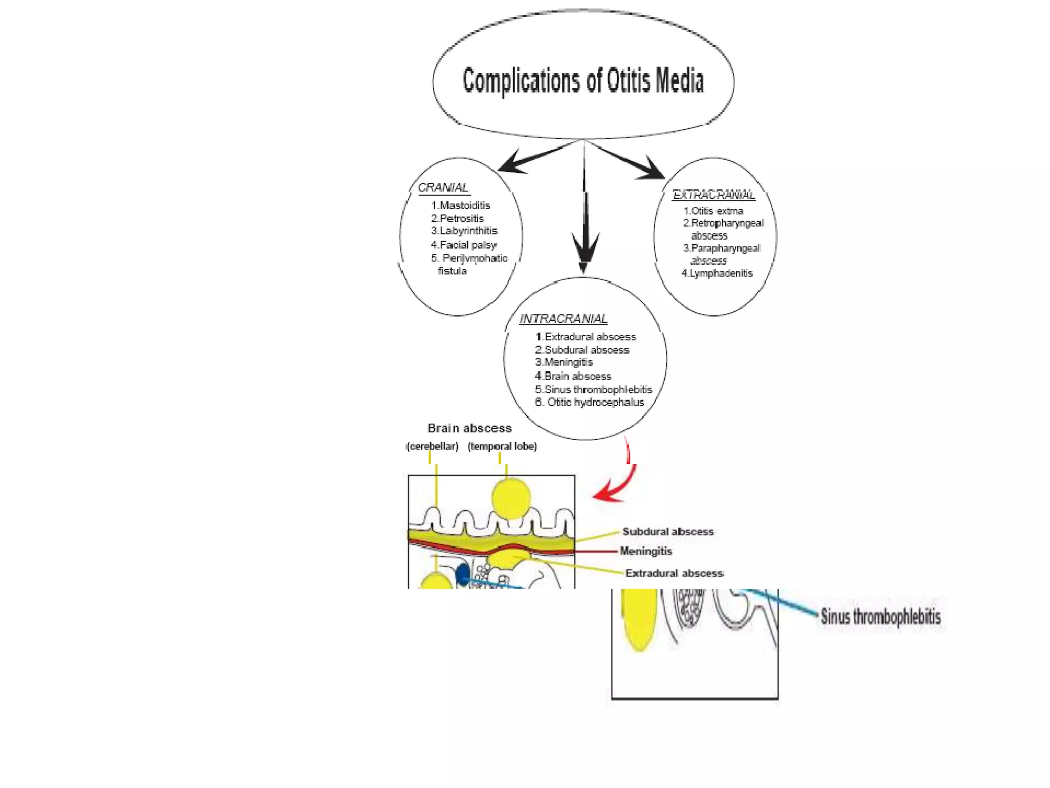
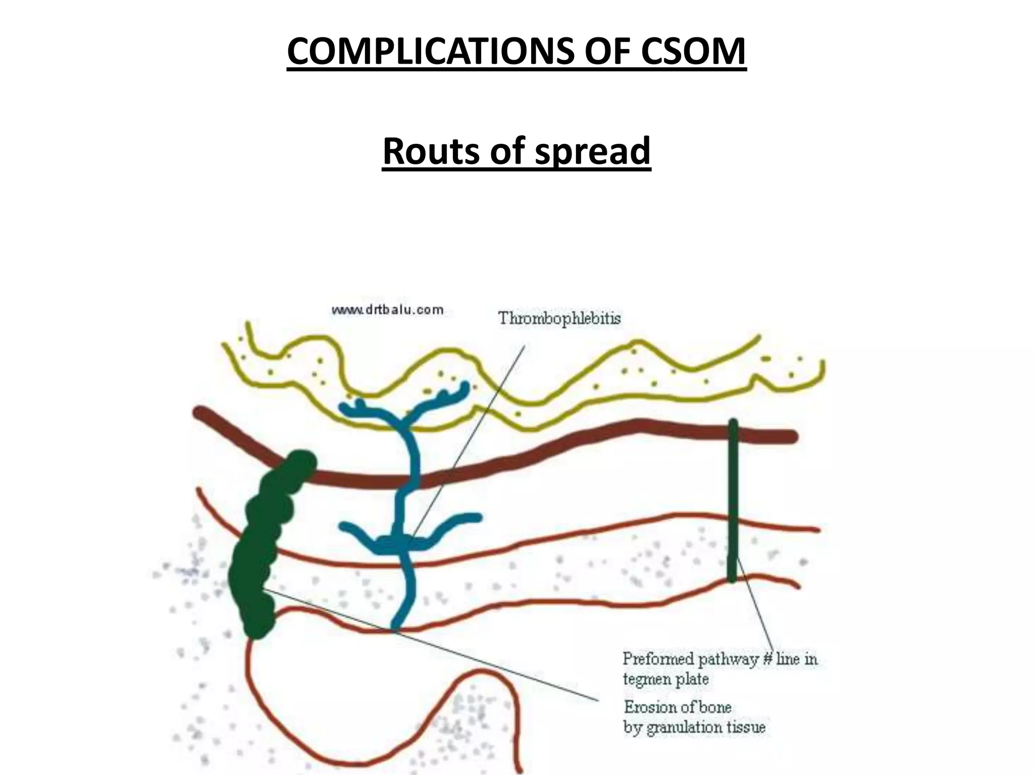
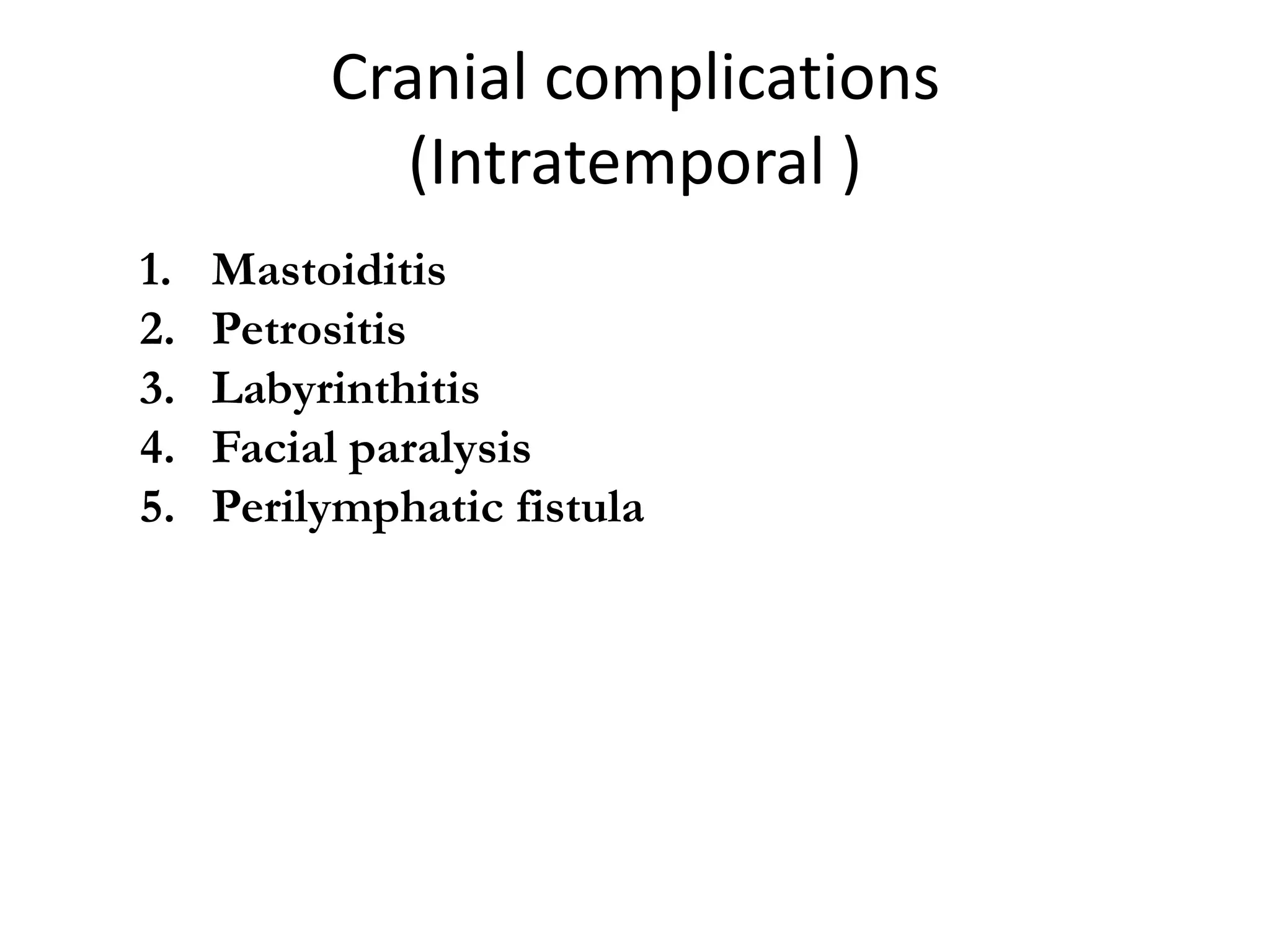
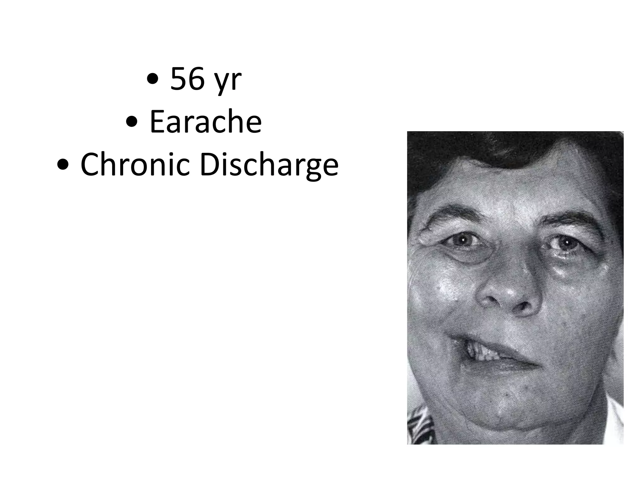
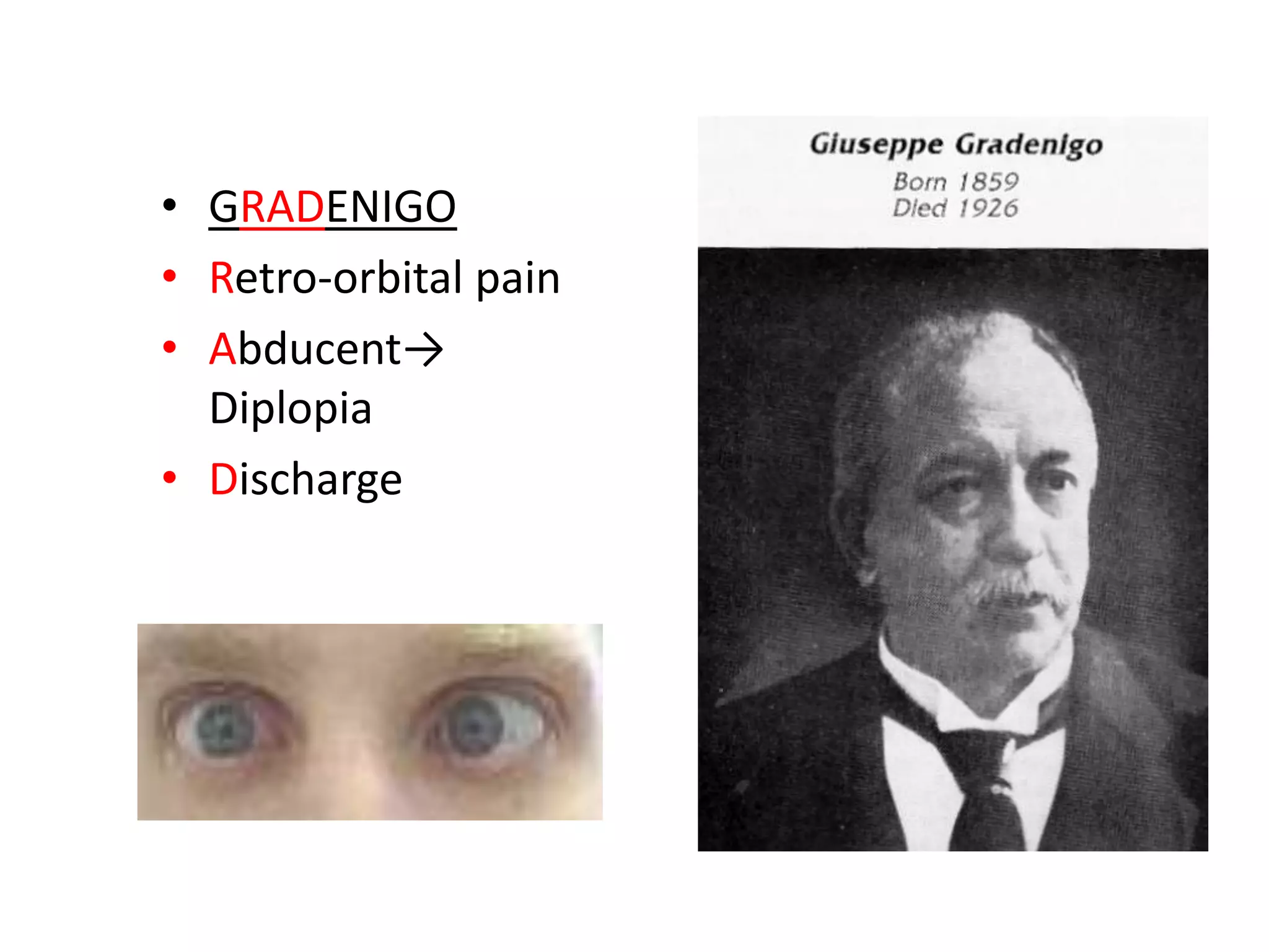
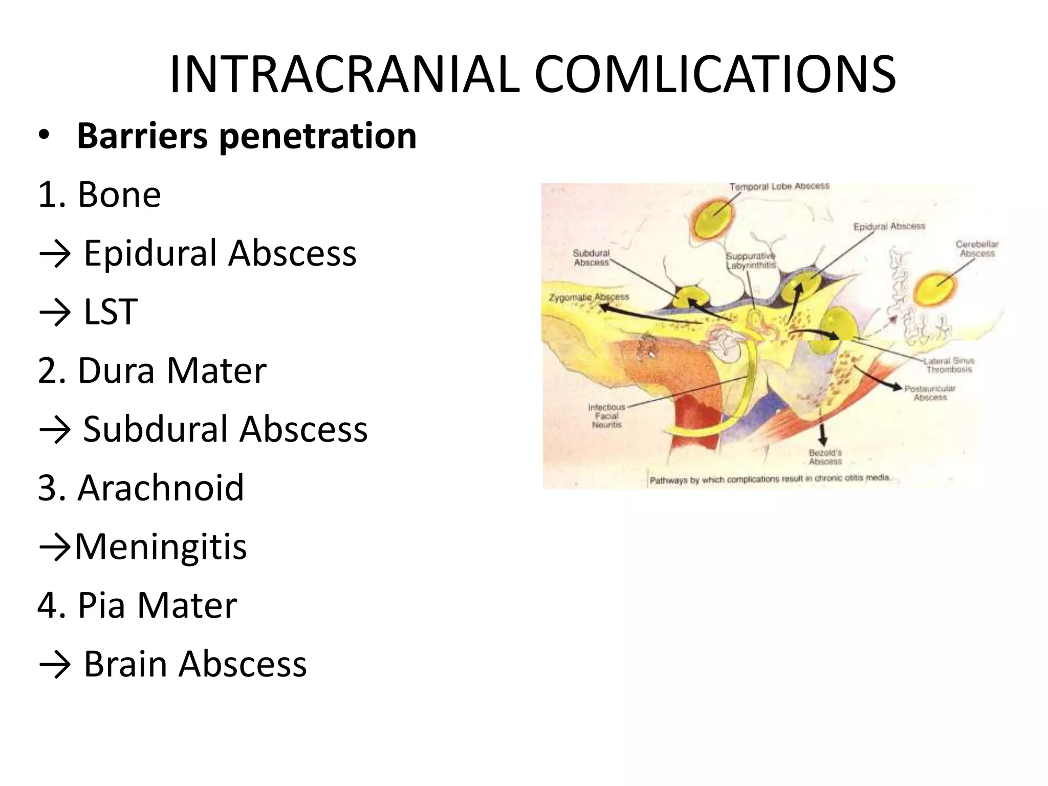
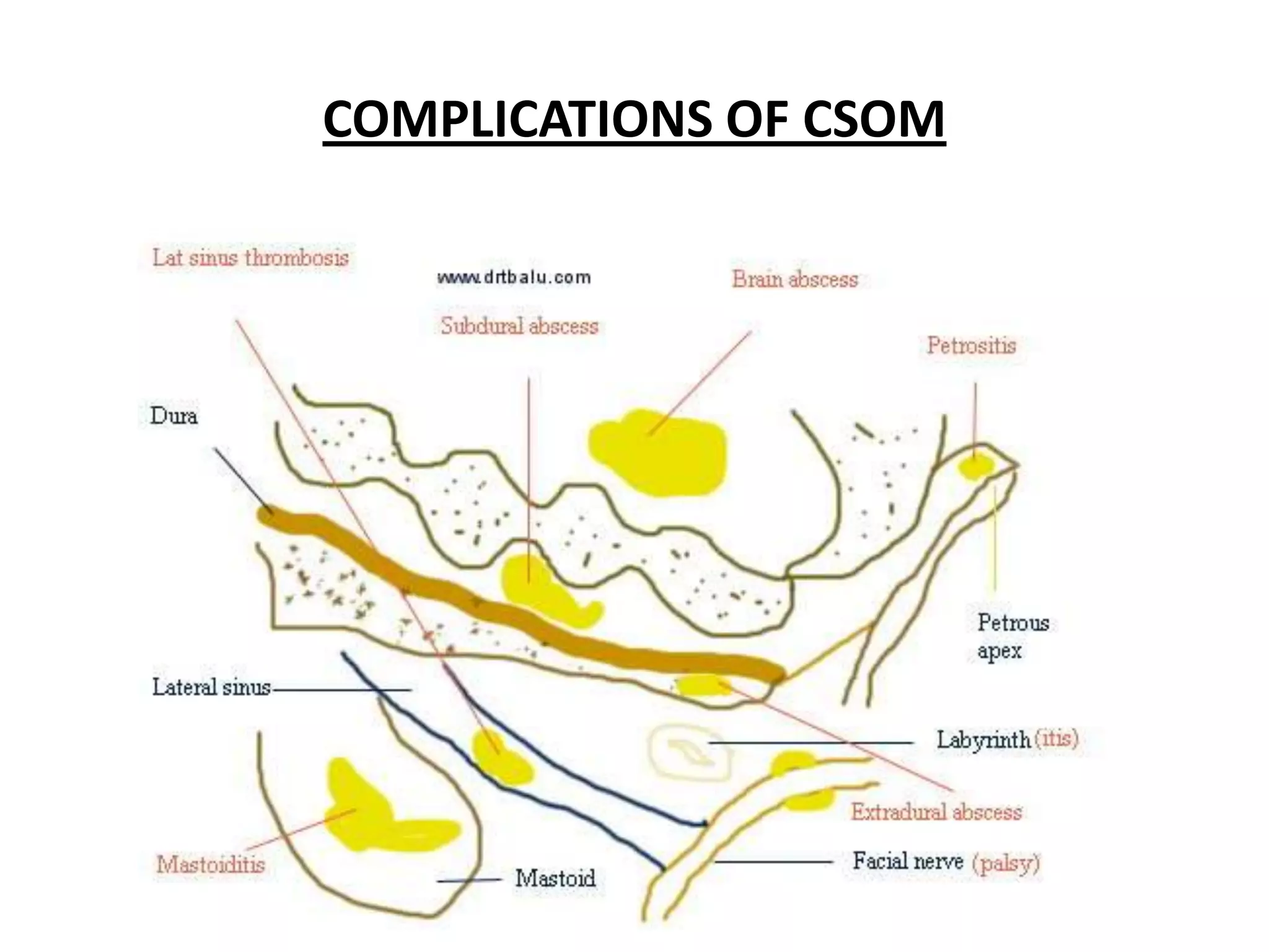
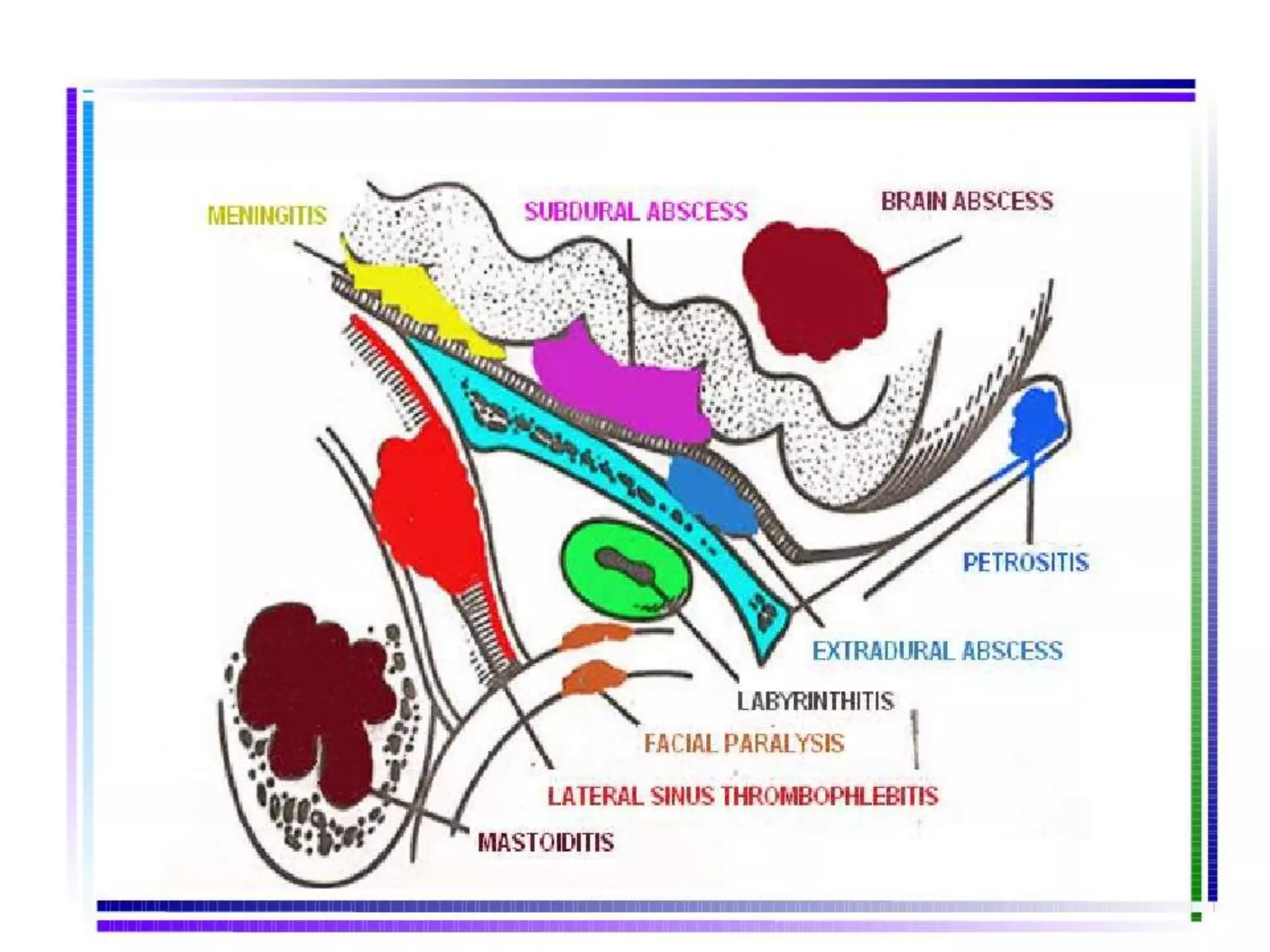
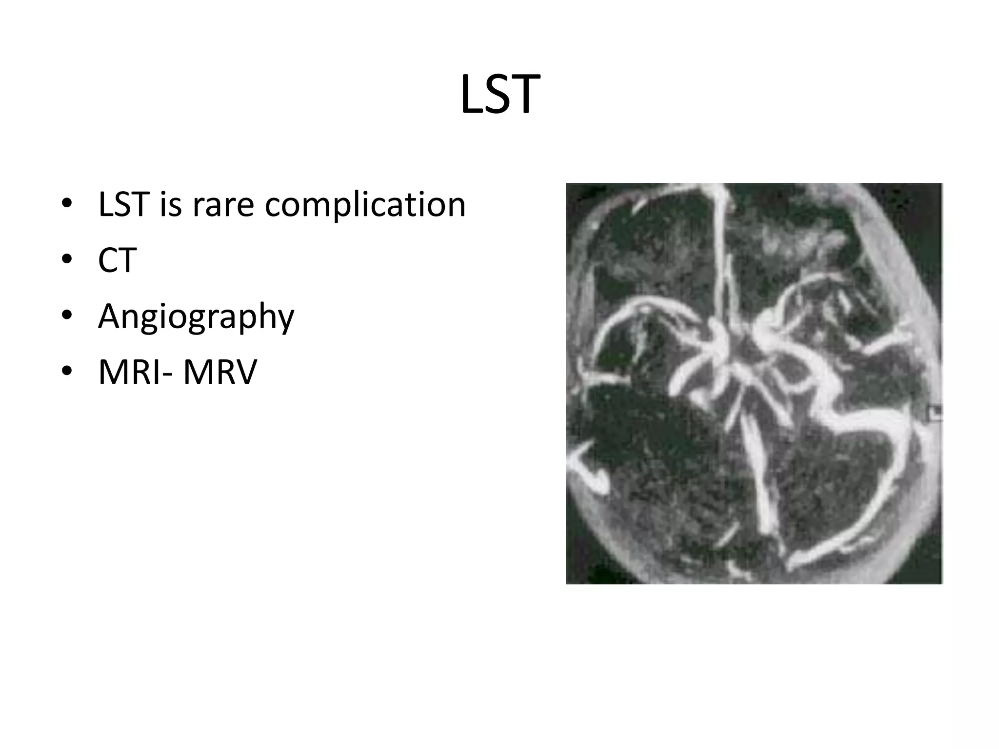
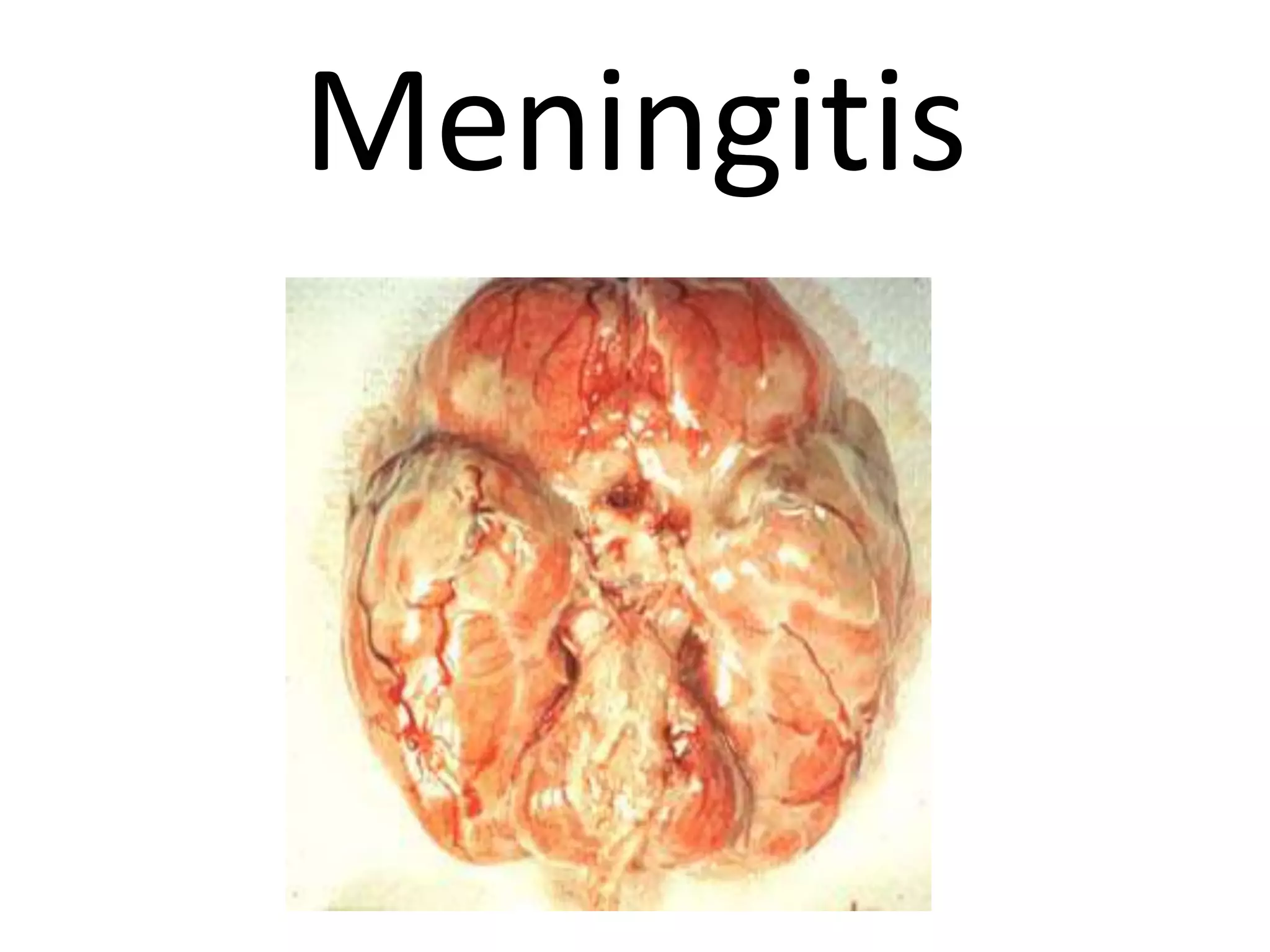
COMPLICATIONS
      C.S.O.M.

Dr. Mohammad Aslam Chaudhry
     Professor E.N.T (BBH)
Anatomical review
“Medial wall of middle ear”
Surgeon’s view
Complications of CSOM
• classification

  1. Cranial (Intra-temporal )

  2. Extra-cranial complications

  3. Intra-cranial complications
Pre-disposing factors
•   Age
•   Virulent organisms
•   Cholesteatoma or Bone erosion
•   Presence of a congenital dehiscence
•   Obstruction of drainage e.g polyp
•   Low resistance of the patient
•   Poor socio-economic status
Routes of spread
Pathways of infections
• The commonest way for extension of
  infection is by bone erosion due to a
  cholesteatoma.
• Vascular extension (retrograde
  thrombophlebitis).
 • Extension along preformed pathways as
– Congenital dehiscences, fracture lines, round
  window membrane, the labyrinth,
– Dehiscences due to previous surgery.
COMPLICATIONS OF CSOM

    Routs of spread
Cranial complications
            (Intratemporal )
1.   Mastoiditis
2.   Petrositis
3.   Labyrinthitis
4.   Facial paralysis
5.   Perilymphatic fistula
Mastoiditis
•    Inflammation of mastoid air cells
•    Acute vs Chronic
•    Fever
•    2 weeks after OM
•   Earache
•   Irritability
Labyrinthitis
•   • Infection of the inner ear
•   • SNHL
•   • Vertigo
•   • Nausea and vomiting
labyrinthitis
Tympanosclerosis
•   Asymptomatic
•   Indicator of OM
•   Ear drum stiffness
•   Ossicular fixation
Tympanosclerosis
• 56 yr
    • Earache
• Chronic Discharge
Facial Paralysis
• Slow – chronic expansion of disease
• Rapid – infected cholesteatoma
• With cholesteatoma requires immediate
  surgery
• CT localizes involved portion
Facial Paralysis
Management

• Mastoidectomy, Remove cholesteatoma and
  infected debris
• IV antibiotics and
  +/- steroids.
Petrositis
•   Only in pneumatised petrous pyramids
•   Infected petrous cells
•   Poor drainage
•   Bony coalescence → symptoms
• GRADENIGO
• Retro-orbital pain
• Abducent→
  Diplopia
• Discharge
Extra-cranial complications
•   Otitis externa
•   Cervical lymphadenitis
•   Retropharyngeal &
•   Parapharyngeal abscesses
Impending sign @ symptoms
• Decreased mental status
• Stiff neck
• Ataxia
• Visual changes
• Seizures
• Other
  Headache, lethargy, fever.
INTRACRANIAL COMLICATIONS
• Barriers penetration
1. Bone
→ Epidural Abscess
→ LST
2. Dura Mater
→ Subdural Abscess
3. Arachnoid
→Meningitis
4. Pia Mater
→ Brain Abscess
COMPLICATIONS OF CSOM
Lateral Sinus Thrombosis (sigmoid sinus)
Spread of infection by direct extension or
via mastoid emissary vein
                    ↓
 Pus and granulation adjacent to sigmoid
sinus
                    ↓
        Reactive thrombophlebitis
                    ↓
        intraluminal thrombus
                    ↓
              CSF obstruction
Signs of LST

• Picket-fence fever 􀃆

• Papilledema

• Torticollis

• Greisinger sign →
LST
•   LST is rare complication
•   CT
•   Angiography
•   MRI- MRV
LST ……Treatment
• Empiric broad coverage until C&S
• antibiotic with good CSF penetration
• Surgery
  – Mastoidectomy
  – Decompression
  – Thrombus evacuation
Intracranial Epidural Abscess
• Localized between dura
  and bone
• dural adherence to bone
  at suture lines
• Focal osteomyelitis
• Management and
  etiology same as
  subdural empyema
Subdural Abscess
• Between the dura and
  the arachnoid.
• Potential space
• Lack of anatomical
  boundaries
→ spread rapidly
• Ear 14%
• (paranasal sinusitis
  75%)
Subdural Abscess - clinical
• Fever
• Focal neurological deficit
• Headache
• Seizures
• Forehead or eye swelling from
  emissary vein thrombosis
• Vomiting
Subdural Abscess - evaluation
• CT of head both with and without contrast
• LP - hazardous - risk of transtentorial
  herniation
Meningitis
Meningitis: Clinical Manifestations
•   Headache
•   Nuchal rigidity
•   Fever and chills
•   Photophobia
•   Vomiting
•   Seizures
•   Focal neurologic symptoms
•   Altered sensorium (confusion, delirium, or
    declining level of consciousness)
Physical Examination


Kernig & Brudzinski signs have low sensitivity but high specificity
Brain Abscess - Clinical
                Presentation
•   non-specific Symptoms for abscess
•   increased intracranial pressure
•   –Headache,
•   –Nausea/Vomiting
•   – Lethargy.
•   – Seizures.
Otic hydrocephalus
Symptoms
• Non-specific
• Headache
• Tinnitus
• Nausea / vomiting
• Visual disturbance
• Others – lethargy, dizziness, mood change
TX
• Decrease the IC pressure
  – Corticosteriods
  – Acetazolamide
  – Lumbar pucture
• I/V Antibiotics
• Mastoidectomy
THANK YOU

Complications of suppurative otitis media

  • 1.
    COMPLICATIONS C.S.O.M. Dr. Mohammad Aslam Chaudhry Professor E.N.T (BBH)
  • 2.
  • 3.
    “Medial wall ofmiddle ear”
  • 4.
  • 5.
    Complications of CSOM •classification 1. Cranial (Intra-temporal ) 2. Extra-cranial complications 3. Intra-cranial complications
  • 7.
    Pre-disposing factors • Age • Virulent organisms • Cholesteatoma or Bone erosion • Presence of a congenital dehiscence • Obstruction of drainage e.g polyp • Low resistance of the patient • Poor socio-economic status
  • 8.
  • 9.
    Pathways of infections •The commonest way for extension of infection is by bone erosion due to a cholesteatoma. • Vascular extension (retrograde thrombophlebitis). • Extension along preformed pathways as – Congenital dehiscences, fracture lines, round window membrane, the labyrinth, – Dehiscences due to previous surgery.
  • 10.
    COMPLICATIONS OF CSOM Routs of spread
  • 11.
    Cranial complications (Intratemporal ) 1. Mastoiditis 2. Petrositis 3. Labyrinthitis 4. Facial paralysis 5. Perilymphatic fistula
  • 12.
    Mastoiditis • Inflammation of mastoid air cells • Acute vs Chronic • Fever • 2 weeks after OM • Earache • Irritability
  • 14.
    Labyrinthitis • • Infection of the inner ear • • SNHL • • Vertigo • • Nausea and vomiting
  • 15.
  • 16.
    Tympanosclerosis • Asymptomatic • Indicator of OM • Ear drum stiffness • Ossicular fixation
  • 17.
  • 18.
    • 56 yr • Earache • Chronic Discharge
  • 19.
    Facial Paralysis • Slow– chronic expansion of disease • Rapid – infected cholesteatoma • With cholesteatoma requires immediate surgery • CT localizes involved portion
  • 20.
    Facial Paralysis Management • Mastoidectomy,Remove cholesteatoma and infected debris • IV antibiotics and +/- steroids.
  • 21.
    Petrositis • Only in pneumatised petrous pyramids • Infected petrous cells • Poor drainage • Bony coalescence → symptoms
  • 22.
    • GRADENIGO • Retro-orbitalpain • Abducent→ Diplopia • Discharge
  • 23.
    Extra-cranial complications • Otitis externa • Cervical lymphadenitis • Retropharyngeal & • Parapharyngeal abscesses
  • 24.
    Impending sign @symptoms • Decreased mental status • Stiff neck • Ataxia • Visual changes • Seizures • Other Headache, lethargy, fever.
  • 25.
    INTRACRANIAL COMLICATIONS • Barrierspenetration 1. Bone → Epidural Abscess → LST 2. Dura Mater → Subdural Abscess 3. Arachnoid →Meningitis 4. Pia Mater → Brain Abscess
  • 26.
  • 28.
    Lateral Sinus Thrombosis(sigmoid sinus) Spread of infection by direct extension or via mastoid emissary vein ↓ Pus and granulation adjacent to sigmoid sinus ↓ Reactive thrombophlebitis ↓ intraluminal thrombus ↓ CSF obstruction
  • 29.
    Signs of LST •Picket-fence fever 􀃆 • Papilledema • Torticollis • Greisinger sign →
  • 30.
    LST • LST is rare complication • CT • Angiography • MRI- MRV
  • 31.
    LST ……Treatment • Empiricbroad coverage until C&S • antibiotic with good CSF penetration • Surgery – Mastoidectomy – Decompression – Thrombus evacuation
  • 32.
    Intracranial Epidural Abscess •Localized between dura and bone • dural adherence to bone at suture lines • Focal osteomyelitis • Management and etiology same as subdural empyema
  • 33.
    Subdural Abscess • Betweenthe dura and the arachnoid. • Potential space • Lack of anatomical boundaries → spread rapidly • Ear 14% • (paranasal sinusitis 75%)
  • 34.
    Subdural Abscess -clinical • Fever • Focal neurological deficit • Headache • Seizures • Forehead or eye swelling from emissary vein thrombosis • Vomiting
  • 35.
    Subdural Abscess -evaluation • CT of head both with and without contrast • LP - hazardous - risk of transtentorial herniation
  • 36.
  • 37.
    Meningitis: Clinical Manifestations • Headache • Nuchal rigidity • Fever and chills • Photophobia • Vomiting • Seizures • Focal neurologic symptoms • Altered sensorium (confusion, delirium, or declining level of consciousness)
  • 38.
    Physical Examination Kernig &Brudzinski signs have low sensitivity but high specificity
  • 39.
    Brain Abscess -Clinical Presentation • non-specific Symptoms for abscess • increased intracranial pressure • –Headache, • –Nausea/Vomiting • – Lethargy. • – Seizures.
  • 40.
    Otic hydrocephalus Symptoms • Non-specific •Headache • Tinnitus • Nausea / vomiting • Visual disturbance • Others – lethargy, dizziness, mood change
  • 41.
    TX • Decrease theIC pressure – Corticosteriods – Acetazolamide – Lumbar pucture • I/V Antibiotics • Mastoidectomy
  • 42.