SlideShare a Scribd company logo
By: Dr. Wudie.M (ORL-HNS R3)
Clinical evaluation and
management of ossicular disorder
12/5/20201
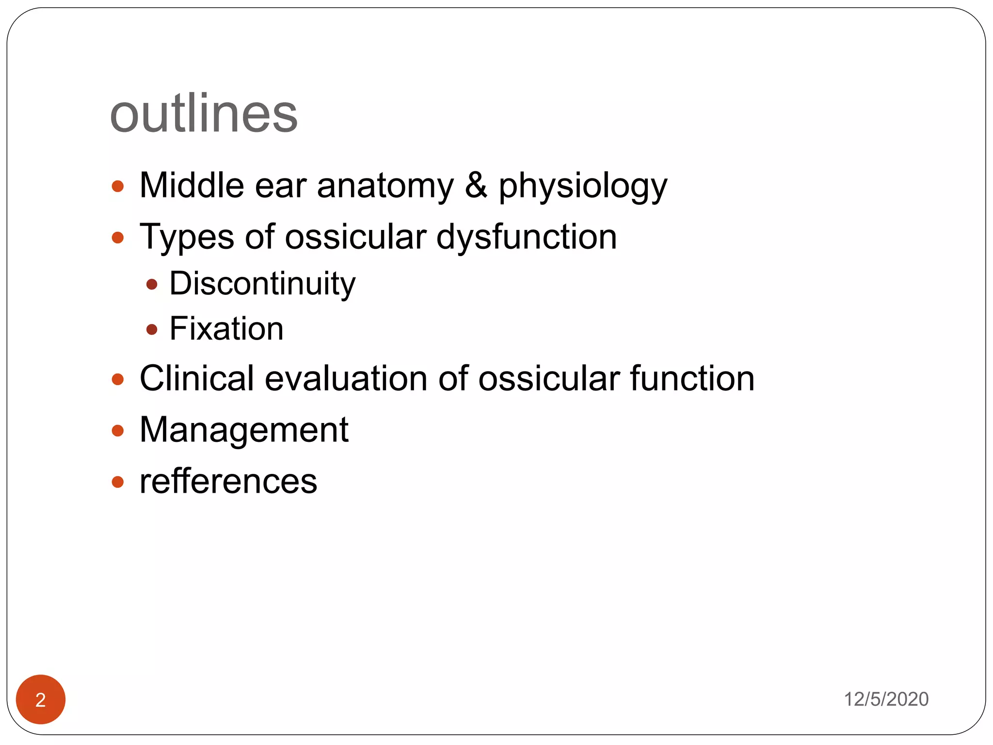
outlines
 Middle ear anatomy & physiology
 Types of ossicular dysfunction
 Discontinuity
 Fixation
 Clinical evaluation of ossicular function
 Management
 refferences
12/5/20202
Anatomy and physiology of middle
ear
 Middle is air filled space
 Divided into hypotympanum,
mesotympanum, and
epitympanum
 The epitympanum, lies above
the level of the malleolar folds.
 The hypotympanum lie below
inferior tympanic sulcus- this
space may contain a
 dehiscent, high-riding jugular
bulb or an aberrant carotid
artery
 The mesotympanum is the
space just medial to the
tympanic membrane,
 Extends from the eustachian
tube opening anteriorly to the
facial nerve posteriorly
12/5/20203
Ossicles
 Malleus
 composed of head
manubrium, neck ,
ant. and lateral
processes
 The anterior process
is attached to the
anterior tympanic
spine by the anterior
mallelar ligament.
 Tensor tympani
muscle attaches to
medial aspect of the
neck and manubrium.
12/5/20204
 The incus is the
largest of the ossicles,
 composed of a body
and three processes
 The short process of
the incus occupies the
incudal fossa,
attached by the
posterior incudal
ligament.
 Body rest to
epitympanuem &
articute with head of
mallues 12/5/20205
 The stapes is the most
medial and smallest of
the ossicles,
 composed of a head
(capitulum), anterior
and posterior crura, and
the footplate.
 The footplate is
encircled by the annular
ligament, which serves
as a “joint,” sealing the
footplate in OW
 Stapedius tendon attach
to the superior aspect of
the posterior crus, just
inferior to the head.
12/5/20206
Blood supply and innervation
 Middle ear is supplied by two arteries are the
main,
 (i) Anterior tympanic branch of maxillary artery
which supplies tympanic membrane.
 (ii) Stylomastoid branch of posterior auricular artery
which supplies middle ear and mastoid air cells
 The main innervation of ME is from the tympanic
plexus and Jacobson’s nerve, which receives a
major contribution from the glossopharyngeal
nerve through the inferior tympanic canaliculus
12/5/20207
12/5/20208
Physiological gain
12/5/20209
 There are two basic pathways by which sound is able
to stimulate the inner ear
1. Ossicular coupling -transmission of sound
pressure through the tympanic membrane and
ossicular chain via the oval window.
hydraulic lever effect -difference between the
surface area of the tympanic membrane relative to
that of the oval window (usually a ratio of nearly 21:1).
 Gain of 26 dB
malleoincudal lever effect – results from length
defference b/n handle of malleus and long process of
incus along its angle of rotation. 1.3:1 … it has gain of
2 dB
2. Acoustic coupling
12/5/202010
 - transmision of sound directley to OW and RW with
out involvement of TM and ossicular chain.
 In the presence of intact ossicular chain and TM , it has
less importance.
 Phase- A sound stimulus to the inner ear is detected
as the net difference in sound pressures applied to
the round and oval windows that results in movement
of intracochlear fluids.
 If these sound pressures are simultaneously applied
to the round and oval windows with equal amplitude
and phase, they will counteract each other and no
resultant intracochlear fluid displacement will occur
Otosclerosis
 Otosclerosis is a disease of bone that is unique to
the otic capsule.
 It may cause a conductive hearing loss, a mixed
conductive-sensorineural hearing loss, or
occasionally a purely sensorineural hearing loss
 It has 2 phase of the disease
 Active phase (cxzed by bone resorption )-
spongiosis
 Remisssion phase – characterized by bone
deposition (sclerosis)
12/5/202011
Types of otosclerosis
1. Clinical otosclerosis – the lesion that affects the
stapdius, stapidiovestibular joint or round wind and
causing CHL
2. Cochlear otosclerosis –the lesion involving cochlear
endostium with out affecting stapidius or
stapidiovestibuar joint, causing pure SNHL
3. Histologic –histopathology of temporal bone shows
otosclerosis but clinically asymptomatic
4. Obliterative -involvement of both oval and round
window and most of bony labyrinthine.
 Results mixed hearing loss
5. Far adevanced otosclerosis- Air or bone
conduction , or at best Ac not better than 95 dB and
bone conduction at 55-60 dB at 1 frequency only.
12/5/202012
Epidemiology
 Most common among Caucasians
 Although occurring in all age groups, usually
presentation is b/n 2nd to 5th decades of age
 F:M 2:1
 In 70 % of cases , it’s bilateral
12/5/202013
Etiology
 Genetic – autosomal dominant
 Infection such as measles viral infection
 Endocrine
 Onset of hearing loss in otosclerosis may
associate pregnancy
12/5/202014
Clinical presentation
History
 Typically, with otosclerosis, hearing loss is
gradual onset and progresses slowly over several
years.
 Disease become apparent in late 10s to 20s of
age
 Most of pts will have CHL with paracusis of
Willis phenomena.
 Unilateral hearing loss may be unoticeble & may
have difficulty with direction of sound.
 Tinnitus is 2nd most common symptoms
12/5/202015
 Positive family hx with negative history for infections
or trauma.
Physical examination
 Otoscopic and microscopic examination – to rule out
other possible causes of CHL
 Schwartze sign - a red blush over the promontory or
the area anterior to the oval window.
 Tuning fork test- with 512 Hz
 Weber's test lateralize to ear which have more CHL
 Rehn’s test BC> AC in the presence of 15 to 20 dB CHL
with 512 Hz fork
12/5/202016
Investigations
 PTA- low frequency CHL with a dip at 2 kHz
known as Carhart’s notch. BC at 2kHZ is 15dB if
it is greater than 15 dB , there may be cochlear
OS.
 Tympanometry-either normal or depressed (As)
 Stapiediul reflex- normal in early stage , and can’t
be elicited in late stage
 Speech reception and perception test are often
normal unless there is choclear involvement.
12/5/202017
CT Scan and MRI
 Not routine workups
 CT scan is more usefull than MRI in OS evalation
 CT scan is indicated in the following cases
 CHL but absent carhart notch
 Equal ABG all over the frequencies
 Mixed hearing loss
 All cases of revision stapes surgery
 Appears as hypodense area, on CT. it has 90%
sensitivity.
12/5/202018
Management
 Surgical- indication is CHL with ABG >20 dB
 stapedioplasty
 Stapedotomy
 Stapedectomy – total vs partial
 Medical
 Hearing aid
12/5/202019
Stapes Mobilization (stapedioplasty)
 performed selected cases who has small point of
fixation of stapes from OS.
 using an endoscope and the argon laser
 potentially avoid the placement of a prosthesis
 Long term follow up show re fixation of stapes.
12/5/202020
Anterior crurotomy with partial
stapedectomy
 removal of only the anterior footplate and crus.
 Done in case of only anterior foot plate fixation
 helpful in a patient with isolated anterior fixation
at the fissula ante fenestram.
 The footplate is fractured in its mid portion, and
only the anterior half is removed
 A connective tissue graft placed over the exposed
area
 The IS join will remain intact and the stapidial
tendon will not cut,
 It has especial benefit for pts who workin noise
env’t. 12/5/202021
Fenestral stapidotomy
 after adequate exposure is established, the
malleus, incus, and stapes are palpated to
ascertain mobility.
 The distance from the incus to the stapes
footplate is measured . The usual distance from
the lateral surface of the incus to the footplate is
4.5 mm.
 Because the piston prosthesis is usually
measured from the medial surface of the incus,
0.25 mm is subtracted to allow for this distance
(incus = 0.5 mm + extension into vestibule of
0.25) .
 The most common piston size is 4.25 mm.
12/5/202022
 A 0.7-mm diamond microdrill is used to create the
fenestra.
 Usually there is adequate room between the
facial nerve and the crus of the stapes to use the
drill on the footplate.
 Despite this, part of the posterior crura is drilled to
warn the patient of the drill noise,
 Weaken the crura for easy fracture, and, at times,
allow better access to the footplate .
 The laser can be employed to begin the fenestra.
 If the footplate is thin, the drill may not be needed
to complete the opening. 12/5/202023
 The fenestra is created with a light touch of the drill .
 The diamond is allowed to create the fenestra without
applying pressure.
 Excess pressure may fracture the footplate.
 When the drill is felt to drop into the vestibule, a
perfect 0.7-mm fenestra is completed.
 The 4.25-mm length, 0.6-mm diameter platinum
Teflon (polytetrafluoroethylene) prosthesis is placed
from the incus to the fenestra .
 It is crimped firmly on the incus .
 The incudostapedial joint is separated, and the
stapedial tendon is sectioned.
12/5/202024
12/5/202025
12/5/202026
 Superstructure of
stapes fractured and
removed
 Test to prostesis
performed to check
position
 Medial lateral
excuresion checked
with light manuplation
 If pt feels vertigo, the
prostesis may be too
long
 If pt does not feel,
check for anterior
posterior excursion & no
 Blood is placed around
the prosthesis as a seal
in the oval window, and
the tympanomeatal flap
is returned to its normal
position.
 Minimal packing is
required on the flap, a
cotton ball is placed in
the meatus,
 and a bandage is
applied to the ear
12/5/202027
stapidectomy
 Total stapedectomy – is
surgical removal of total
foot plate of stapes with
pick and hook
 replace it with a vein
graft and polyethylene
strut.
 partial stapedectomy,
removing only the
posterior half of the
footplate.
 The footplate is opened
with either picks or a
microdril
 The most common
tissue types used for
grafting are the dorsal
hand vein, tragal
perichondrium, or
fascia.
12/5/202028
Post operative care
 Immediately after surgary the pt head should be
elvated to 30 degree for an Hr.
 In the immediate postoperative period, the patient
is asked to avoid lifting and straining for about 1
month.
 Nose blowing should be discouraged.
 The patient should also cough or sneeze with the
mouth open, to reduce the risk of increased
middle ear pressure and displacement of the TM .
 The patient is kept on dry ear precautions until
the TM flap has completely healed.
12/5/202029
 A postoperative audiogram is obtained 2 to 3
months after surgery.
 In 90 % of cases there will be closure of ABG
within 10 dB of the preoperative bone-conduction.
 About 10% of patients experience either
worsening hearing or no improvement
 About 2% of patients suffer persistent and
profound SNHL.
12/5/202030
Out come
 small fenestra
techniques argue that
limited opening of the
vestibule
 lower risk of damage
to the inner ear and
resultant
sensorineural hearing
loss
 The most common
cause for failure of
stapedectomy has
been prosthesis
displacement, with or
without incus erosion.
 Other causes of
failure are
 footplate refixation,
 perilymph fistula.
 otosclerotic regrowth,
and lateralization of
the OW membrane
12/5/202031
 Persistent or progressive CHL can follow
stapedectomy.
 Loosening or displacement of the prosthesis,
 resorption of the incus long process,
 adhesions around the prosthesis, and further OS
lesions can produce postoperative CHL
12/5/202032
complication
Immediate complication
 Complication occurs
intra op
 Facial nerve injury
 Vertigo- usually
resolves with in short
period of time
 Hearing loss
 Persistant perilymphatic
leakage from OW
 Change in taste- in 9 %
of cases , improve with
in 3 to 4 mnths
 Labrynitis
 Tympanic menbrane
perforation- use paper
patch
Delayed complications
 Fistula formation – most
common cause is
poststapedectomy ( 3 %
to 9%),rare in
stapedotomy
 Granuloma
 Prosthesis dislocation-
needs revision surgary
12/5/202033
Alternative treatment
 Hearing aid- CHL caused by OS may use hearing
aid as alternative for surgical mgt
 Pts who have mixed HL may need Hearing aid after
surgical mngt.
 Fluoride therapy- NaF
 Pt with progressive hearing loss may benefit
 It changes active otospongiotic lesion to more
otosclerotic lesion
 Reduce the progression of hearing loss
 8mg po TID until the hearing loss stablized
12/5/202034
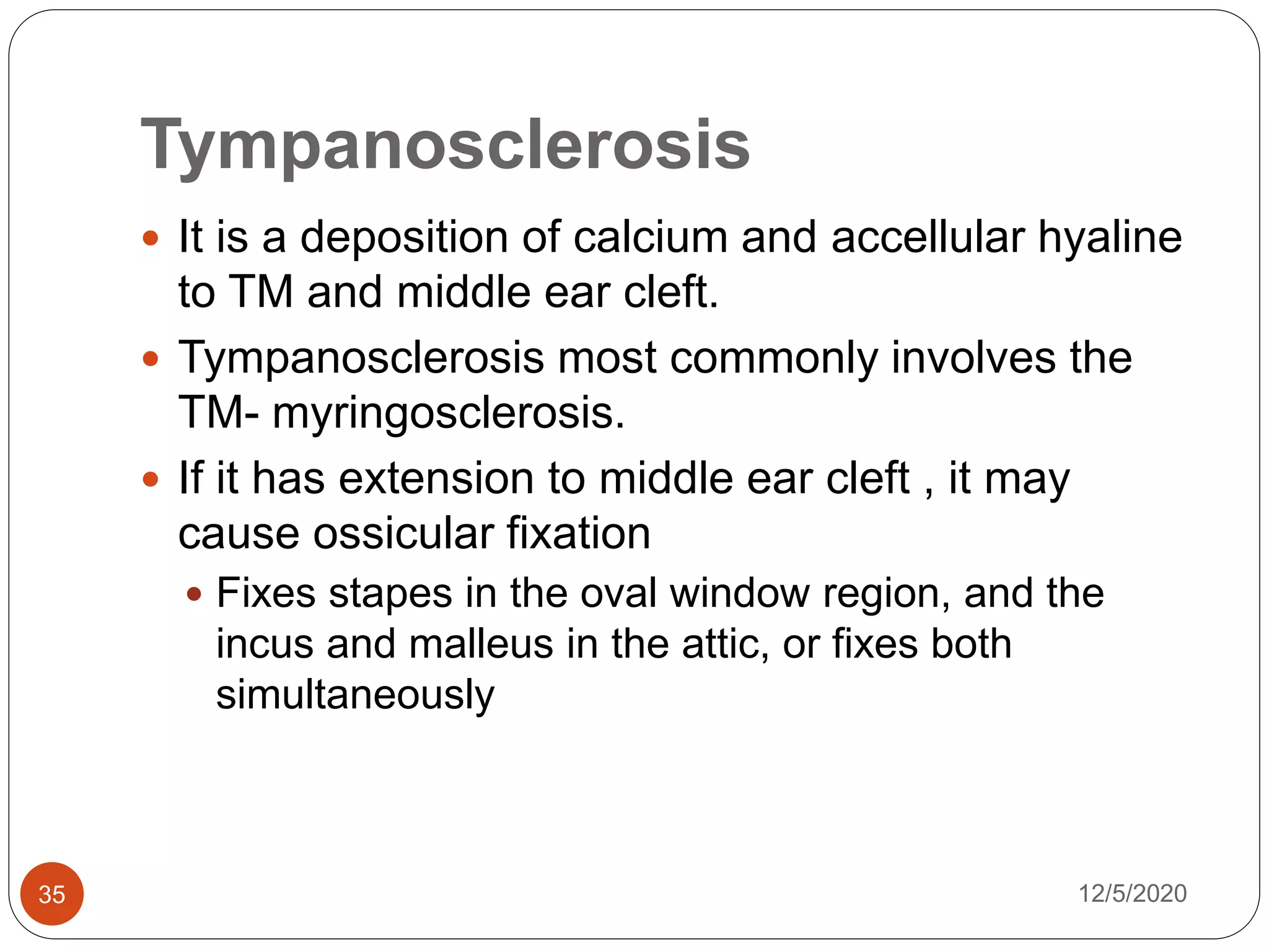
Tympanosclerosis
 It is a deposition of calcium and accellular hyaline
to TM and middle ear cleft.
 Tympanosclerosis most commonly involves the
TM- myringosclerosis.
 If it has extension to middle ear cleft , it may
cause ossicular fixation
 Fixes stapes in the oval window region, and the
incus and malleus in the attic, or fixes both
simultaneously
12/5/202035
 Pathogenesis. Tympanosclerosis results as a
consequence of recurrent AOM, COM, or
tympanostomy-tube placement.
 The exact pathogenesis of tympanosclerosis
remains unclear.
 One possible mechanism is degeneration of
fibroblasts which are known to accumulate in
these plaques progressively
12/5/202036
 cytosolic matrix vesicles rich in calcium,
phosphate, and alkaline phosphatase that
eventually merge with the cell membrane and are
released extracellularly upon fibroblast-cell death
 Another possible mechanism is that of dystrophic
calcification of degenerated collagen fibers after
an infectious or inflammatory insult
12/5/202037
Diagnosis
 Usually it’s asymptomatic and incidental finding
 horseshoe-shaped white plaque over TM
 If there is ossicular chain involvement, will have
significant CHL
 Management
 Tympanoplasty with ossiculoplasty
12/5/202038
12/5/202039
Ossicular chain discontinuity
disorder
12/5/202040
Ossicular trauma
 Most ossicular injuries are dislocation
 Dislocation of incuse being the most frequent
dislocation
 Incudo stapidial joint dislocation with minimal
incus displacement or complete separation may
occur.
 Ossicular fracture are much less frequent. If it
occurs the long process of ossicle is commonest
part.
 Isolated fructure of stapial foot plate is very rare
and almost always associated with penetrating
trauma. 12/5/202041
 Mechanism of injury –
 Skull trauma with or without # of the temporal bone
is the main cause
 About 60% occur with out concomitant TB #
 Barotrauma to middle ear
 Surgical trauma
 Drill-induced trauma to the incus during tympanomastoid
surgery usually causes incudostapedial joint dislocation
12/5/202042
 Evaluation
 PTA
 Initial audiometric evaluation should be done as
soon as after the injury.
 If there is CHL at initial evaluation, , Repeat PTA
after 3 months
 CHL in the presence of intact or healed TM and
with out any sign of hemotympanum ossicular
chain disruption
 Ossicular-chain disruption
 without perforation may result ABG up to 60 dB
 With perforated TM ABG 40 to 50 dB
12/5/202043
 Tympanometry- helps to identify cases of
complete ossicular disruption
 Discontinuous ossicular chain allows a wide
excursion of the tympanic membrane in response to
changing ear canal pressure, AD
 CT scan-
12/5/202044
Management of ossicular trauma
Conservative management
 surprisingly good hearing recovery may take
place with fibrous healing when the
incudostapedial joint is disrupted, even if the
incus is entirely displaced, a 3-month period is
recommended prior to surgical exploration
12/5/202045
Surgical management
 Indication for ossicular reconstruction is
conductive hearing loss of more than 30 dB that
persists for more than 2 months after injury.
 The most conducive injury for ossicular
reconstruction is a dislocation of the
incudostapedial joint.
 In this situation, an Applebaum hydroxyapatite
prosthesis is inserted between the long process
of the incus and the capitulum of the stapes .
 Dislocation of the entire incus requires bridging
the gap between the stapes suprastructure and
the manubrium of the malleus. A sculpted incus
interposition graft is preferred.
12/5/202046
 when the stapes suprastructure is fractured but
the incus remains connected to the malleus;
these patients are good candidates for a laser
stapedotomy.
12/5/202047
Ossicular erosion
 Chronic otitis media in almost any form can result in
the disruption of the integrity of the ossicular chain.
 Chronic eustachian tube insufficiency and tympanic
membrane retraction that results in prolonged contact
of the tympanic membrane with the tip of the incus
can cause this type of erosion, even without active
infection.
 Cholesteatoma is the most common cause of erosion
of the ossicles.
12/5/202048
 It can result
 Loss of lenticular process with preservation of soft
tissue connection
 Erosion of long process of incuse with stapes
suprastructure
 Incudostapidial erosion with partial or total malleus
fixation
12/5/202049
Ossiculoplasty
MATERIALS FOR RECONSTRUCTION
 The ideal material for ossicular reconstruction
should be biologically stable (resistant to resorption
and non-reactive),
 correct mass and stiffness,
 be easy to handle, and
 ideally low cost
 can be broadly divided into autografts,
homografts and alloplastic materials
12/5/202050
 Autografts are tissues that are harvested from the
same patient on which they are to be used, and
can include ossicles, cortical bone and cartilage.
 Homograft material is derived from human donor
tissue. A wide choice of pre-prepared graft
material is available with options including cortical
bone, cartilage, ossicles, and en bloc ossicular
chain with tympanic membrane attached
12/5/202051
 Alloplastic material
 solid plastics: polytetrafluoroethylene, polyethylene
 solid metals: stainless steel, gold, titanium
 porous sponge-like plastics: ProplastR, Plasti-Pore
 ceramics: aluminium oxide, hydroxyapatite.
 PORP = partial ossicular replacement prosthesis.
Generally used to mean a prosthesis that is
designed for situations with an intact stapes
superstructure.
 TORP = total ossicular replacement prosthesis.
For use in situations where there is no stapes
superstructure and the prosthesis restores a
connection with the stapes footplate.
12/5/202052
 A major decision to be made during surgery for
chronic otitis media (especially for cholesteatoma) is
whether to perform ossiculoplasty at the time of the
cholesteatoma excision or to perform ossiculoplasty
as a planned procedure some months later.
 Important factors relevant to this decision include the
following:
1. Status of the middle ear mucosa,
2. Amount of bleeding,
3. Advisability of reoperation for possible cholesteatoma
recurrence, and
 Middle ear mucosa that is thickened, infected,
traumatized, or partially missing is likely to heal with
fibrous tissue formation that may displace a
perfectly placed prosthesis 12/5/202053
SURGICAL OPTIONS TO
CORRECT SPECIFIC DEFECTS
Incus Erosion
 Erosion of long process of incus is the most
frequent ossicular defect
 There are 2 option to reconstruct incus erosion
1. Reconstruct the incudostapidial joint with type II
tympanoplasty
 If the defect between the incus and stapes is very
small, this can be accomplished by wedging a piece
of cartilage or bone between the incus and malleus
12/5/202054
 If the defect is large, we can use various prosthesis
engaged b/n stapes capitulum and the eroded
lentirular process
2. removal of the incus remnant and reconstruction
between the stapes and malleus (or tympanic
membrane),
 a type Ill minor columella tympanoplasty.
 Incus interposition
 PORP- stapes to malleus
12/5/202055
12/5/202056
Malleus present, stapes absent
12/5/202057
 When both the incus and stapes superstructure
are absent, common options for reconstruction
include a type III major columella mechanism or a
type IV tympanoplasty,
 TORP
 Stapes foot plate to malleus
 Stapes foot plate to TM
Malleus absent, stapes present
12/5/202058
 Absence of the malleus handle to be a major
independent prognostic factor resulting in
poorer hearing outcomes following
reconstruction.
 Reconstructed by using
Either by stapes columella or
recreate the malleus handle, either with
autologous material or with a prosthesis
Homograft tympanic membrane with
attached malleus handles
Malleus and stapes absent
12/5/202059
 The most challenging ossicular defect and lead to
the poorest outcomes
 reconstruction may be footplate-totympanic
membrane, or alternatively a neo–malleus or
malleus replacement prosthesis may be used to
improve stability
Reference
12/5/202060
 Cummings otolaryngology–head and neck
surgery, 5th edition
 Scott-brown’s otorhinolaryngology head and neck
surgery,8th edition
 Ballenger’s otorhinolaryngology , head and neck
surgery 18th edition
 Bailey otorhinolaryngology head and neck
surgery, 5th edition
 Otosclerosis Diagnosis, Evaluation, Pathology,
Surgical Techniques, and Outcomes
12/5/202061

More Related Content

What's hot

Glomus Tumour
Glomus TumourGlomus Tumour
Glomus Tumour
Utkal Mishra
 
Endoscopic ear surgery
Endoscopic ear surgeryEndoscopic ear surgery
Endoscopic ear surgery
Ausaf Khan
 
Menieres disease
Menieres diseaseMenieres disease
Menieres disease
Dr. Rajendra Singh Lakhawat
 
Audiometry Ashly
Audiometry  AshlyAudiometry  Ashly
Audiometry Ashly
ashlyalexanderkiran
 
Otoacoustic Emission & BERA
Otoacoustic Emission & BERAOtoacoustic Emission & BERA
Otoacoustic Emission & BERA
Prasanna Datta
 
Chloesteatoma surgery mukace
Chloesteatoma surgery mukaceChloesteatoma surgery mukace
Chloesteatoma surgery mukace
Mukesh Sah
 
Superior Semicircular Canal Dehiscence Syndrome
Superior Semicircular Canal Dehiscence SyndromeSuperior Semicircular Canal Dehiscence Syndrome
Superior Semicircular Canal Dehiscence Syndrome
Ade Wijaya
 
Glomus Tumour
Glomus TumourGlomus Tumour
Glomus Tumour
drvineeth g
 
Otosclerosis
OtosclerosisOtosclerosis
Otosclerosis
ashish gupta
 
Ent essentials
Ent essentialsEnt essentials
Ent essentials
Antriksh Wahi
 
Middle ear ventilatory pathway and Mucosal folds.pptx
Middle ear ventilatory pathway and Mucosal folds.pptxMiddle ear ventilatory pathway and Mucosal folds.pptx
Middle ear ventilatory pathway and Mucosal folds.pptx
SaneeshDamodaran
 
Cavity obliteration @ sayan
Cavity obliteration  @ sayanCavity obliteration  @ sayan
Cavity obliteration @ sayan
IPGMER
 
Tympanoplasty and ossiculoplasty
Tympanoplasty and ossiculoplastyTympanoplasty and ossiculoplasty
Tympanoplasty and ossiculoplasty
Prashant Zade
 
Thyroplasty
ThyroplastyThyroplasty
Measurements of nasal airway(dr ravindra daggupati),rhinomanometry,acoustic r...
Measurements of nasal airway(dr ravindra daggupati),rhinomanometry,acoustic r...Measurements of nasal airway(dr ravindra daggupati),rhinomanometry,acoustic r...
Measurements of nasal airway(dr ravindra daggupati),rhinomanometry,acoustic r...
Ravindra Daggupati
 
Nasal Cavity and Sphenoid Sinus Anatomy
Nasal Cavity and Sphenoid Sinus AnatomyNasal Cavity and Sphenoid Sinus Anatomy
Nasal Cavity and Sphenoid Sinus Anatomy
Farrukh Javeed
 
Spaces of middle ear and their surgical importance
Spaces of middle ear  and their surgical importanceSpaces of middle ear  and their surgical importance
Spaces of middle ear and their surgical importance
Dr Soumya Singh
 
Congenital anomalies of inner ear
Congenital anomalies of inner earCongenital anomalies of inner ear
Congenital anomalies of inner ear
Chanmiki Sayoo
 
anatomy and physiology of middle ear spaces
anatomy and physiology of middle ear spacesanatomy and physiology of middle ear spaces
anatomy and physiology of middle ear spaces
Ram Raju
 
OME-Otitis Media with effusion in children
OME-Otitis Media with effusion in childrenOME-Otitis Media with effusion in children
OME-Otitis Media with effusion in children
Razal M
 

What's hot (20)

Glomus Tumour
Glomus TumourGlomus Tumour
Glomus Tumour
 
Endoscopic ear surgery
Endoscopic ear surgeryEndoscopic ear surgery
Endoscopic ear surgery
 
Menieres disease
Menieres diseaseMenieres disease
Menieres disease
 
Audiometry Ashly
Audiometry  AshlyAudiometry  Ashly
Audiometry Ashly
 
Otoacoustic Emission & BERA
Otoacoustic Emission & BERAOtoacoustic Emission & BERA
Otoacoustic Emission & BERA
 
Chloesteatoma surgery mukace
Chloesteatoma surgery mukaceChloesteatoma surgery mukace
Chloesteatoma surgery mukace
 
Superior Semicircular Canal Dehiscence Syndrome
Superior Semicircular Canal Dehiscence SyndromeSuperior Semicircular Canal Dehiscence Syndrome
Superior Semicircular Canal Dehiscence Syndrome
 
Glomus Tumour
Glomus TumourGlomus Tumour
Glomus Tumour
 
Otosclerosis
OtosclerosisOtosclerosis
Otosclerosis
 
Ent essentials
Ent essentialsEnt essentials
Ent essentials
 
Middle ear ventilatory pathway and Mucosal folds.pptx
Middle ear ventilatory pathway and Mucosal folds.pptxMiddle ear ventilatory pathway and Mucosal folds.pptx
Middle ear ventilatory pathway and Mucosal folds.pptx
 
Cavity obliteration @ sayan
Cavity obliteration  @ sayanCavity obliteration  @ sayan
Cavity obliteration @ sayan
 
Tympanoplasty and ossiculoplasty
Tympanoplasty and ossiculoplastyTympanoplasty and ossiculoplasty
Tympanoplasty and ossiculoplasty
 
Thyroplasty
ThyroplastyThyroplasty
Thyroplasty
 
Measurements of nasal airway(dr ravindra daggupati),rhinomanometry,acoustic r...
Measurements of nasal airway(dr ravindra daggupati),rhinomanometry,acoustic r...Measurements of nasal airway(dr ravindra daggupati),rhinomanometry,acoustic r...
Measurements of nasal airway(dr ravindra daggupati),rhinomanometry,acoustic r...
 
Nasal Cavity and Sphenoid Sinus Anatomy
Nasal Cavity and Sphenoid Sinus AnatomyNasal Cavity and Sphenoid Sinus Anatomy
Nasal Cavity and Sphenoid Sinus Anatomy
 
Spaces of middle ear and their surgical importance
Spaces of middle ear  and their surgical importanceSpaces of middle ear  and their surgical importance
Spaces of middle ear and their surgical importance
 
Congenital anomalies of inner ear
Congenital anomalies of inner earCongenital anomalies of inner ear
Congenital anomalies of inner ear
 
anatomy and physiology of middle ear spaces
anatomy and physiology of middle ear spacesanatomy and physiology of middle ear spaces
anatomy and physiology of middle ear spaces
 
OME-Otitis Media with effusion in children
OME-Otitis Media with effusion in childrenOME-Otitis Media with effusion in children
OME-Otitis Media with effusion in children
 

Similar to Ossicular disorder

Surgical mx. of meneiers disease
Surgical mx. of meneiers diseaseSurgical mx. of meneiers disease
Surgical mx. of meneiers disease
Manipal college of medical sciences
 
External auditory canal anatomy pathologies & management
External auditory canal anatomy pathologies & managementExternal auditory canal anatomy pathologies & management
External auditory canal anatomy pathologies & management
Vikas Jorwal
 
ear surgery ppt.pptx
ear surgery ppt.pptxear surgery ppt.pptx
ear surgery ppt.pptx
nr_amilah
 
Vestibular schwannoma (acoustic neuroma) surgical anatomy and microsurgeries,...
Vestibular schwannoma (acoustic neuroma) surgical anatomy and microsurgeries,...Vestibular schwannoma (acoustic neuroma) surgical anatomy and microsurgeries,...
Vestibular schwannoma (acoustic neuroma) surgical anatomy and microsurgeries,...
Dr Raja Preetham Betha
 
Complications of Stapes surgery.pptx
Complications of Stapes surgery.pptxComplications of Stapes surgery.pptx
Complications of Stapes surgery.pptx
Dr Safika Zaman
 
3)mucosal com
3)mucosal com3)mucosal com
6)active squamous com(cholesteatoma)
6)active squamous com(cholesteatoma)6)active squamous com(cholesteatoma)
6)active squamous com(cholesteatoma)
Shekhar Krishna Debnath
 
Tympanoplasty; Indications, types, anesthesia, surgical procedure.
Tympanoplasty; Indications, types, anesthesia, surgical procedure.Tympanoplasty; Indications, types, anesthesia, surgical procedure.
Tympanoplasty; Indications, types, anesthesia, surgical procedure.
Prasanna Datta
 
Lesions of the temporal bone & petrous ppt
Lesions of the  temporal bone & petrous  pptLesions of the  temporal bone & petrous  ppt
Lesions of the temporal bone & petrous ppt
DEBKUMAR BISWAS
 
Cholesteatoma etiology, theories, clinical features and management
Cholesteatoma  etiology, theories, clinical features and managementCholesteatoma  etiology, theories, clinical features and management
Cholesteatoma etiology, theories, clinical features and management
Divya Raana
 
Otosclersis and stapes surgical management
Otosclersis and stapes surgical managementOtosclersis and stapes surgical management
Otosclersis and stapes surgical management
DRRamendrakumarSingh
 
surgical management of sinusitis
surgical management of sinusitissurgical management of sinusitis
surgical management of sinusitis
hitesh verma
 
Mastoidectomy; Types, Indications, Procedure
Mastoidectomy; Types, Indications, ProcedureMastoidectomy; Types, Indications, Procedure
Mastoidectomy; Types, Indications, Procedure
Prasanna Datta
 
Mastoidectomy
MastoidectomyMastoidectomy
Mastoidectomy
Daria Otgonbayar
 
Traumatic ear
Traumatic earTraumatic ear
Traumatic ear
Marei Elkadeki
 
Congenital nasal obstruction
Congenital nasal obstructionCongenital nasal obstruction
Congenital nasal obstruction
Nassr ALBarhi
 
The septum
The septumThe septum
Nasal fracture ent ppt
Nasal fracture ent ppt Nasal fracture ent ppt
Nasal fracture ent ppt
Ayushree Sharma
 
Otitis part 2
Otitis part 2Otitis part 2
Otitis part 2
Kapil Dhital
 
External approaches to sinus surgery
External approaches to sinus surgeryExternal approaches to sinus surgery
External approaches to sinus surgery
Balasubramanian Thiagarajan
 

Similar to Ossicular disorder (20)

Surgical mx. of meneiers disease
Surgical mx. of meneiers diseaseSurgical mx. of meneiers disease
Surgical mx. of meneiers disease
 
External auditory canal anatomy pathologies & management
External auditory canal anatomy pathologies & managementExternal auditory canal anatomy pathologies & management
External auditory canal anatomy pathologies & management
 
ear surgery ppt.pptx
ear surgery ppt.pptxear surgery ppt.pptx
ear surgery ppt.pptx
 
Vestibular schwannoma (acoustic neuroma) surgical anatomy and microsurgeries,...
Vestibular schwannoma (acoustic neuroma) surgical anatomy and microsurgeries,...Vestibular schwannoma (acoustic neuroma) surgical anatomy and microsurgeries,...
Vestibular schwannoma (acoustic neuroma) surgical anatomy and microsurgeries,...
 
Complications of Stapes surgery.pptx
Complications of Stapes surgery.pptxComplications of Stapes surgery.pptx
Complications of Stapes surgery.pptx
 
3)mucosal com
3)mucosal com3)mucosal com
3)mucosal com
 
6)active squamous com(cholesteatoma)
6)active squamous com(cholesteatoma)6)active squamous com(cholesteatoma)
6)active squamous com(cholesteatoma)
 
Tympanoplasty; Indications, types, anesthesia, surgical procedure.
Tympanoplasty; Indications, types, anesthesia, surgical procedure.Tympanoplasty; Indications, types, anesthesia, surgical procedure.
Tympanoplasty; Indications, types, anesthesia, surgical procedure.
 
Lesions of the temporal bone & petrous ppt
Lesions of the  temporal bone & petrous  pptLesions of the  temporal bone & petrous  ppt
Lesions of the temporal bone & petrous ppt
 
Cholesteatoma etiology, theories, clinical features and management
Cholesteatoma  etiology, theories, clinical features and managementCholesteatoma  etiology, theories, clinical features and management
Cholesteatoma etiology, theories, clinical features and management
 
Otosclersis and stapes surgical management
Otosclersis and stapes surgical managementOtosclersis and stapes surgical management
Otosclersis and stapes surgical management
 
surgical management of sinusitis
surgical management of sinusitissurgical management of sinusitis
surgical management of sinusitis
 
Mastoidectomy; Types, Indications, Procedure
Mastoidectomy; Types, Indications, ProcedureMastoidectomy; Types, Indications, Procedure
Mastoidectomy; Types, Indications, Procedure
 
Mastoidectomy
MastoidectomyMastoidectomy
Mastoidectomy
 
Traumatic ear
Traumatic earTraumatic ear
Traumatic ear
 
Congenital nasal obstruction
Congenital nasal obstructionCongenital nasal obstruction
Congenital nasal obstruction
 
The septum
The septumThe septum
The septum
 
Nasal fracture ent ppt
Nasal fracture ent ppt Nasal fracture ent ppt
Nasal fracture ent ppt
 
Otitis part 2
Otitis part 2Otitis part 2
Otitis part 2
 
External approaches to sinus surgery
External approaches to sinus surgeryExternal approaches to sinus surgery
External approaches to sinus surgery
 

Recently uploaded

Physical demands in sports - WCSPT Oslo 2024
Physical demands in sports - WCSPT Oslo 2024Physical demands in sports - WCSPT Oslo 2024
Physical demands in sports - WCSPT Oslo 2024
Torstein Dalen-Lorentsen
 
Journal Article Review on Rasamanikya
Journal Article Review on RasamanikyaJournal Article Review on Rasamanikya
Journal Article Review on Rasamanikya
Dr. Jyothirmai Paindla
 
OCT Training Course for clinical practice Part 1
OCT Training Course for clinical practice Part 1OCT Training Course for clinical practice Part 1
OCT Training Course for clinical practice Part 1
KafrELShiekh University
 
Role of Mukta Pishti in the Management of Hyperthyroidism
Role of Mukta Pishti in the Management of HyperthyroidismRole of Mukta Pishti in the Management of Hyperthyroidism
Role of Mukta Pishti in the Management of Hyperthyroidism
Dr. Jyothirmai Paindla
 
Integrating Ayurveda into Parkinson’s Management: A Holistic Approach
Integrating Ayurveda into Parkinson’s Management: A Holistic ApproachIntegrating Ayurveda into Parkinson’s Management: A Holistic Approach
Integrating Ayurveda into Parkinson’s Management: A Holistic Approach
Ayurveda ForAll
 
Hemodialysis: Chapter 5, Dialyzers Overview - Dr.Gawad
Hemodialysis: Chapter 5, Dialyzers Overview - Dr.GawadHemodialysis: Chapter 5, Dialyzers Overview - Dr.Gawad
Hemodialysis: Chapter 5, Dialyzers Overview - Dr.Gawad
NephroTube - Dr.Gawad
 
Medical Quiz ( Online Quiz for API Meet 2024 ).pdf
Medical Quiz ( Online Quiz for API Meet 2024 ).pdfMedical Quiz ( Online Quiz for API Meet 2024 ).pdf
Medical Quiz ( Online Quiz for API Meet 2024 ).pdf
Jim Jacob Roy
 
Efficacy of Avartana Sneha in Ayurveda
Efficacy of Avartana Sneha in AyurvedaEfficacy of Avartana Sneha in Ayurveda
Efficacy of Avartana Sneha in Ayurveda
Dr. Jyothirmai Paindla
 
Local Advanced Lung Cancer: Artificial Intelligence, Synergetics, Complex Sys...
Local Advanced Lung Cancer: Artificial Intelligence, Synergetics, Complex Sys...Local Advanced Lung Cancer: Artificial Intelligence, Synergetics, Complex Sys...
Local Advanced Lung Cancer: Artificial Intelligence, Synergetics, Complex Sys...
Oleg Kshivets
 
Identifying Major Symptoms of Slip Disc.
 Identifying Major Symptoms of Slip Disc. Identifying Major Symptoms of Slip Disc.
Identifying Major Symptoms of Slip Disc.
Gokuldas Hospital
 
CHEMOTHERAPY_RDP_CHAPTER 4_ANTI VIRAL DRUGS.pdf
CHEMOTHERAPY_RDP_CHAPTER 4_ANTI VIRAL DRUGS.pdfCHEMOTHERAPY_RDP_CHAPTER 4_ANTI VIRAL DRUGS.pdf
CHEMOTHERAPY_RDP_CHAPTER 4_ANTI VIRAL DRUGS.pdf
rishi2789
 
REGULATION FOR COMBINATION PRODUCTS AND MEDICAL DEVICES.pptx
REGULATION FOR COMBINATION PRODUCTS AND MEDICAL DEVICES.pptxREGULATION FOR COMBINATION PRODUCTS AND MEDICAL DEVICES.pptx
REGULATION FOR COMBINATION PRODUCTS AND MEDICAL DEVICES.pptx
LaniyaNasrink
 
Does Over-Masturbation Contribute to Chronic Prostatitis.pptx
Does Over-Masturbation Contribute to Chronic Prostatitis.pptxDoes Over-Masturbation Contribute to Chronic Prostatitis.pptx
Does Over-Masturbation Contribute to Chronic Prostatitis.pptx
walterHu5
 
CHEMOTHERAPY_RDP_CHAPTER 6_Anti Malarial Drugs.pdf
CHEMOTHERAPY_RDP_CHAPTER 6_Anti Malarial Drugs.pdfCHEMOTHERAPY_RDP_CHAPTER 6_Anti Malarial Drugs.pdf
CHEMOTHERAPY_RDP_CHAPTER 6_Anti Malarial Drugs.pdf
rishi2789
 
NARCOTICS- POLICY AND PROCEDURES FOR ITS USE
NARCOTICS- POLICY AND PROCEDURES FOR ITS USENARCOTICS- POLICY AND PROCEDURES FOR ITS USE
NARCOTICS- POLICY AND PROCEDURES FOR ITS USE
Dr. Ahana Haroon
 
Ketone bodies and metabolism-biochemistry
Ketone bodies and metabolism-biochemistryKetone bodies and metabolism-biochemistry
Ketone bodies and metabolism-biochemistry
Dhayanithi C
 
Ear and its clinical correlations By Dr. Rabia Inam Gandapore.pptx
Ear and its clinical correlations By Dr. Rabia Inam Gandapore.pptxEar and its clinical correlations By Dr. Rabia Inam Gandapore.pptx
Ear and its clinical correlations By Dr. Rabia Inam Gandapore.pptx
Dr. Rabia Inam Gandapore
 
Artificial Intelligence Symposium (THAIS)
Artificial Intelligence Symposium (THAIS)Artificial Intelligence Symposium (THAIS)
Artificial Intelligence Symposium (THAIS)
Josep Vidal-Alaball
 
pathology MCQS introduction to pathology general pathology
pathology MCQS introduction to pathology general pathologypathology MCQS introduction to pathology general pathology
pathology MCQS introduction to pathology general pathology
ZayedKhan38
 
K CỔ TỬ CUNG.pdf tự ghi chép, chữ hơi xấu
K CỔ TỬ CUNG.pdf tự ghi chép, chữ hơi xấuK CỔ TỬ CUNG.pdf tự ghi chép, chữ hơi xấu
K CỔ TỬ CUNG.pdf tự ghi chép, chữ hơi xấu
HongBiThi1
 

Recently uploaded (20)

Physical demands in sports - WCSPT Oslo 2024
Physical demands in sports - WCSPT Oslo 2024Physical demands in sports - WCSPT Oslo 2024
Physical demands in sports - WCSPT Oslo 2024
 
Journal Article Review on Rasamanikya
Journal Article Review on RasamanikyaJournal Article Review on Rasamanikya
Journal Article Review on Rasamanikya
 
OCT Training Course for clinical practice Part 1
OCT Training Course for clinical practice Part 1OCT Training Course for clinical practice Part 1
OCT Training Course for clinical practice Part 1
 
Role of Mukta Pishti in the Management of Hyperthyroidism
Role of Mukta Pishti in the Management of HyperthyroidismRole of Mukta Pishti in the Management of Hyperthyroidism
Role of Mukta Pishti in the Management of Hyperthyroidism
 
Integrating Ayurveda into Parkinson’s Management: A Holistic Approach
Integrating Ayurveda into Parkinson’s Management: A Holistic ApproachIntegrating Ayurveda into Parkinson’s Management: A Holistic Approach
Integrating Ayurveda into Parkinson’s Management: A Holistic Approach
 
Hemodialysis: Chapter 5, Dialyzers Overview - Dr.Gawad
Hemodialysis: Chapter 5, Dialyzers Overview - Dr.GawadHemodialysis: Chapter 5, Dialyzers Overview - Dr.Gawad
Hemodialysis: Chapter 5, Dialyzers Overview - Dr.Gawad
 
Medical Quiz ( Online Quiz for API Meet 2024 ).pdf
Medical Quiz ( Online Quiz for API Meet 2024 ).pdfMedical Quiz ( Online Quiz for API Meet 2024 ).pdf
Medical Quiz ( Online Quiz for API Meet 2024 ).pdf
 
Efficacy of Avartana Sneha in Ayurveda
Efficacy of Avartana Sneha in AyurvedaEfficacy of Avartana Sneha in Ayurveda
Efficacy of Avartana Sneha in Ayurveda
 
Local Advanced Lung Cancer: Artificial Intelligence, Synergetics, Complex Sys...
Local Advanced Lung Cancer: Artificial Intelligence, Synergetics, Complex Sys...Local Advanced Lung Cancer: Artificial Intelligence, Synergetics, Complex Sys...
Local Advanced Lung Cancer: Artificial Intelligence, Synergetics, Complex Sys...
 
Identifying Major Symptoms of Slip Disc.
 Identifying Major Symptoms of Slip Disc. Identifying Major Symptoms of Slip Disc.
Identifying Major Symptoms of Slip Disc.
 
CHEMOTHERAPY_RDP_CHAPTER 4_ANTI VIRAL DRUGS.pdf
CHEMOTHERAPY_RDP_CHAPTER 4_ANTI VIRAL DRUGS.pdfCHEMOTHERAPY_RDP_CHAPTER 4_ANTI VIRAL DRUGS.pdf
CHEMOTHERAPY_RDP_CHAPTER 4_ANTI VIRAL DRUGS.pdf
 
REGULATION FOR COMBINATION PRODUCTS AND MEDICAL DEVICES.pptx
REGULATION FOR COMBINATION PRODUCTS AND MEDICAL DEVICES.pptxREGULATION FOR COMBINATION PRODUCTS AND MEDICAL DEVICES.pptx
REGULATION FOR COMBINATION PRODUCTS AND MEDICAL DEVICES.pptx
 
Does Over-Masturbation Contribute to Chronic Prostatitis.pptx
Does Over-Masturbation Contribute to Chronic Prostatitis.pptxDoes Over-Masturbation Contribute to Chronic Prostatitis.pptx
Does Over-Masturbation Contribute to Chronic Prostatitis.pptx
 
CHEMOTHERAPY_RDP_CHAPTER 6_Anti Malarial Drugs.pdf
CHEMOTHERAPY_RDP_CHAPTER 6_Anti Malarial Drugs.pdfCHEMOTHERAPY_RDP_CHAPTER 6_Anti Malarial Drugs.pdf
CHEMOTHERAPY_RDP_CHAPTER 6_Anti Malarial Drugs.pdf
 
NARCOTICS- POLICY AND PROCEDURES FOR ITS USE
NARCOTICS- POLICY AND PROCEDURES FOR ITS USENARCOTICS- POLICY AND PROCEDURES FOR ITS USE
NARCOTICS- POLICY AND PROCEDURES FOR ITS USE
 
Ketone bodies and metabolism-biochemistry
Ketone bodies and metabolism-biochemistryKetone bodies and metabolism-biochemistry
Ketone bodies and metabolism-biochemistry
 
Ear and its clinical correlations By Dr. Rabia Inam Gandapore.pptx
Ear and its clinical correlations By Dr. Rabia Inam Gandapore.pptxEar and its clinical correlations By Dr. Rabia Inam Gandapore.pptx
Ear and its clinical correlations By Dr. Rabia Inam Gandapore.pptx
 
Artificial Intelligence Symposium (THAIS)
Artificial Intelligence Symposium (THAIS)Artificial Intelligence Symposium (THAIS)
Artificial Intelligence Symposium (THAIS)
 
pathology MCQS introduction to pathology general pathology
pathology MCQS introduction to pathology general pathologypathology MCQS introduction to pathology general pathology
pathology MCQS introduction to pathology general pathology
 
K CỔ TỬ CUNG.pdf tự ghi chép, chữ hơi xấu
K CỔ TỬ CUNG.pdf tự ghi chép, chữ hơi xấuK CỔ TỬ CUNG.pdf tự ghi chép, chữ hơi xấu
K CỔ TỬ CUNG.pdf tự ghi chép, chữ hơi xấu
 

Ossicular disorder

  • 1. By: Dr. Wudie.M (ORL-HNS R3) Clinical evaluation and management of ossicular disorder 12/5/20201
  • 2. outlines  Middle ear anatomy & physiology  Types of ossicular dysfunction  Discontinuity  Fixation  Clinical evaluation of ossicular function  Management  refferences 12/5/20202
  • 3. Anatomy and physiology of middle ear  Middle is air filled space  Divided into hypotympanum, mesotympanum, and epitympanum  The epitympanum, lies above the level of the malleolar folds.  The hypotympanum lie below inferior tympanic sulcus- this space may contain a  dehiscent, high-riding jugular bulb or an aberrant carotid artery  The mesotympanum is the space just medial to the tympanic membrane,  Extends from the eustachian tube opening anteriorly to the facial nerve posteriorly 12/5/20203
  • 4. Ossicles  Malleus  composed of head manubrium, neck , ant. and lateral processes  The anterior process is attached to the anterior tympanic spine by the anterior mallelar ligament.  Tensor tympani muscle attaches to medial aspect of the neck and manubrium. 12/5/20204
  • 5.  The incus is the largest of the ossicles,  composed of a body and three processes  The short process of the incus occupies the incudal fossa, attached by the posterior incudal ligament.  Body rest to epitympanuem & articute with head of mallues 12/5/20205
  • 6.  The stapes is the most medial and smallest of the ossicles,  composed of a head (capitulum), anterior and posterior crura, and the footplate.  The footplate is encircled by the annular ligament, which serves as a “joint,” sealing the footplate in OW  Stapedius tendon attach to the superior aspect of the posterior crus, just inferior to the head. 12/5/20206
  • 7. Blood supply and innervation  Middle ear is supplied by two arteries are the main,  (i) Anterior tympanic branch of maxillary artery which supplies tympanic membrane.  (ii) Stylomastoid branch of posterior auricular artery which supplies middle ear and mastoid air cells  The main innervation of ME is from the tympanic plexus and Jacobson’s nerve, which receives a major contribution from the glossopharyngeal nerve through the inferior tympanic canaliculus 12/5/20207
  • 9. Physiological gain 12/5/20209  There are two basic pathways by which sound is able to stimulate the inner ear 1. Ossicular coupling -transmission of sound pressure through the tympanic membrane and ossicular chain via the oval window. hydraulic lever effect -difference between the surface area of the tympanic membrane relative to that of the oval window (usually a ratio of nearly 21:1).  Gain of 26 dB malleoincudal lever effect – results from length defference b/n handle of malleus and long process of incus along its angle of rotation. 1.3:1 … it has gain of 2 dB 2. Acoustic coupling
  • 10. 12/5/202010  - transmision of sound directley to OW and RW with out involvement of TM and ossicular chain.  In the presence of intact ossicular chain and TM , it has less importance.  Phase- A sound stimulus to the inner ear is detected as the net difference in sound pressures applied to the round and oval windows that results in movement of intracochlear fluids.  If these sound pressures are simultaneously applied to the round and oval windows with equal amplitude and phase, they will counteract each other and no resultant intracochlear fluid displacement will occur
  • 11. Otosclerosis  Otosclerosis is a disease of bone that is unique to the otic capsule.  It may cause a conductive hearing loss, a mixed conductive-sensorineural hearing loss, or occasionally a purely sensorineural hearing loss  It has 2 phase of the disease  Active phase (cxzed by bone resorption )- spongiosis  Remisssion phase – characterized by bone deposition (sclerosis) 12/5/202011
  • 12. Types of otosclerosis 1. Clinical otosclerosis – the lesion that affects the stapdius, stapidiovestibular joint or round wind and causing CHL 2. Cochlear otosclerosis –the lesion involving cochlear endostium with out affecting stapidius or stapidiovestibuar joint, causing pure SNHL 3. Histologic –histopathology of temporal bone shows otosclerosis but clinically asymptomatic 4. Obliterative -involvement of both oval and round window and most of bony labyrinthine.  Results mixed hearing loss 5. Far adevanced otosclerosis- Air or bone conduction , or at best Ac not better than 95 dB and bone conduction at 55-60 dB at 1 frequency only. 12/5/202012
  • 13. Epidemiology  Most common among Caucasians  Although occurring in all age groups, usually presentation is b/n 2nd to 5th decades of age  F:M 2:1  In 70 % of cases , it’s bilateral 12/5/202013
  • 14. Etiology  Genetic – autosomal dominant  Infection such as measles viral infection  Endocrine  Onset of hearing loss in otosclerosis may associate pregnancy 12/5/202014
  • 15. Clinical presentation History  Typically, with otosclerosis, hearing loss is gradual onset and progresses slowly over several years.  Disease become apparent in late 10s to 20s of age  Most of pts will have CHL with paracusis of Willis phenomena.  Unilateral hearing loss may be unoticeble & may have difficulty with direction of sound.  Tinnitus is 2nd most common symptoms 12/5/202015
  • 16.  Positive family hx with negative history for infections or trauma. Physical examination  Otoscopic and microscopic examination – to rule out other possible causes of CHL  Schwartze sign - a red blush over the promontory or the area anterior to the oval window.  Tuning fork test- with 512 Hz  Weber's test lateralize to ear which have more CHL  Rehn’s test BC> AC in the presence of 15 to 20 dB CHL with 512 Hz fork 12/5/202016
  • 17. Investigations  PTA- low frequency CHL with a dip at 2 kHz known as Carhart’s notch. BC at 2kHZ is 15dB if it is greater than 15 dB , there may be cochlear OS.  Tympanometry-either normal or depressed (As)  Stapiediul reflex- normal in early stage , and can’t be elicited in late stage  Speech reception and perception test are often normal unless there is choclear involvement. 12/5/202017
  • 18. CT Scan and MRI  Not routine workups  CT scan is more usefull than MRI in OS evalation  CT scan is indicated in the following cases  CHL but absent carhart notch  Equal ABG all over the frequencies  Mixed hearing loss  All cases of revision stapes surgery  Appears as hypodense area, on CT. it has 90% sensitivity. 12/5/202018
  • 19. Management  Surgical- indication is CHL with ABG >20 dB  stapedioplasty  Stapedotomy  Stapedectomy – total vs partial  Medical  Hearing aid 12/5/202019
  • 20. Stapes Mobilization (stapedioplasty)  performed selected cases who has small point of fixation of stapes from OS.  using an endoscope and the argon laser  potentially avoid the placement of a prosthesis  Long term follow up show re fixation of stapes. 12/5/202020
  • 21. Anterior crurotomy with partial stapedectomy  removal of only the anterior footplate and crus.  Done in case of only anterior foot plate fixation  helpful in a patient with isolated anterior fixation at the fissula ante fenestram.  The footplate is fractured in its mid portion, and only the anterior half is removed  A connective tissue graft placed over the exposed area  The IS join will remain intact and the stapidial tendon will not cut,  It has especial benefit for pts who workin noise env’t. 12/5/202021
  • 22. Fenestral stapidotomy  after adequate exposure is established, the malleus, incus, and stapes are palpated to ascertain mobility.  The distance from the incus to the stapes footplate is measured . The usual distance from the lateral surface of the incus to the footplate is 4.5 mm.  Because the piston prosthesis is usually measured from the medial surface of the incus, 0.25 mm is subtracted to allow for this distance (incus = 0.5 mm + extension into vestibule of 0.25) .  The most common piston size is 4.25 mm. 12/5/202022
  • 23.  A 0.7-mm diamond microdrill is used to create the fenestra.  Usually there is adequate room between the facial nerve and the crus of the stapes to use the drill on the footplate.  Despite this, part of the posterior crura is drilled to warn the patient of the drill noise,  Weaken the crura for easy fracture, and, at times, allow better access to the footplate .  The laser can be employed to begin the fenestra.  If the footplate is thin, the drill may not be needed to complete the opening. 12/5/202023
  • 24.  The fenestra is created with a light touch of the drill .  The diamond is allowed to create the fenestra without applying pressure.  Excess pressure may fracture the footplate.  When the drill is felt to drop into the vestibule, a perfect 0.7-mm fenestra is completed.  The 4.25-mm length, 0.6-mm diameter platinum Teflon (polytetrafluoroethylene) prosthesis is placed from the incus to the fenestra .  It is crimped firmly on the incus .  The incudostapedial joint is separated, and the stapedial tendon is sectioned. 12/5/202024
  • 27.  Superstructure of stapes fractured and removed  Test to prostesis performed to check position  Medial lateral excuresion checked with light manuplation  If pt feels vertigo, the prostesis may be too long  If pt does not feel, check for anterior posterior excursion & no  Blood is placed around the prosthesis as a seal in the oval window, and the tympanomeatal flap is returned to its normal position.  Minimal packing is required on the flap, a cotton ball is placed in the meatus,  and a bandage is applied to the ear 12/5/202027
  • 28. stapidectomy  Total stapedectomy – is surgical removal of total foot plate of stapes with pick and hook  replace it with a vein graft and polyethylene strut.  partial stapedectomy, removing only the posterior half of the footplate.  The footplate is opened with either picks or a microdril  The most common tissue types used for grafting are the dorsal hand vein, tragal perichondrium, or fascia. 12/5/202028
  • 29. Post operative care  Immediately after surgary the pt head should be elvated to 30 degree for an Hr.  In the immediate postoperative period, the patient is asked to avoid lifting and straining for about 1 month.  Nose blowing should be discouraged.  The patient should also cough or sneeze with the mouth open, to reduce the risk of increased middle ear pressure and displacement of the TM .  The patient is kept on dry ear precautions until the TM flap has completely healed. 12/5/202029
  • 30.  A postoperative audiogram is obtained 2 to 3 months after surgery.  In 90 % of cases there will be closure of ABG within 10 dB of the preoperative bone-conduction.  About 10% of patients experience either worsening hearing or no improvement  About 2% of patients suffer persistent and profound SNHL. 12/5/202030
  • 31. Out come  small fenestra techniques argue that limited opening of the vestibule  lower risk of damage to the inner ear and resultant sensorineural hearing loss  The most common cause for failure of stapedectomy has been prosthesis displacement, with or without incus erosion.  Other causes of failure are  footplate refixation,  perilymph fistula.  otosclerotic regrowth, and lateralization of the OW membrane 12/5/202031
  • 32.  Persistent or progressive CHL can follow stapedectomy.  Loosening or displacement of the prosthesis,  resorption of the incus long process,  adhesions around the prosthesis, and further OS lesions can produce postoperative CHL 12/5/202032
  • 33. complication Immediate complication  Complication occurs intra op  Facial nerve injury  Vertigo- usually resolves with in short period of time  Hearing loss  Persistant perilymphatic leakage from OW  Change in taste- in 9 % of cases , improve with in 3 to 4 mnths  Labrynitis  Tympanic menbrane perforation- use paper patch Delayed complications  Fistula formation – most common cause is poststapedectomy ( 3 % to 9%),rare in stapedotomy  Granuloma  Prosthesis dislocation- needs revision surgary 12/5/202033
  • 34. Alternative treatment  Hearing aid- CHL caused by OS may use hearing aid as alternative for surgical mgt  Pts who have mixed HL may need Hearing aid after surgical mngt.  Fluoride therapy- NaF  Pt with progressive hearing loss may benefit  It changes active otospongiotic lesion to more otosclerotic lesion  Reduce the progression of hearing loss  8mg po TID until the hearing loss stablized 12/5/202034
  • 35. Tympanosclerosis  It is a deposition of calcium and accellular hyaline to TM and middle ear cleft.  Tympanosclerosis most commonly involves the TM- myringosclerosis.  If it has extension to middle ear cleft , it may cause ossicular fixation  Fixes stapes in the oval window region, and the incus and malleus in the attic, or fixes both simultaneously 12/5/202035
  • 36.  Pathogenesis. Tympanosclerosis results as a consequence of recurrent AOM, COM, or tympanostomy-tube placement.  The exact pathogenesis of tympanosclerosis remains unclear.  One possible mechanism is degeneration of fibroblasts which are known to accumulate in these plaques progressively 12/5/202036
  • 37.  cytosolic matrix vesicles rich in calcium, phosphate, and alkaline phosphatase that eventually merge with the cell membrane and are released extracellularly upon fibroblast-cell death  Another possible mechanism is that of dystrophic calcification of degenerated collagen fibers after an infectious or inflammatory insult 12/5/202037
  • 38. Diagnosis  Usually it’s asymptomatic and incidental finding  horseshoe-shaped white plaque over TM  If there is ossicular chain involvement, will have significant CHL  Management  Tympanoplasty with ossiculoplasty 12/5/202038
  • 41. Ossicular trauma  Most ossicular injuries are dislocation  Dislocation of incuse being the most frequent dislocation  Incudo stapidial joint dislocation with minimal incus displacement or complete separation may occur.  Ossicular fracture are much less frequent. If it occurs the long process of ossicle is commonest part.  Isolated fructure of stapial foot plate is very rare and almost always associated with penetrating trauma. 12/5/202041
  • 42.  Mechanism of injury –  Skull trauma with or without # of the temporal bone is the main cause  About 60% occur with out concomitant TB #  Barotrauma to middle ear  Surgical trauma  Drill-induced trauma to the incus during tympanomastoid surgery usually causes incudostapedial joint dislocation 12/5/202042
  • 43.  Evaluation  PTA  Initial audiometric evaluation should be done as soon as after the injury.  If there is CHL at initial evaluation, , Repeat PTA after 3 months  CHL in the presence of intact or healed TM and with out any sign of hemotympanum ossicular chain disruption  Ossicular-chain disruption  without perforation may result ABG up to 60 dB  With perforated TM ABG 40 to 50 dB 12/5/202043
  • 44.  Tympanometry- helps to identify cases of complete ossicular disruption  Discontinuous ossicular chain allows a wide excursion of the tympanic membrane in response to changing ear canal pressure, AD  CT scan- 12/5/202044
  • 45. Management of ossicular trauma Conservative management  surprisingly good hearing recovery may take place with fibrous healing when the incudostapedial joint is disrupted, even if the incus is entirely displaced, a 3-month period is recommended prior to surgical exploration 12/5/202045
  • 46. Surgical management  Indication for ossicular reconstruction is conductive hearing loss of more than 30 dB that persists for more than 2 months after injury.  The most conducive injury for ossicular reconstruction is a dislocation of the incudostapedial joint.  In this situation, an Applebaum hydroxyapatite prosthesis is inserted between the long process of the incus and the capitulum of the stapes .  Dislocation of the entire incus requires bridging the gap between the stapes suprastructure and the manubrium of the malleus. A sculpted incus interposition graft is preferred. 12/5/202046
  • 47.  when the stapes suprastructure is fractured but the incus remains connected to the malleus; these patients are good candidates for a laser stapedotomy. 12/5/202047
  • 48. Ossicular erosion  Chronic otitis media in almost any form can result in the disruption of the integrity of the ossicular chain.  Chronic eustachian tube insufficiency and tympanic membrane retraction that results in prolonged contact of the tympanic membrane with the tip of the incus can cause this type of erosion, even without active infection.  Cholesteatoma is the most common cause of erosion of the ossicles. 12/5/202048
  • 49.  It can result  Loss of lenticular process with preservation of soft tissue connection  Erosion of long process of incuse with stapes suprastructure  Incudostapidial erosion with partial or total malleus fixation 12/5/202049
  • 50. Ossiculoplasty MATERIALS FOR RECONSTRUCTION  The ideal material for ossicular reconstruction should be biologically stable (resistant to resorption and non-reactive),  correct mass and stiffness,  be easy to handle, and  ideally low cost  can be broadly divided into autografts, homografts and alloplastic materials 12/5/202050
  • 51.  Autografts are tissues that are harvested from the same patient on which they are to be used, and can include ossicles, cortical bone and cartilage.  Homograft material is derived from human donor tissue. A wide choice of pre-prepared graft material is available with options including cortical bone, cartilage, ossicles, and en bloc ossicular chain with tympanic membrane attached 12/5/202051
  • 52.  Alloplastic material  solid plastics: polytetrafluoroethylene, polyethylene  solid metals: stainless steel, gold, titanium  porous sponge-like plastics: ProplastR, Plasti-Pore  ceramics: aluminium oxide, hydroxyapatite.  PORP = partial ossicular replacement prosthesis. Generally used to mean a prosthesis that is designed for situations with an intact stapes superstructure.  TORP = total ossicular replacement prosthesis. For use in situations where there is no stapes superstructure and the prosthesis restores a connection with the stapes footplate. 12/5/202052
  • 53.  A major decision to be made during surgery for chronic otitis media (especially for cholesteatoma) is whether to perform ossiculoplasty at the time of the cholesteatoma excision or to perform ossiculoplasty as a planned procedure some months later.  Important factors relevant to this decision include the following: 1. Status of the middle ear mucosa, 2. Amount of bleeding, 3. Advisability of reoperation for possible cholesteatoma recurrence, and  Middle ear mucosa that is thickened, infected, traumatized, or partially missing is likely to heal with fibrous tissue formation that may displace a perfectly placed prosthesis 12/5/202053
  • 54. SURGICAL OPTIONS TO CORRECT SPECIFIC DEFECTS Incus Erosion  Erosion of long process of incus is the most frequent ossicular defect  There are 2 option to reconstruct incus erosion 1. Reconstruct the incudostapidial joint with type II tympanoplasty  If the defect between the incus and stapes is very small, this can be accomplished by wedging a piece of cartilage or bone between the incus and malleus 12/5/202054
  • 55.  If the defect is large, we can use various prosthesis engaged b/n stapes capitulum and the eroded lentirular process 2. removal of the incus remnant and reconstruction between the stapes and malleus (or tympanic membrane),  a type Ill minor columella tympanoplasty.  Incus interposition  PORP- stapes to malleus 12/5/202055
  • 57. Malleus present, stapes absent 12/5/202057  When both the incus and stapes superstructure are absent, common options for reconstruction include a type III major columella mechanism or a type IV tympanoplasty,  TORP  Stapes foot plate to malleus  Stapes foot plate to TM
  • 58. Malleus absent, stapes present 12/5/202058  Absence of the malleus handle to be a major independent prognostic factor resulting in poorer hearing outcomes following reconstruction.  Reconstructed by using Either by stapes columella or recreate the malleus handle, either with autologous material or with a prosthesis Homograft tympanic membrane with attached malleus handles
  • 59. Malleus and stapes absent 12/5/202059  The most challenging ossicular defect and lead to the poorest outcomes  reconstruction may be footplate-totympanic membrane, or alternatively a neo–malleus or malleus replacement prosthesis may be used to improve stability
  • 60. Reference 12/5/202060  Cummings otolaryngology–head and neck surgery, 5th edition  Scott-brown’s otorhinolaryngology head and neck surgery,8th edition  Ballenger’s otorhinolaryngology , head and neck surgery 18th edition  Bailey otorhinolaryngology head and neck surgery, 5th edition  Otosclerosis Diagnosis, Evaluation, Pathology, Surgical Techniques, and Outcomes

Editor's Notes

  1. the medial pull of the tensor tympani muscle is opposed by the tympanic membrane. In cases of long-standing perforation,
  2. Ossification of this ligament occurs in otosclerosis Stapedius originates from pyramidal prominance attached to superior part of pos crus