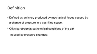
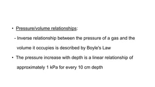
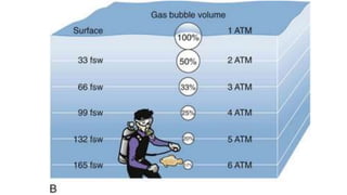
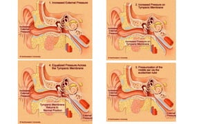
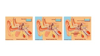
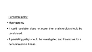
This document summarizes otitic barotrauma, which refers to injuries to the ear caused by pressure changes during diving or flying. It discusses the history of diving and describes the three main manifestations of barotrauma: sinus/middle ear issues, decompression sickness, and arterial gas emboli. It then covers the physiological effects of pressure changes on descent and ascent, outlines different types of ear barotrauma injuries, and provides treatment recommendations.
2. Introduction
• Diving as a profession can be traced back more than 5000
years, yet diving-related disease was not described until Paul
Bert wrote about caisson disease in 1878.
• Diving barotrauma can present with: Pain in ear, face or mouth
and headaches to major joint pain, paralysis, coma and death
3.
4. • Three major manifestations:
(1) Sinus or middle ear
(2) Decompression sickness
(3) Arterial gas emboli
Introduction
5. History
• Diving bell:
- Alexander the Great at the siege of Tyre in 323 Bc
• Modern diving bell:
- A form of caisson, was invented by John Smeaton in 1788
• SCUBA: self contained underwater breathing apparatus
- Invented by Emile Gagnon and Jaques Cousteau in 1943
6. • In 1897, Alt described middle and inner ear injuries caused by
compression and decompression in caisson workers
• Alt and Vail:
- Animal experiment
- Inner ear compression barotrauma was related to the failure of
middle ear pressure equalization
7. • Vail: also hypothesized that decompression injury was caused
by an effect on the inner ear by nitrogen bubbles
8. Definition
• Defined as an injury produced by mechanical forces caused by
a change of pressure in a gas-filled space.
• Otitic barotrauma: pathological conditions of the ear
induced by pressure changes.
9. • Pressure/volume relationships:
- Inverse relationship between the pressure of a gas and the
volume it occupies is described by Boyle's Law
• The pressure increase with depth is a linear relationship of
approximately 1 kPa for every 10 cm depth
10.
11. • Descent in air from 5500 m to sea level represents a pressure
increase of only 0.5 atmospheres (50.5 kPa)
• When diving, the largest percentage change in gas volume
occurs during the first 10 metres of descent
• For aviators during the first 1000m of altitude
12. Ambient pressure change
• The external pressure is uniformly distributed over an object
• In a system which has no air spaces within it, this pressure is
transmitted equally throughout the structure
• Head-out immersion causes a significant squeezing of blood
volume from the lower limbs into the chest and into the head
causing an increasing cardiac output and raised intracranial
pressure and headaches respectively
13. • In the submerged diver, or aviator, the pressure is equally applied to
all body structures, there is no differential pressure across any
membranes or systems so function is essentially unchanged
• The exception is an air-filled cavity within the body
• Middle ear is a bony cavity, no distension or expansion is able to
take place
Ambient pressure change
14. • Failure to equalize via the Eustachian tube -> negative middle
ear pressure
• Difference between the intraluminal vascular pressure and the
middle ear air pressure
• Oedema and even rupture of those vessels within the mucosal
lining will occur
Ambient pressure change
15. • Physiological Consequences of compression (Descent):
• As the diver descends, the ambient pressure increases
• The middle ear pressure becomes increasingly negative
compared to the ambient pressure unless equalization via the
Eustachian tube takes place
• This occurs by the voluntary action of the tensor and levator
palati muscles opening the usually closed tube
16. • If Eustachian tube cannot be opened -> pressure equalization in
middle ear space, with that of the external pressure, cannot
occur
• Further descent -> resulting in a pressure differential across the
tympanic membrane, which is therefore pushed inwards by the
external water pressure
Descent:
17. • This stretching is perceived as a sensation of external pressure
and discomfort, which stimulates further attempts at
equalization
• Diver should return towards the surface
• If descent continues, then middle and inner ear barotrauma
Descent:
18. • The perception of the requirement to equalize occurs at a
maximum of 86 cm (2.6 ft) reflecting a pressure change of only
8.7 kPa (60 mmHg).
• At this pressure, if no equalizing manoeuvre -> increases the
transmural pressures, thus increasing transudation -> mucosal
congestion and oedema
• Continued descent results in 'Eustachian locking' at 13 kPa (90
mmHg), an equivalent depth of approximately 1.3 m (3.9 ft)
Descent:
19. • Levator palati muscles is insufficient to voluntarily overcome the
external closing pressures -> middle ear barotrauma
• If the pressure gradient across the tympanic membrane is large
and occurs before the middle ear fills with an effusion or blood,
then the tympanic membrane may rupture
Descent:
20.
21.
22. • Physiological consequences of decompressin (Ascent):
• During normal ascent, as the middle ear pressure exceeds that
of the ambient pressure, passive ventilation through the
Eustachian tube into the pharynx occurs.
• When flying, this normally occurs every 13.25 m (43.5 ft) of
ascent, regardless of the speed of decompression.
23. • If both Eustachian tubes are functioning correctly then the only
conditions experienced on ascent are decompression illness
• During a diver's descent mild middle ear barotrauma may occur
due to suboptimal Eustachian tube function
• To continue ascending -> positive middle ear pressure ->
outward bulging of the tympanic membrane
24. • Tympanic membrane is pushed outwards
• Inward force on the round window membrane
• Pressure gradient is increased further as the perilymphatic pressure,
reflecting the ambient pressure, has decreased with ascent.
• It is possible, therefore for perilymphatic fistulae to occur during this
phase of a dive.
25. Manifestations of otitic barotrauma divided into:
1. Compression injuries
a) External ear barotrauma
b) Middle ear barotrauma
c) Inner ear barotrauma
2. Injuries at stable pressure
3. Decompression injuries
27. Clinical presentation
• Main symptom is of pain, increasing with depth
• The ear canal skin and tympanic membrane:
- hyperemic, petechial haemorrhages and even bleeding
28. B) Middle ear barotrauma
Clinical Features:
1. The initial symptom is the sensation of a blocked ear with a strong
desire to equalize
2. Otalgia, which worsens with increased compression
3. Minimal initial conductive loss (decreased compliance)
to the Larger conductive loss (due to damage to the stapes foot
plate, incus dislocation and fractured malleus handle)
29. 4. Tympanic membrane perforation
5. Caloric vertigo - sudden ingress of water into the middle ear
Tympanic membrane appearance in middle ear barotrauma (Modified
Teeds grading):
Grade Symptoms and signs
O Symptoms, no signs
1 Redness and retraction
32. • Equalization tests:
1. Valsalva's manoeuvre - poor predictive test for potential
barotrauma
2 Toynbee's test (positive predictive value of 25 percent)
3 Nine-step Eustachian tube test (positive predictive value, 25
percent; negative predictive value, 75 percent).
4. Combining the nine-step test with Toynbee's test -Reliable (100
percent)
33. 5. Sonotuberometry and tubotympanometry: accurately predict
• Degree of mastoid pneumatization:
- Pneumatization of greater than 34.7 cm2-> decreased
barotrauma
- Pneumatization less than 13.6 cm2 -> inceased
barotrauma
34. Treatment of ME Barotrauma
• Treatment regimen (on basis of 3 level clinical classification of
severity):
• Type 1. symptoms, but no (or minimal) signs
- requires no specific treatment
Type 2. significant signs, but no perforation
- conservative with oral or topical nasal decongestants
35. Type 3. with perforation
- initial observation
- myringoplasty (fail to heal)
36. Prevention of middle ear barotrauma
Medical:
1. Nasopharyngeal irradiation
- Poor success rates and radiation induced tumors, stopped
this practice
2. Topical nasal decongestants and oral pseudoephedrine
37. • 3. Nasal balloon inflation (auto-Politzerization) using Otovents
Surgical:
• Myringotomies with or without the insertion of ventilation
tubes
38. Diving and flying after Middle ear surgery:
• Restriction of any activity for at least six weeks
• Stapedotomy technique:
- As the potential consequences of a perilymphatic fistula when
diving are so extreme, postoperative wait of 12 months is
recommended
39. Caloric Vertigo:
• Rupture of tympanic membrane
• Severe acute caloric vertigo
• Colder the water, more severe the effect
• Vertigo is self-correcting if caloric stimulus is removed
45. • Inner ear haemorrhage:
- Transient, or minimal, vestibular symptoms
- Mild to moderate sensorineural hearing loss
- Good recovery
Inner ear barotrauma
46. • Labyrinthine membrane tears:
- Vertigo and tinnitus
- Characteristic low frequency hearing loss
- Inner ear damage: haemorrhage around Reissner's and the
round window membranes
- Rupture of the utricle and saccule
Inner ear barotrauma
47. • Temporal bone:
- Reissner's membrane rupture
• In normally ventilated ears, the inner ear is more susceptible to
decompression trauma
• Implosive forces to the inner ear causes middle ear
overpressure (ascent with a locked ET), than compression
(descent)
Inner ear barotrauma
48. Perilymphatic fistulae
• Sudden onset of vertigo
• Sensorineural hearing loss
• Tinnitus
• Positional nystagmus
• Tullio phenomenon
49. • Tympanic membrane: signs of middle ear barotrauma
• Nystagmus usually towards the opposite side
• Romberg's test is normal in more mild cases
• Unterberger's step test
• Side-step test
Perilymphatic fistulae
50. • Inner ear decompression sickness:
• Commercial and military divers who breathe a compressed
mixture of helium and oxygen
• Decompression (ascent), or shortly after surfacing from a dive
• Hyperbaric chamber for recompression
Perilymphatic fistulae
51. Treatment:
• Initial conservative approach (bed rest for 05 days)
• Exploration reserved for:
(1) Those with progressive hearing deterioration observed
on daily, or more frequent, audiometry
(2) If the vestibular symptoms fail to improve after 05 days
(3) Failure of complete resolution after one month
Perilymphatic fistulae
52. • Moderate to Severe hearing loss:
- Emergency exploration in severe acute cases with both a
significant hearing loss and vertigo (40 dB HL)
Perilymphatic fistulae
53. • Minimal Hearing loss:
- Mild vestibular symptoms and hearing losses ( high frequency only,
or < 20 dB HL)
- Initial conservative (bed rest with elevation of the bed head to 30-40
degree)
Perilymphatic fistulae
55. • Mild to moderate hearing loss:
• Persistant vestibular symptoms without hearing loss:
- Conservatively for upto 04 weeks
- Hearing loss with vestibular component presenting more than
24 hours following the incident -> conservative
Perilymphatic fistulae
56. • Surgical exploration:
- Earlier the presentation with hearing loss after the event, and
worse the vestibular component-> urgent surgery
- Evidence of deteriorating hearing
- if vestibular symptoms and hearing loss persist (exploration
after 05 days)
Perilymphatic fistulae
57. • Surgery for perilymphatic fistula:
• Exploratory tympanotomy
- Look at the round, oval windows and Eustachian tube orifice
for a source of the leak
58. - Tissue plug and absorbable middle ear packing material
- If no fistula is observed, then tissue graft in the round window
and over the foot plate
- Vein (sticky nature of its adventitial surface)
- Fluid collection for beta 2-transferrin
59. Injuries at Stable Depth
High-pressure nervous syndrome:
Clinical presentation:
• General dizziness and tremors
• Ataxia and myoclonus
• Sudden onset of vertigo and nausea with nystagmus
has been reported as part of this syndrome.
60. • Permanent vestibular damage has been described, but there is
no hearing loss
• Prevention:
- Minimized by ensuring slow descents and by avoiding inert gas
changes at depth
High-pressure nervous syndrome
61. • If symptoms are encountered when a helium-oxygen mix
is changed to air during ascent, then the treatment is to
recompress back on helium-oxygen
High-pressure nervous syndrome
62. Decompression injuries
• Alternobaric vertigo:
• Condition of asymmetrical middle ear overpressure stimulation
occurring in both divers and aircrew.
• Pathoetiology:
- On ascent, failure of equalization due to minor middle ear
congestion and oedema -> relatively higher pressure in one middle
ear than the other.
63. • Condition occurs on ascent or within two minutes of surfacing.
• Episodes are short lived, with a maximum duration of ten
minutes.
• Divers may also describe a tumbling sensation or a tilting of
their surroundings
Alternobaric vertigo
64. Barotraumatic facial palsy
• Patho-aetiology:
• Pressure-induced neuropraxia (most widely accepted
explanation for transient unilateral facial palsy .
• Dehiscence of the facial nerve in its intramastoid portion (0.5 to
57%)
65. • Animal studies have demonstrated that blood flow in the vasa
nervora of the facial nerve decreases if increased middle ear
pressure is transmitted through a dehiscence of the facial canal
• Alternative hypothesis:
- In a non-dehiscent facial canal, pressure may be transmitted
through the fenestra of the chorda tympani.
66. Persistent palsy:
• Myringotomy
• If rapid resolution does not occur, then oral steroids should be
considered.
• A persisting palsy should be investigated and treated as for a
decompression illness.
Caissons are bells that are pressurized to keep the water out, enabling workers to work for many hours at depth , caisson disease were noted among bridge workers after finishing their shifts underwater and coming back to the surface. These symptoms included dizzy spells, difficulty breathing, and sharp pain in the joints or abdomen. severe back pain that left them bent over-> bends
By N2 bubbles in body at deep pressure
nitrogen bubbles: obstruction in the internal auditory artery system
slap injuries , flying and subaqua diving, water skiing, high board diving and from blast injuries
relative bubble volume decrease experienced with increased pressure
If a large vein, e.g. the internal jugular, has pressure applied from the outside then, provided that the whole body is submerged, it will transmit this pressure
onwards.
EAC separated from the middle ear by TM, ET connects middle ear cavity with the nasopharynx
2. Nasopharynx is filled with inspired air from a compressed air cylinder via a regulator during breathing. This air pressure is equilibrated by the regulator in the mouthpiece to be equal with the ambient external pressure, provided the diver continues to breath normally.
Equalization should be done
sudden influx of cold water into the middle ear may result in unequal thermal stimulation inducing a rigorous caloric vertigo.
traction force is applied to the oval window via the ossicles.
occurs when a pocket of air is trapped within the external auditory meatus. This may be because of cerumen, earplugs, foreign bodies or exostoses,
air-containing space that can change in volume in response to changes in ambient pressure. During descent, the volume of this space decreases causing the tympanic membrane to bulge outward
nine-step tympanometry : 1. The tympanogram records resting middle-ear pressure. 2. EAC pressure is increased to +200 mm H2 O with medial deflection of TM and a corresponding increase in middle-ear pressure. The subject swallows to equilibrate ME overpressure. 3. While the subject refrains from swallowing, EAC pressure is returned to normal, thus establishing a slight negative ME pressure (as the TM moves outward). The tympanogram documents the established ME underpressure. 4. The subject swallows in an attempt to equilibrate negative ME pressure. If equilibration is successful, airflow is from the nasopharynx to the middle ear. 5. The tympanogram records the extent of equilibration. 6. EAC pressure is decreased to -200 mm H2 O, causing a lateral deflection of the TM and a corresponding decrease in ME pressure. The subject swallows to equilibrate negative ME pressure; airflow is from the nasopharynx to the middle ear. 7. The subject refrains from swallowing while EAC pressure is returned to normal, thus establishing a slight positive pressure in the middle ear as the TM moves medially. The tympanogram records the overpressure established. 8. The subject swallows to reduce overpressure. If equilibration is successful, airflow is from the middle ear to the nasopharynx. 9. The final tympanogram documents the extent of equilibration.
Surgical: for flying, This procedure is clearly contraindicated for diving
1 asymmetric exposure to cold water
2. Vomiting may occur under water, perhaps necessitating the removal of the regulator from the mouth (although modern regulators are designed to
enable some vomiting to take place without their removal)
Failure of equalization of the middle ear pressure on the affected side. Sudden, large pressure changes in the middle ear can be transmitted to the inner ear, resulting in damage to the inner ear. This can cause severe vertigo and even deafness
Clearing of the middle ear in descent. Inward bulging TM->, ossicles toward the inner ear at the oval window-> pressure wave through the inner ear and outward bulging of the round window If forceful Politzer maneuver: Inflation of ET and tympanum by forcing air into the nasal cavity at swallowing.
and ET suddenly opens,->a rapid increase in middle ear pressure ->ossicles to suddenly return to normal positions, causing the round window to implode.
attempts clear a blocked middle ear by Politzer maneuver and ET blocked and locked, a dramatic increase in ICT. Since the fluids surrounding the brain communicate freely with the inner ear fluids, this pressure may be transmitted to the inner ear-> sudden rise in the inner ear pressure -> round or oval window membrane to explode.
Siimilar to Acute Meniere's disease attack,
perilymphatic fistula should be suspected in an otherwise healthy ear (or in one with evidence of middle ear barotrauma) if there is a SNHL of rapid onset, constant
Dysequilibrium, symptoms of a chronic perilymphatic fistula. Dysequilibrium which worsens, or changes to momentary vertigo when performing manoeuvres which increase
the intracranial (and hence perilymphatic) pressure
Side step test: eyes closed, the patient takes two consecutive broad steps sideways, and then stops with both feet together. sway or overshoot in the same direction as the steps were taken is regarded as a positive result, the affected ear being on the side towards the direction of movement.
1. IEDCS is an injury that closely resembles inner ear barotrauma
2. contrast, barotrauma most often occurs during compression (descent)
3. recompression in ME and Inner Bro cause further middle and inner ear barotrauma. Conversely, not recompressing a patient with a decompression
illness may result in permanent cochleovestibular damage, further progression of the decompression illness symptoms and even death.
Treatment options are centred on hearing and balance preservation
to minimize the intracranial, cerebrospinal fluid, and hence perilymphatic, pressures. Raising the intracranial pressure by coughing, straining, vomiting or
performing Valsalva's manoeuvre should be avoided
may not have had fistulae, but intralabyrinthine pathology
if after five days there has been no hearing improvement and if any vestibular symptoms persist (or at any time at the first sign of further hearing deterioration), then a tympanotomy is recommended.
confirmatory test for the presence of perilymph
2. If the tympanotomy then it is probably unwise to perform ossicular surgery at the same procedure ->cochlear concussion due to minor further trauma.
3. Repair of TM can be performed at the time, but ossicular surgery should be staged
High-pressure nervous syndrome occurs when divers are, exposed to extremely high pressures
tremors (both postural and intentional hands and even of the whole body) which may progress, with increasing pressure
usually occurs in those who describe unilateral equalization problem
condition occurs on ascent or within two minutes of surfacing.
given to a facial nerve palsy occurring as a result of high middle ear pressure during ascent.
Both would explain the transient nature of the condition, as the blood flow would rapidly return to normal when the middle ear pressure is
relieved.