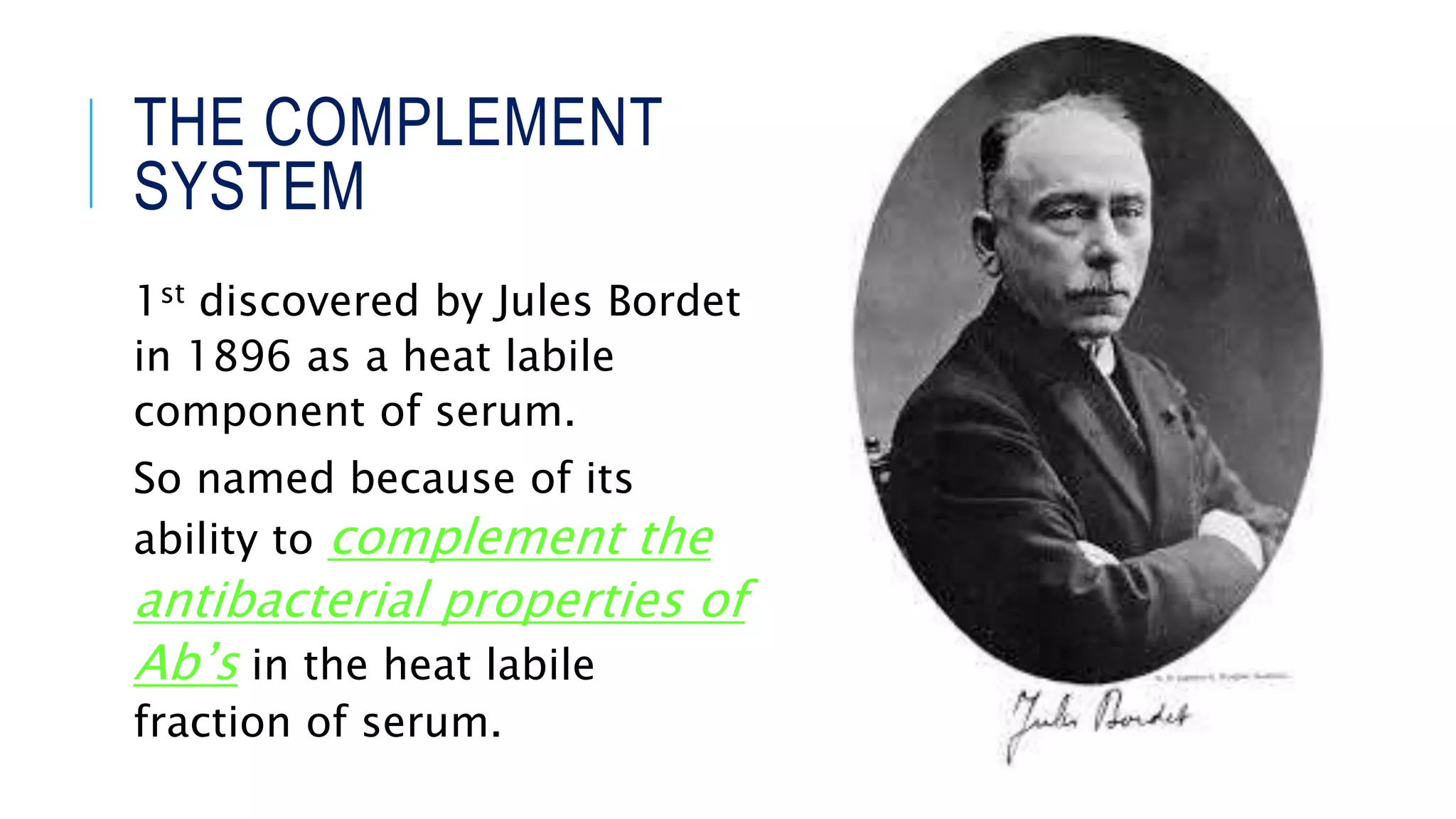
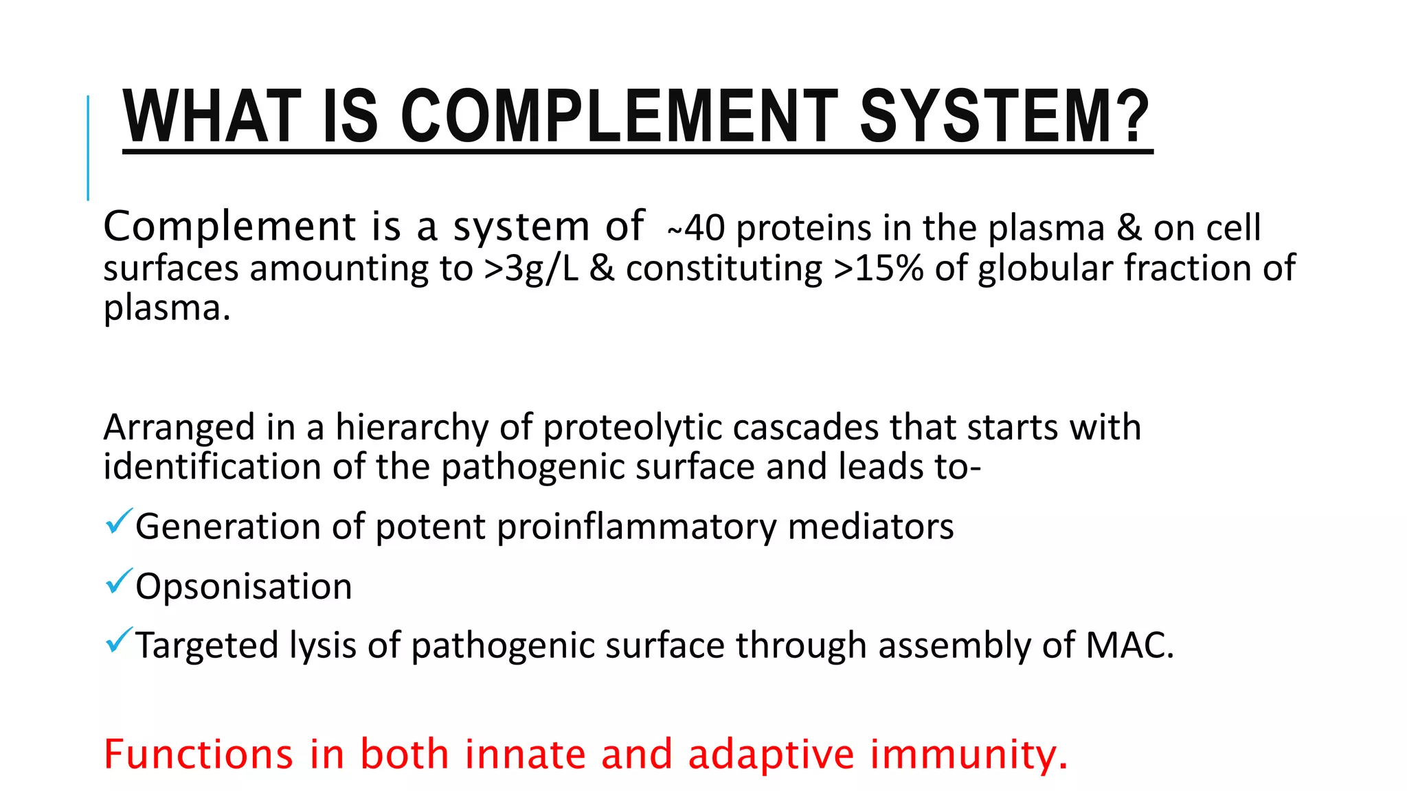
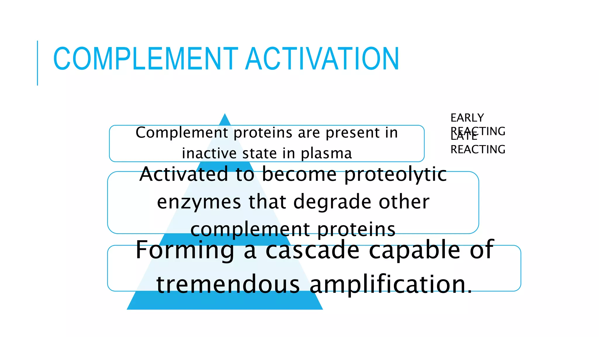
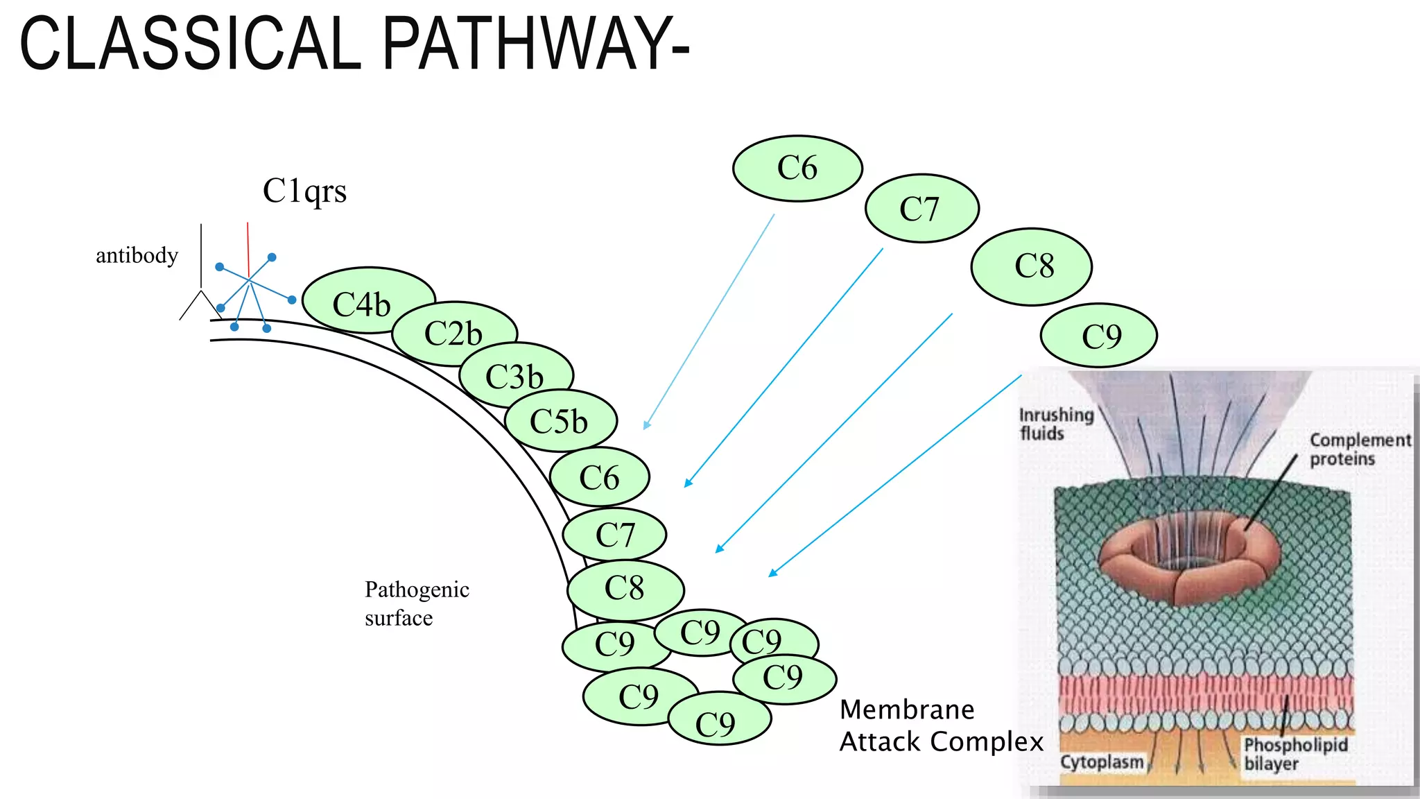
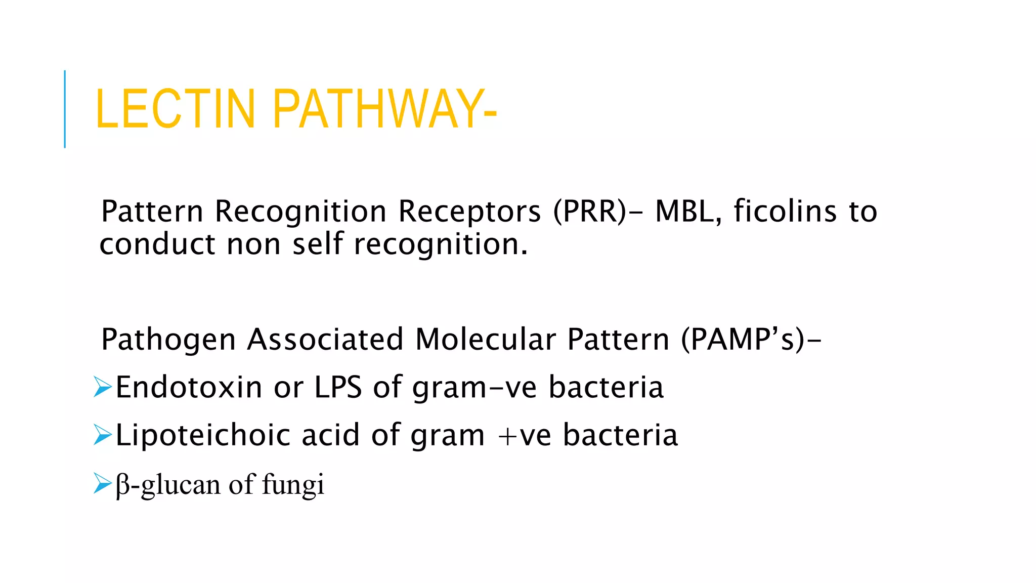
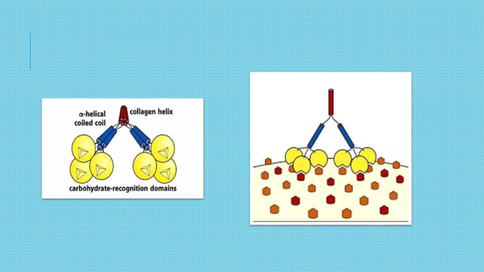
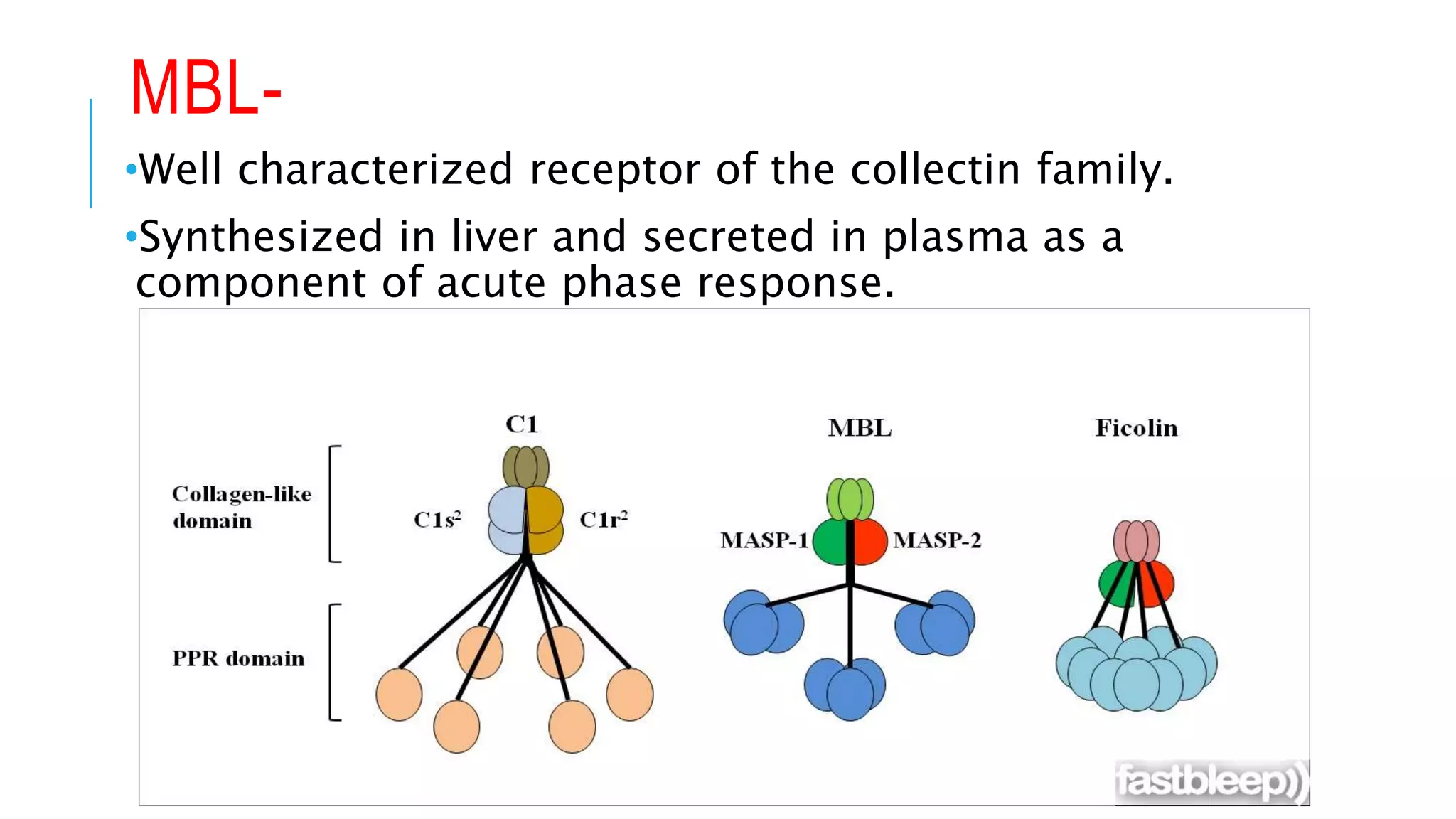
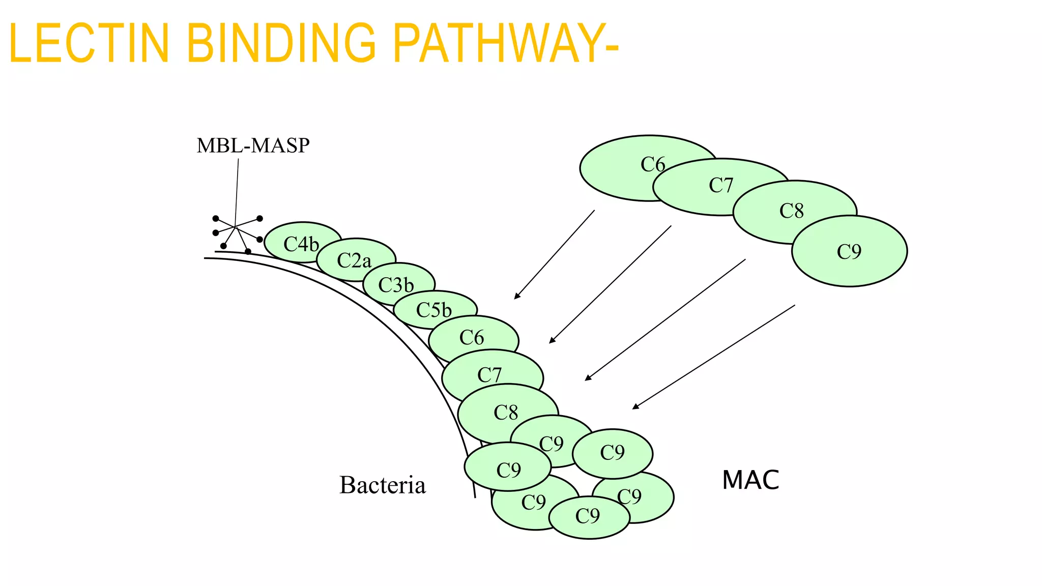
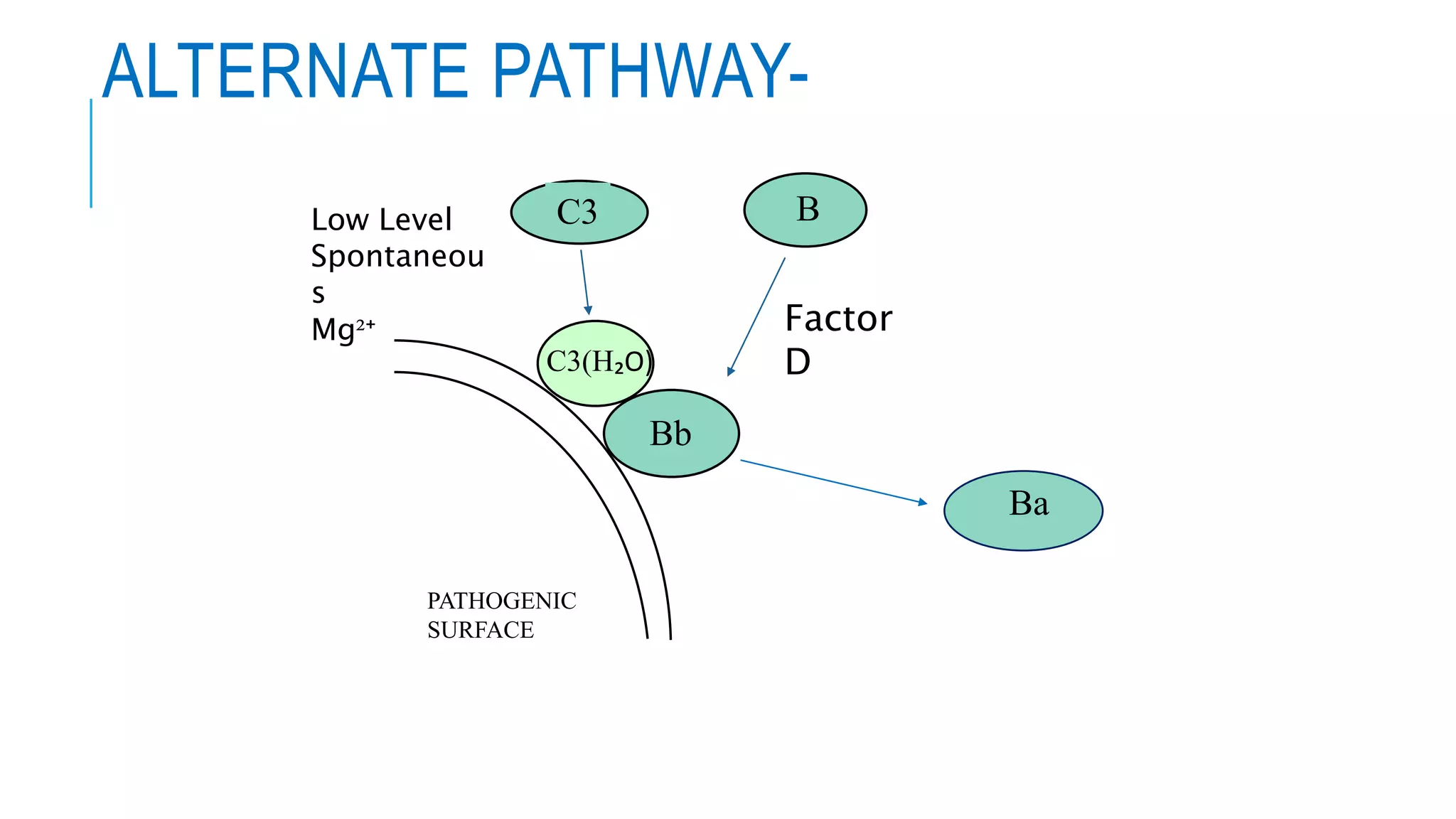
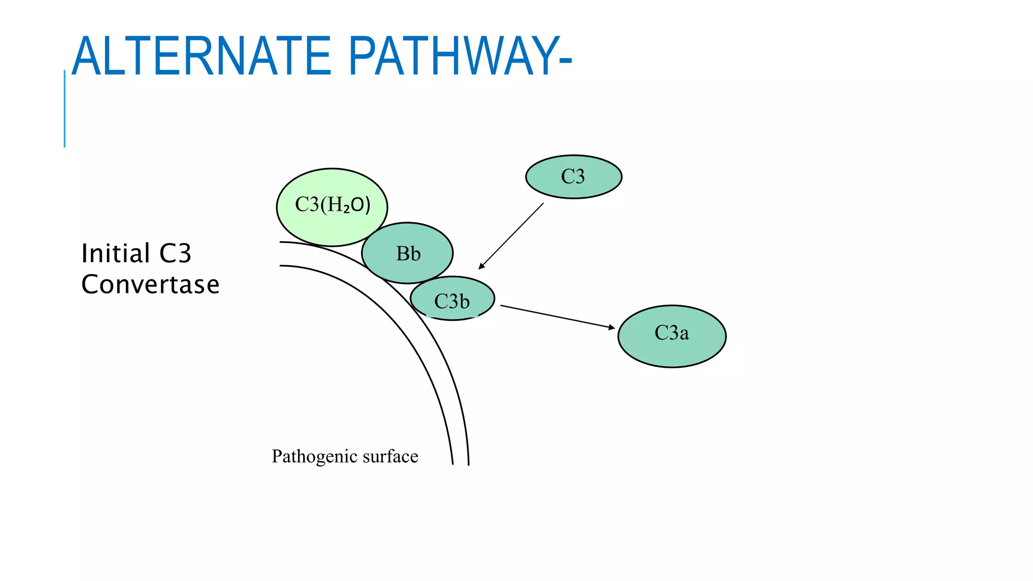
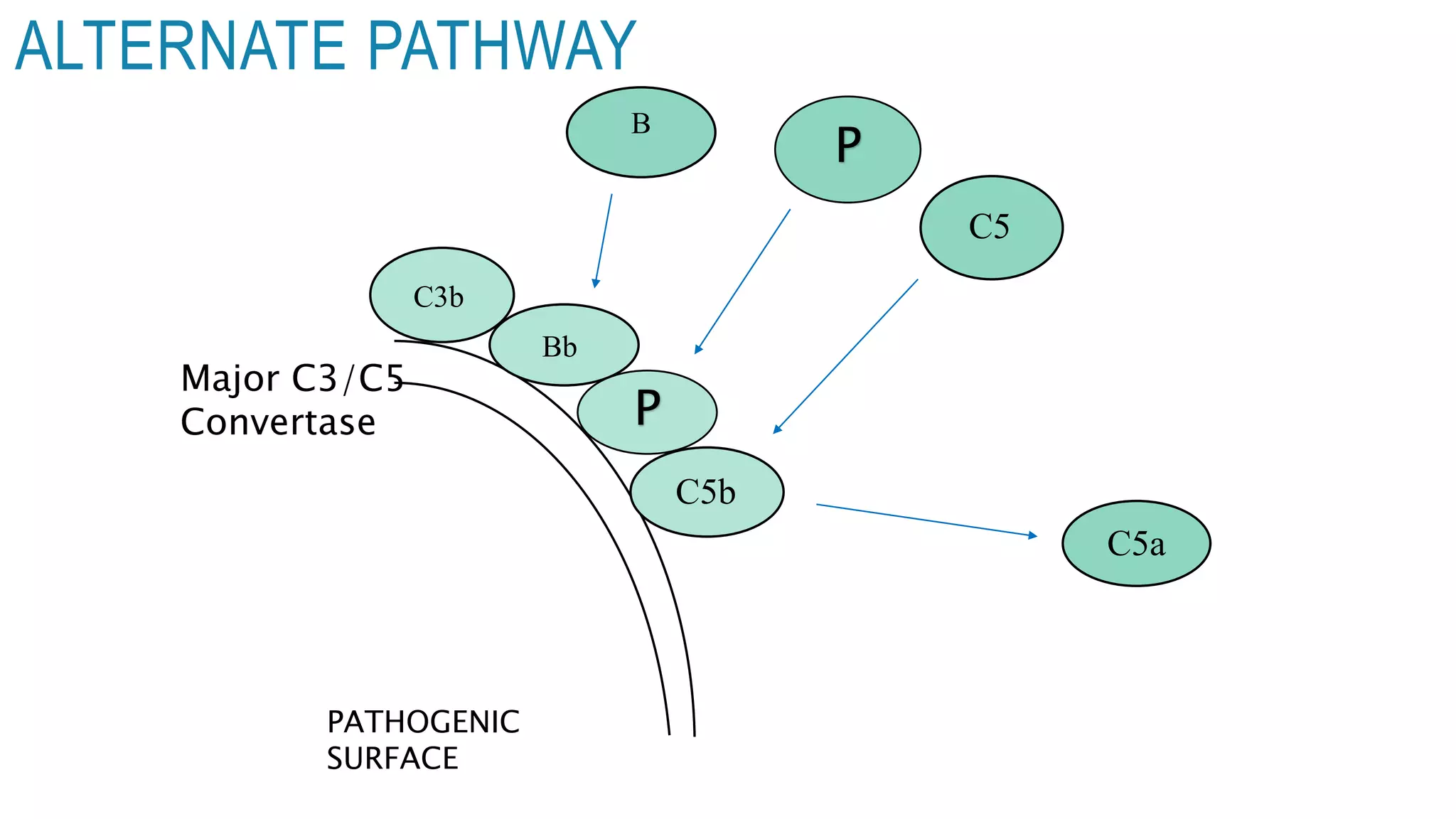
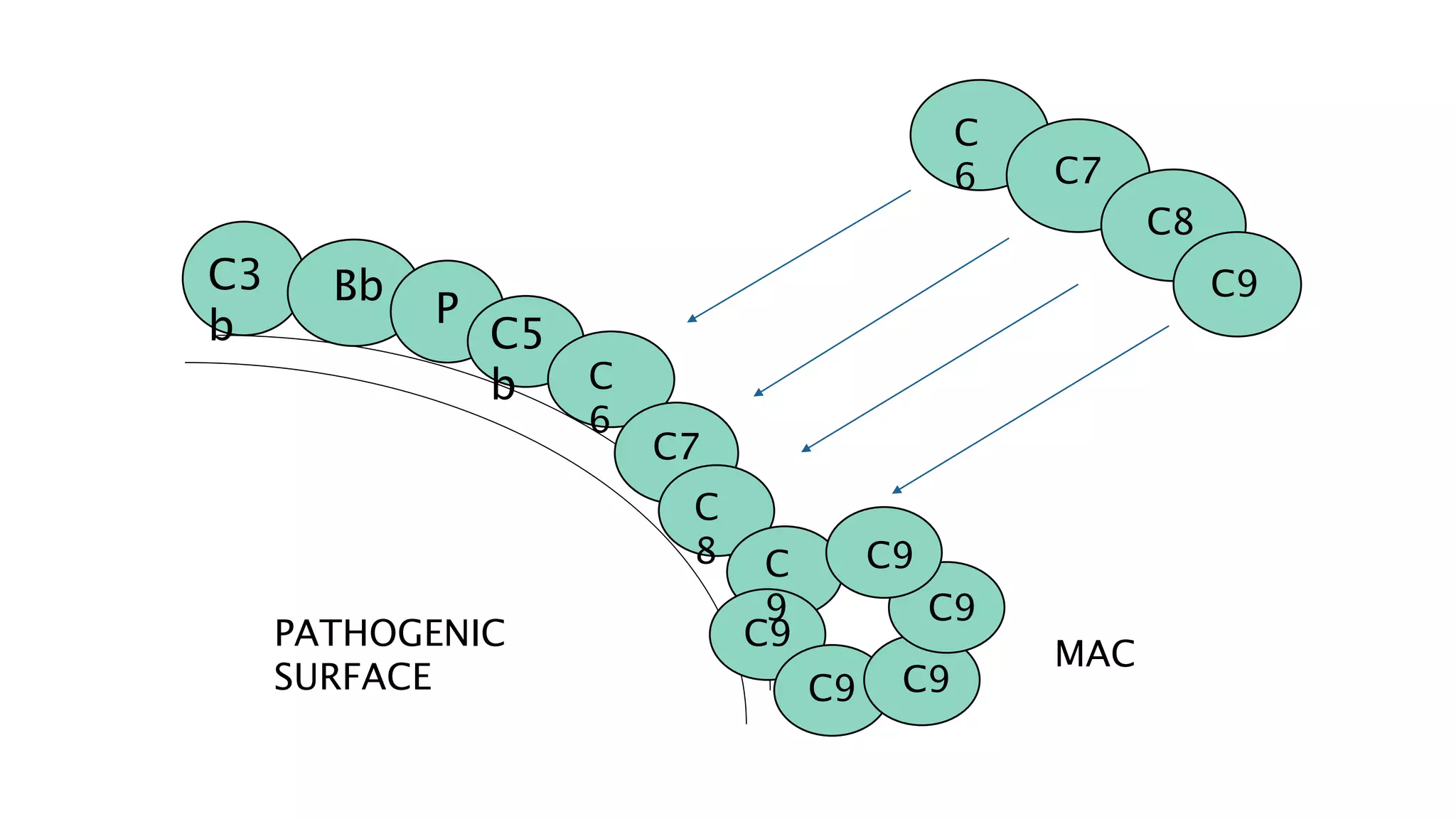
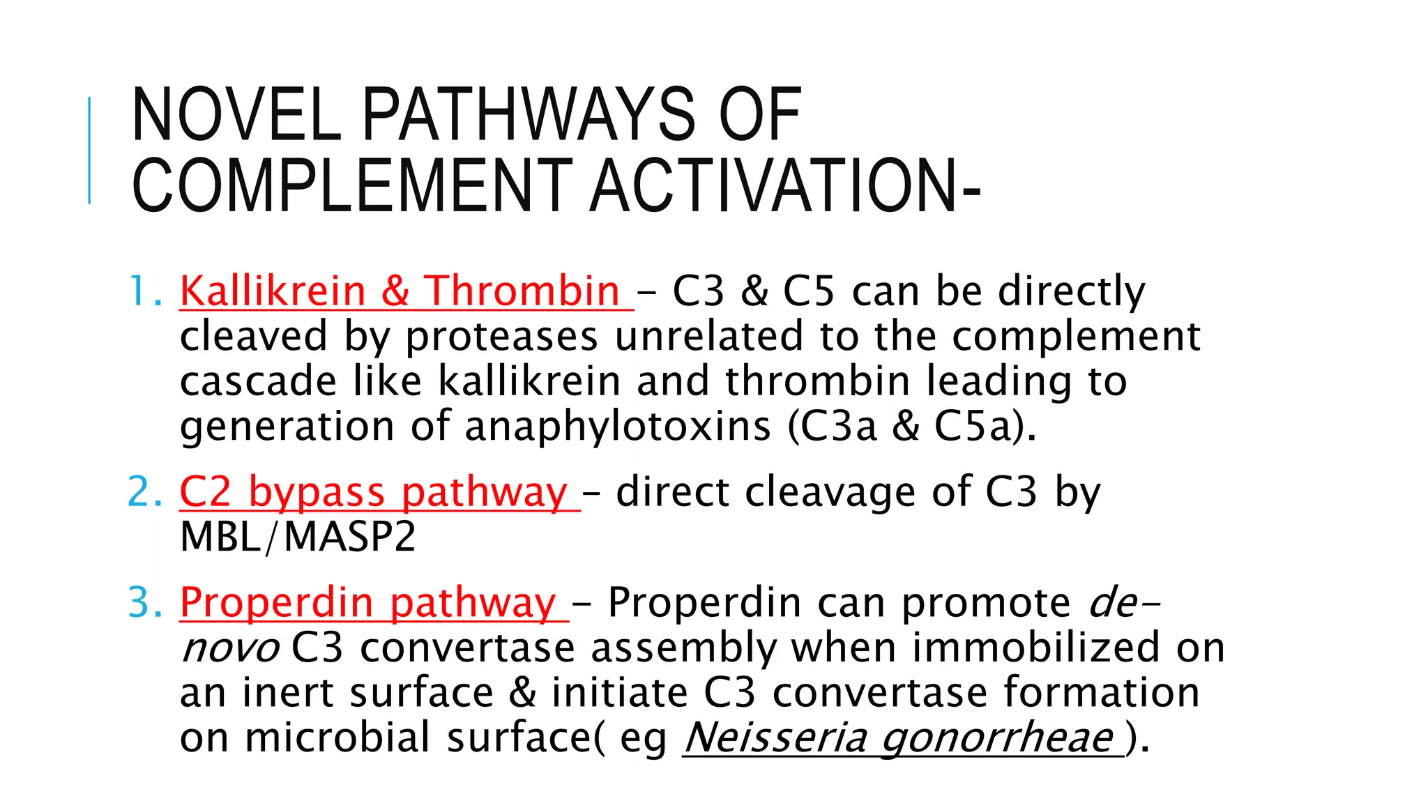
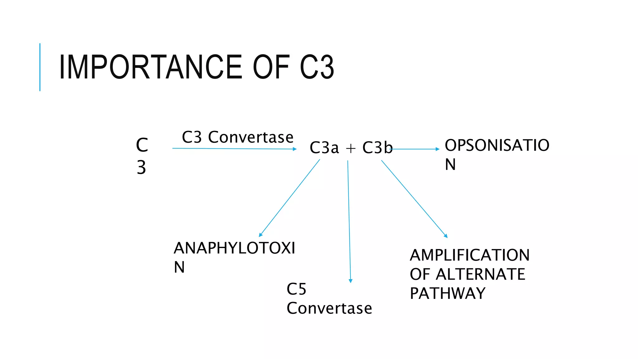
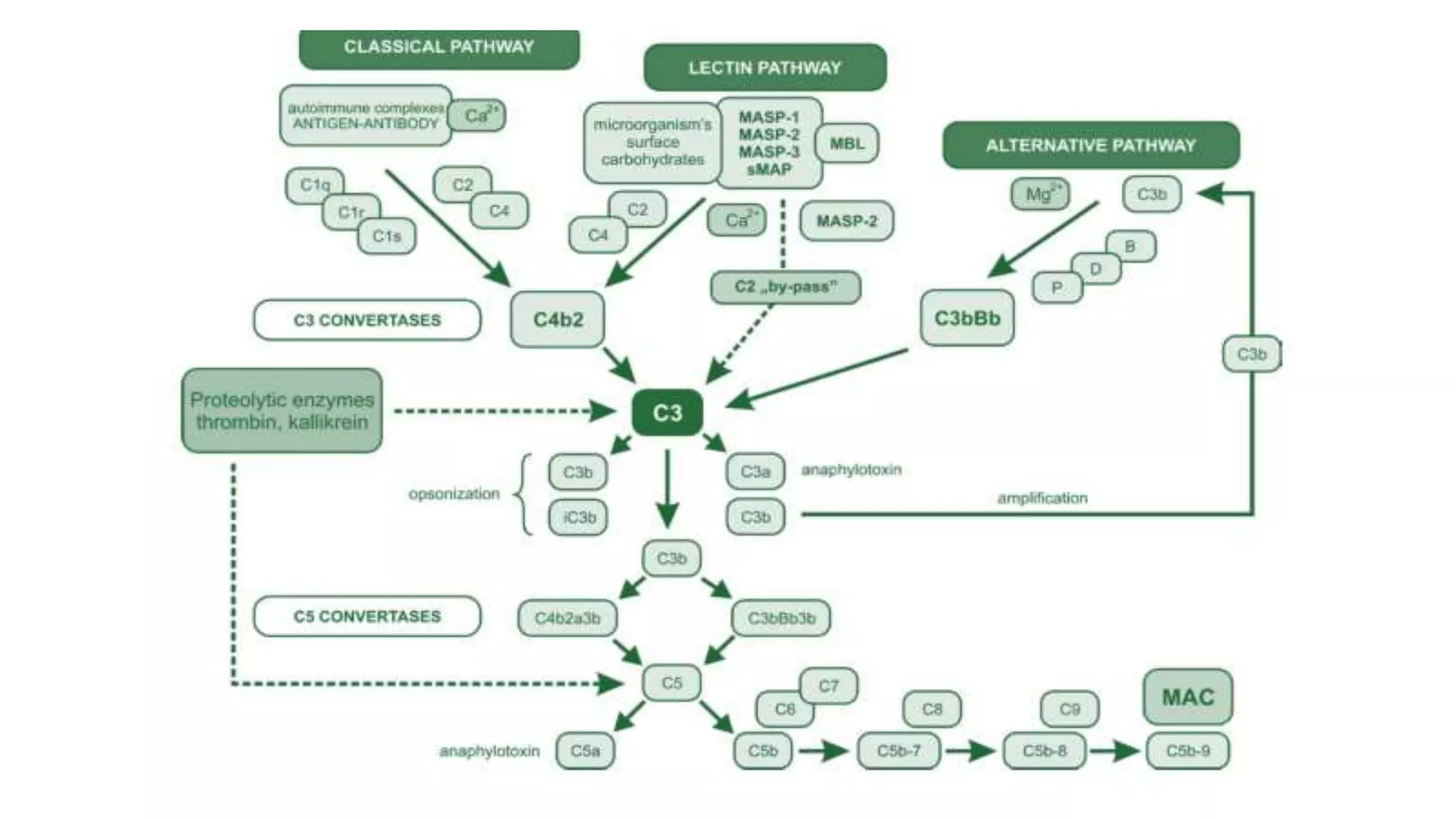
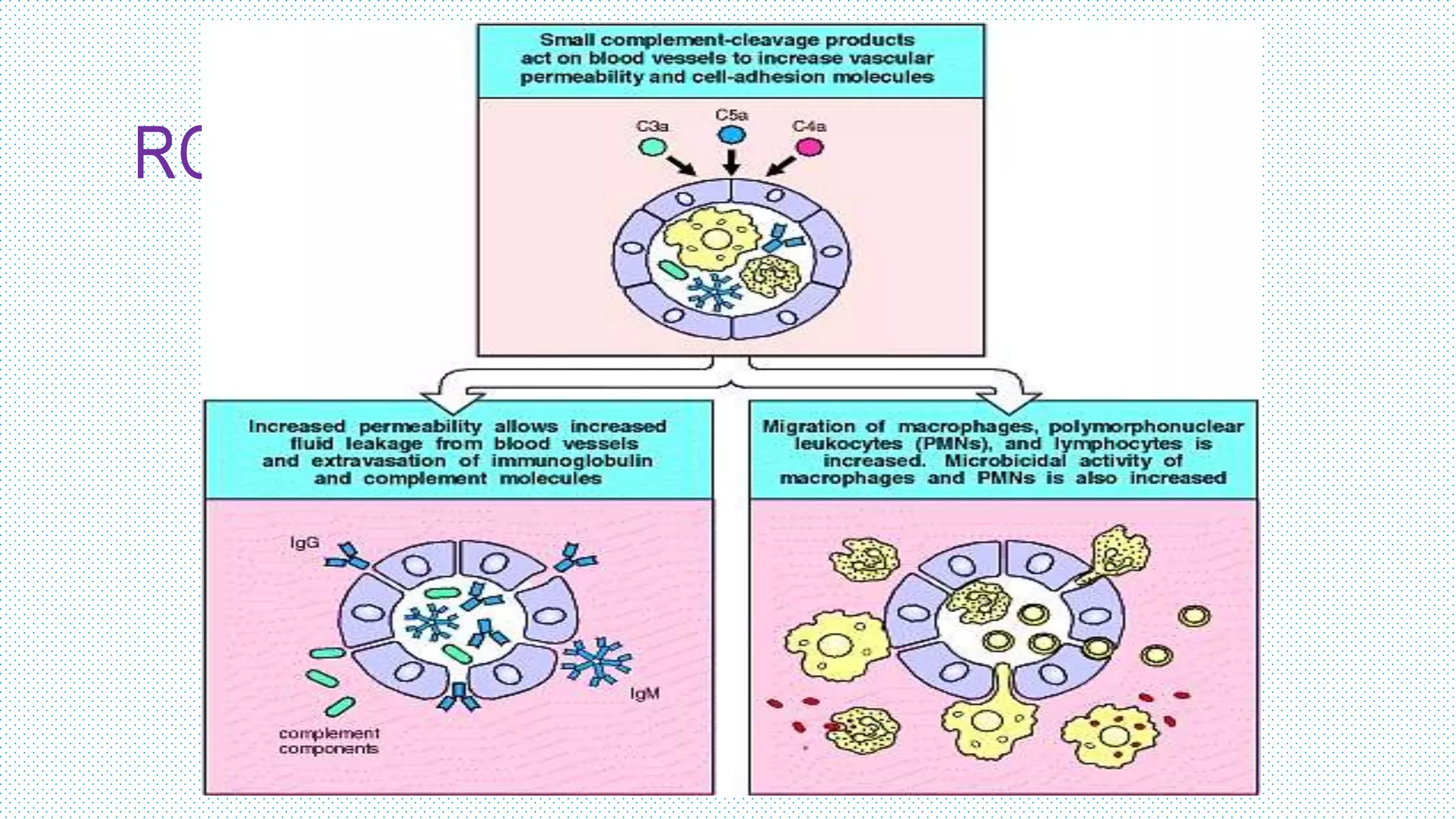
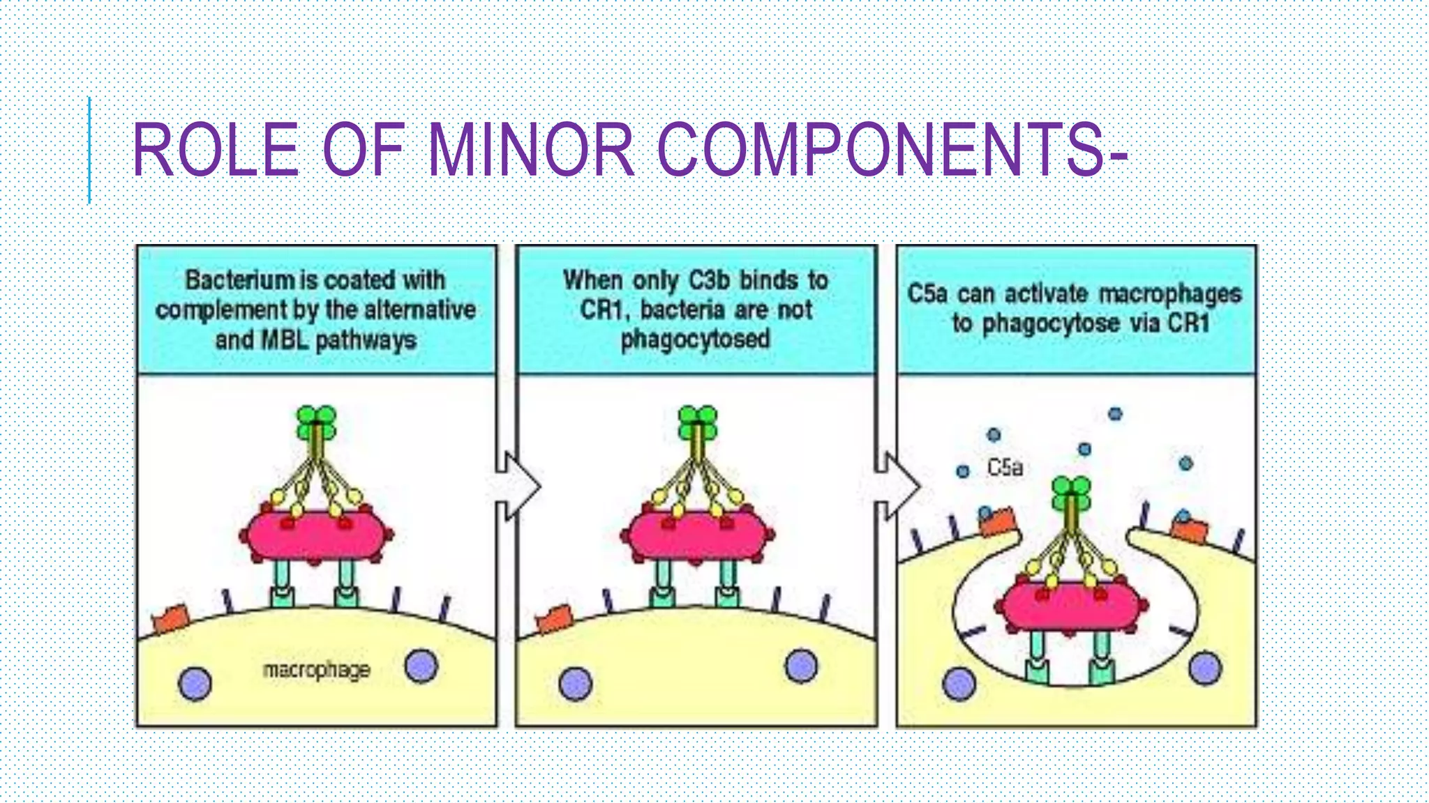
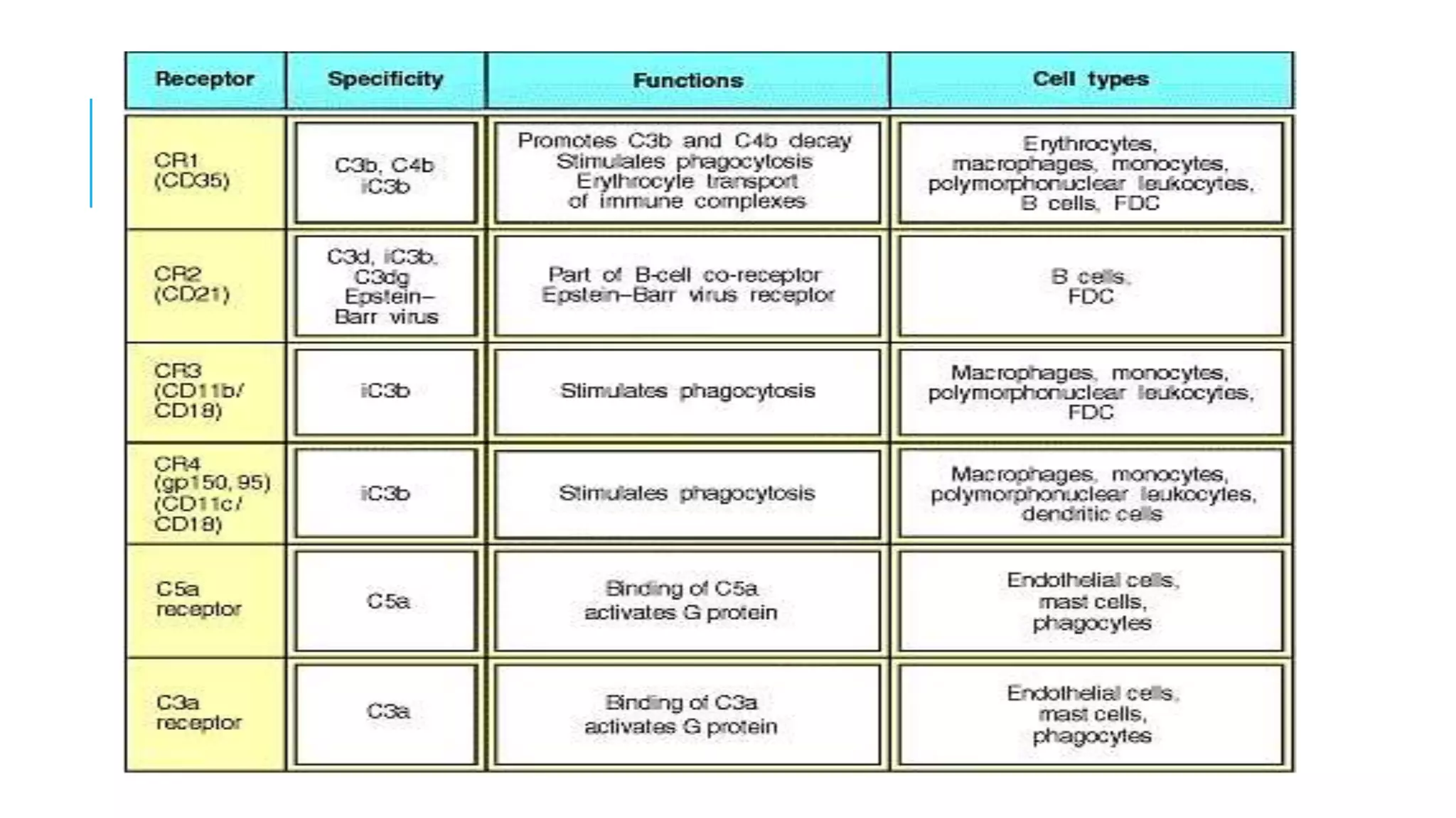
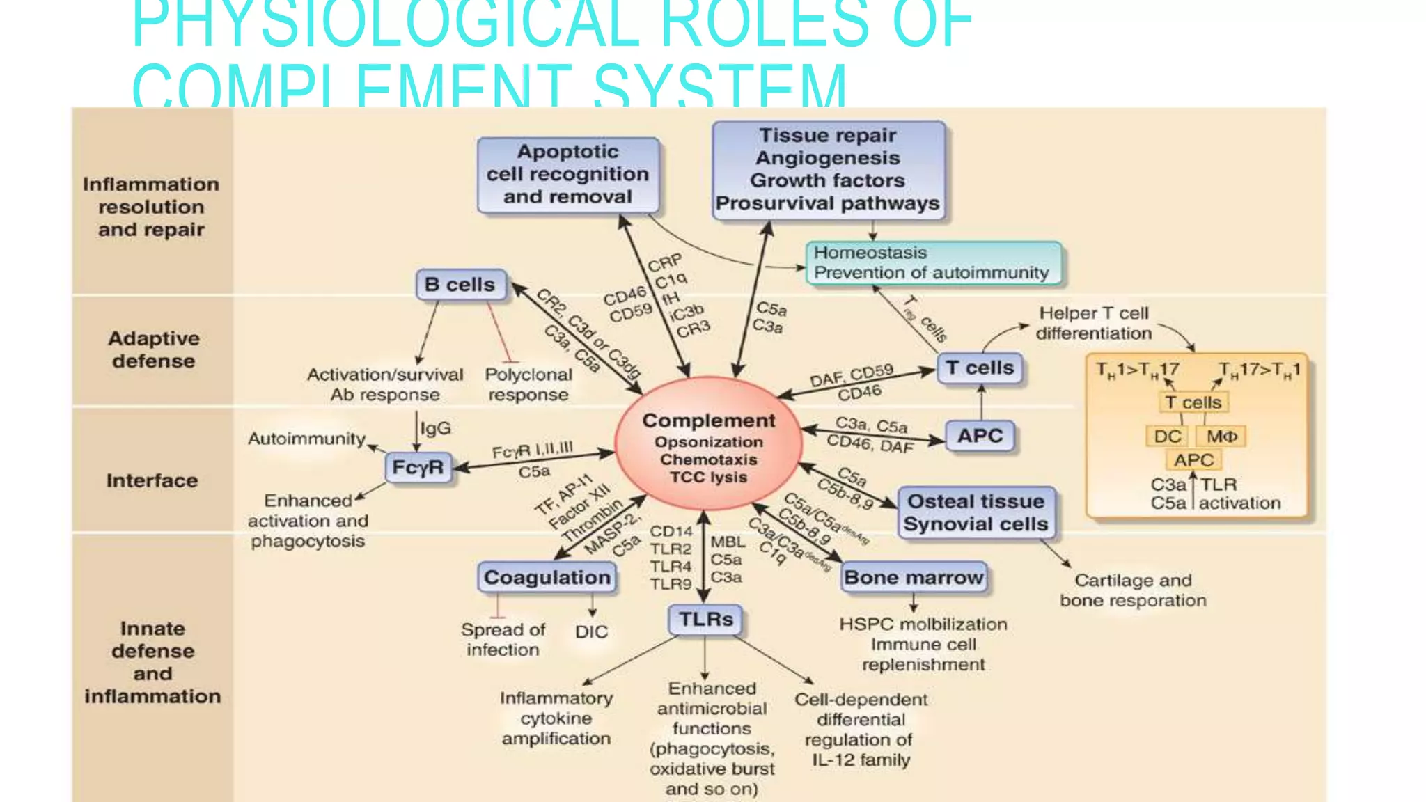
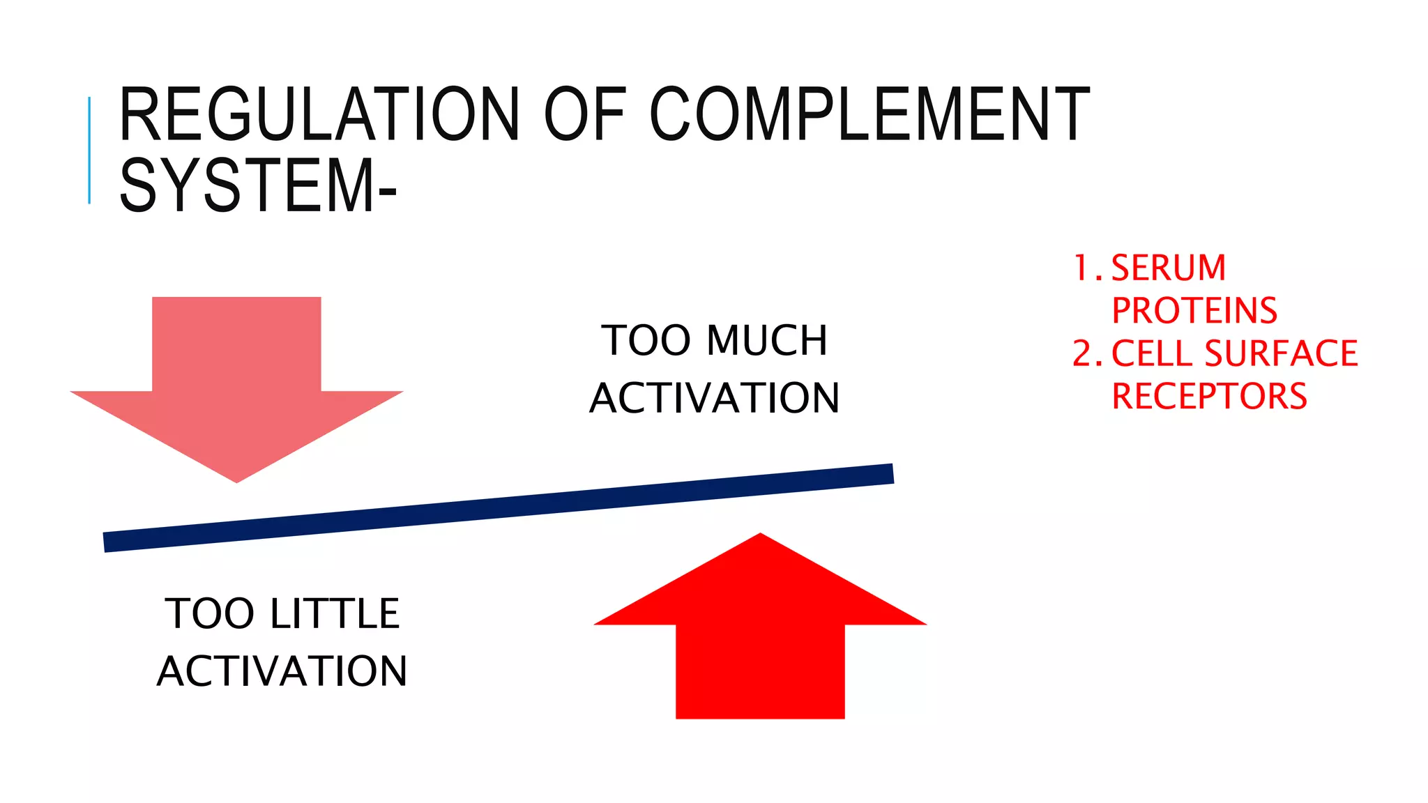
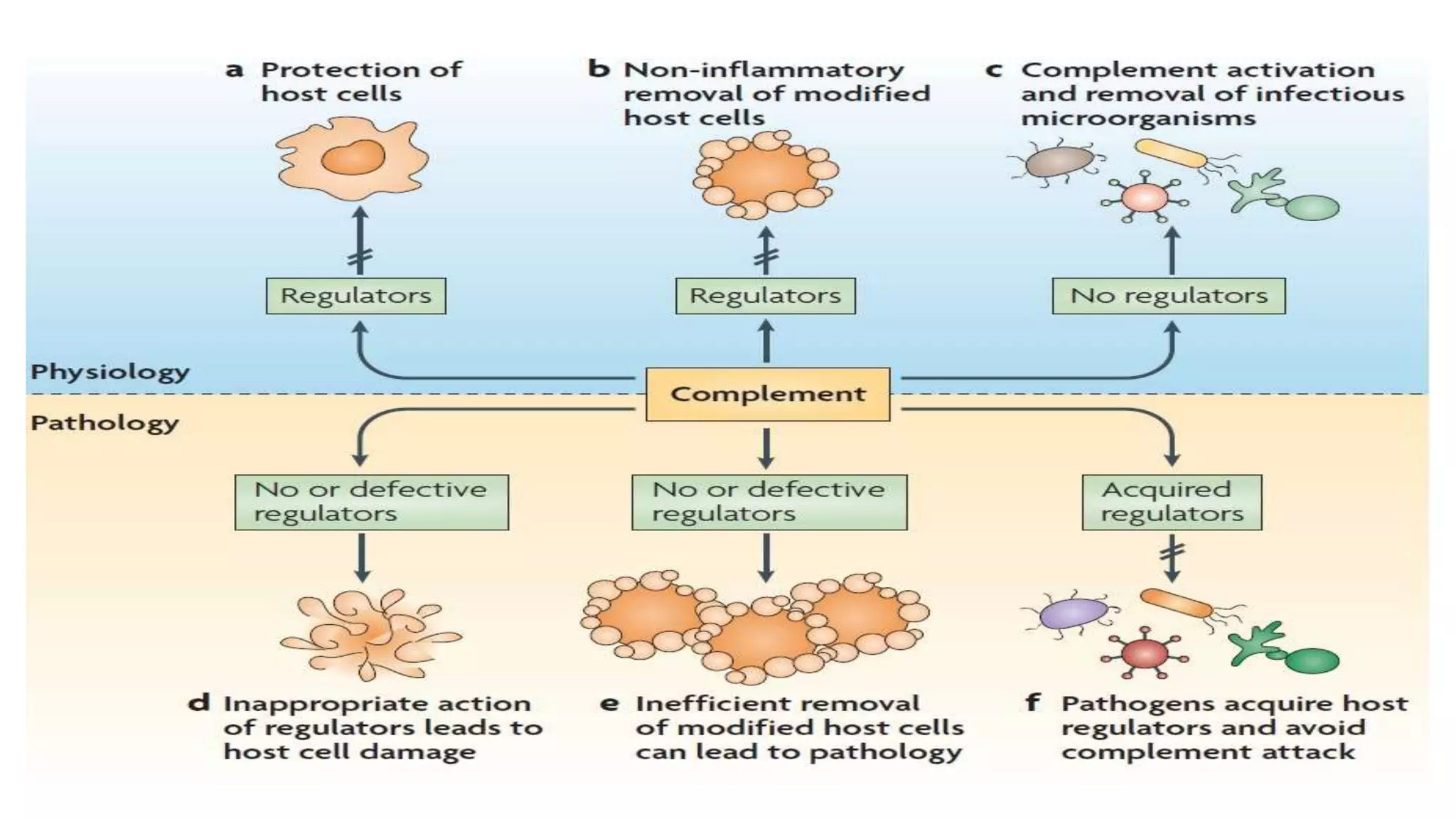
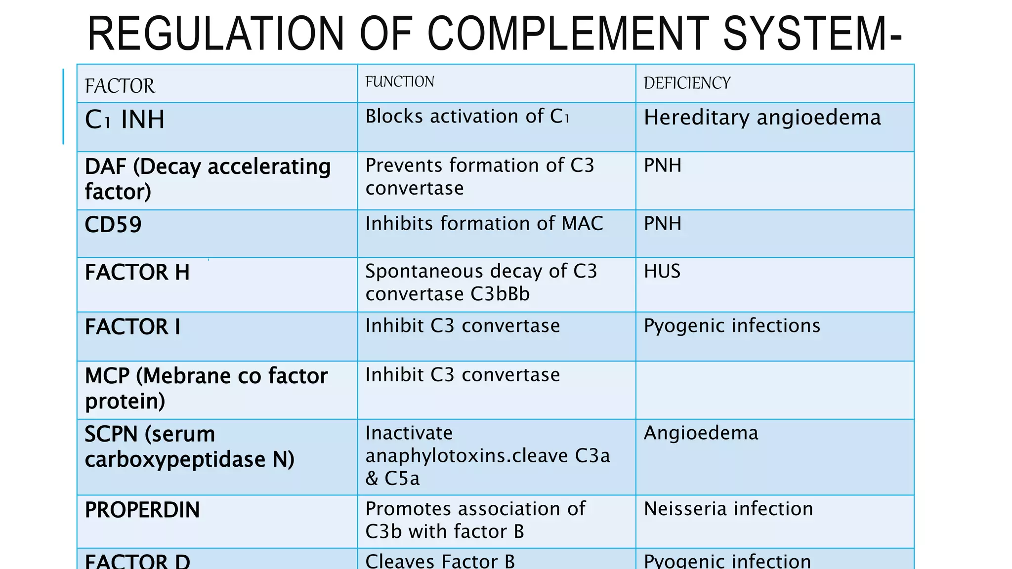
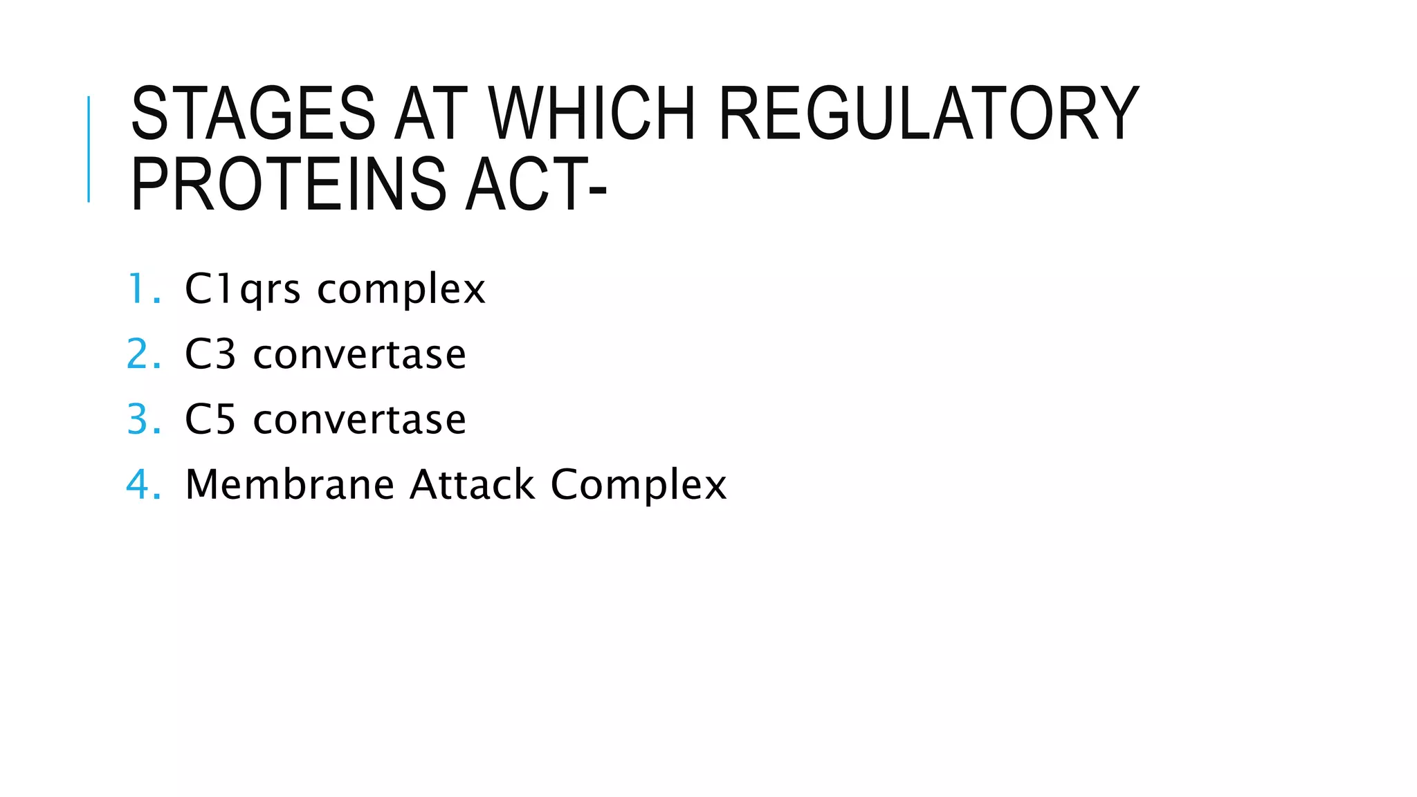
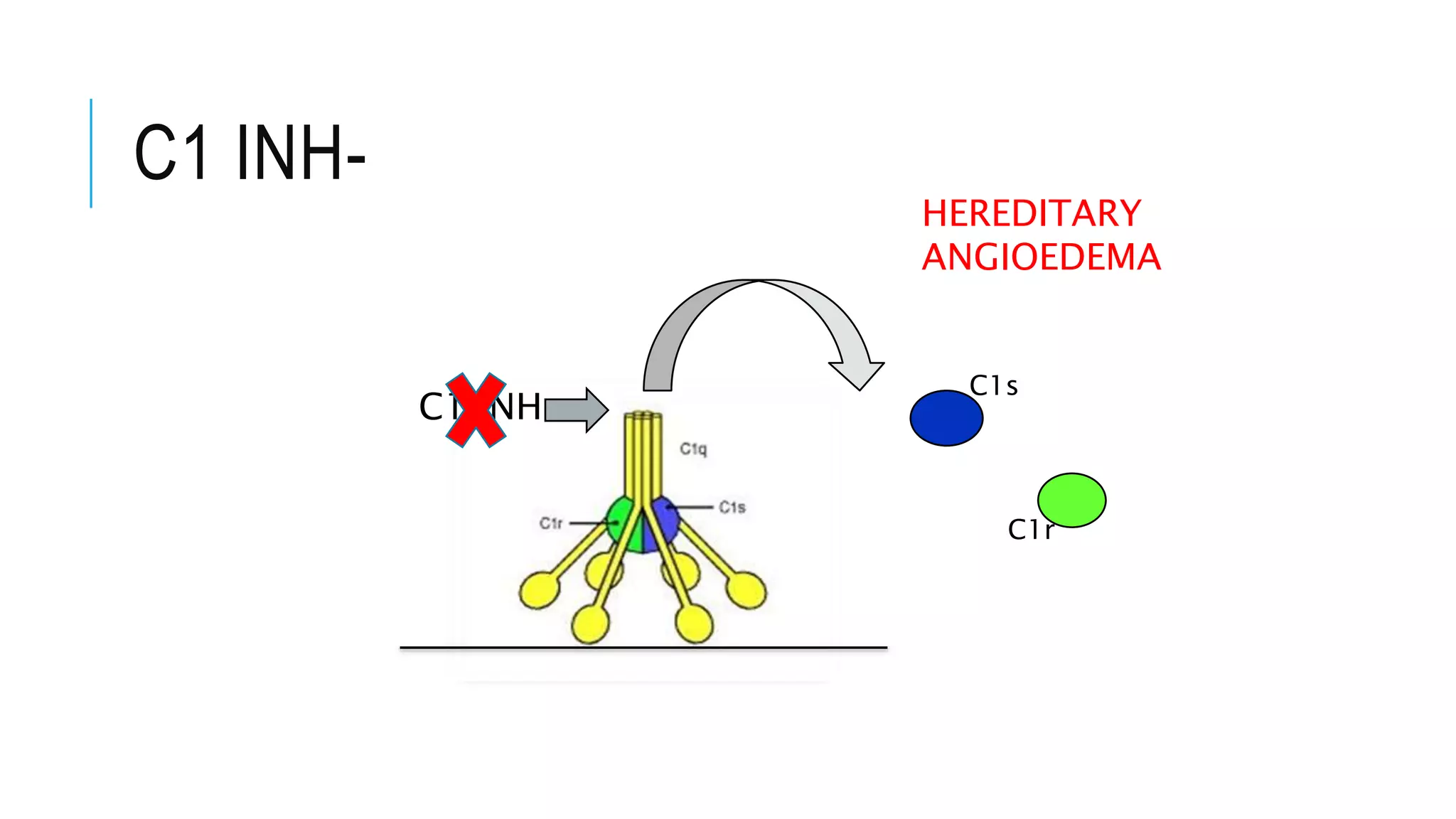
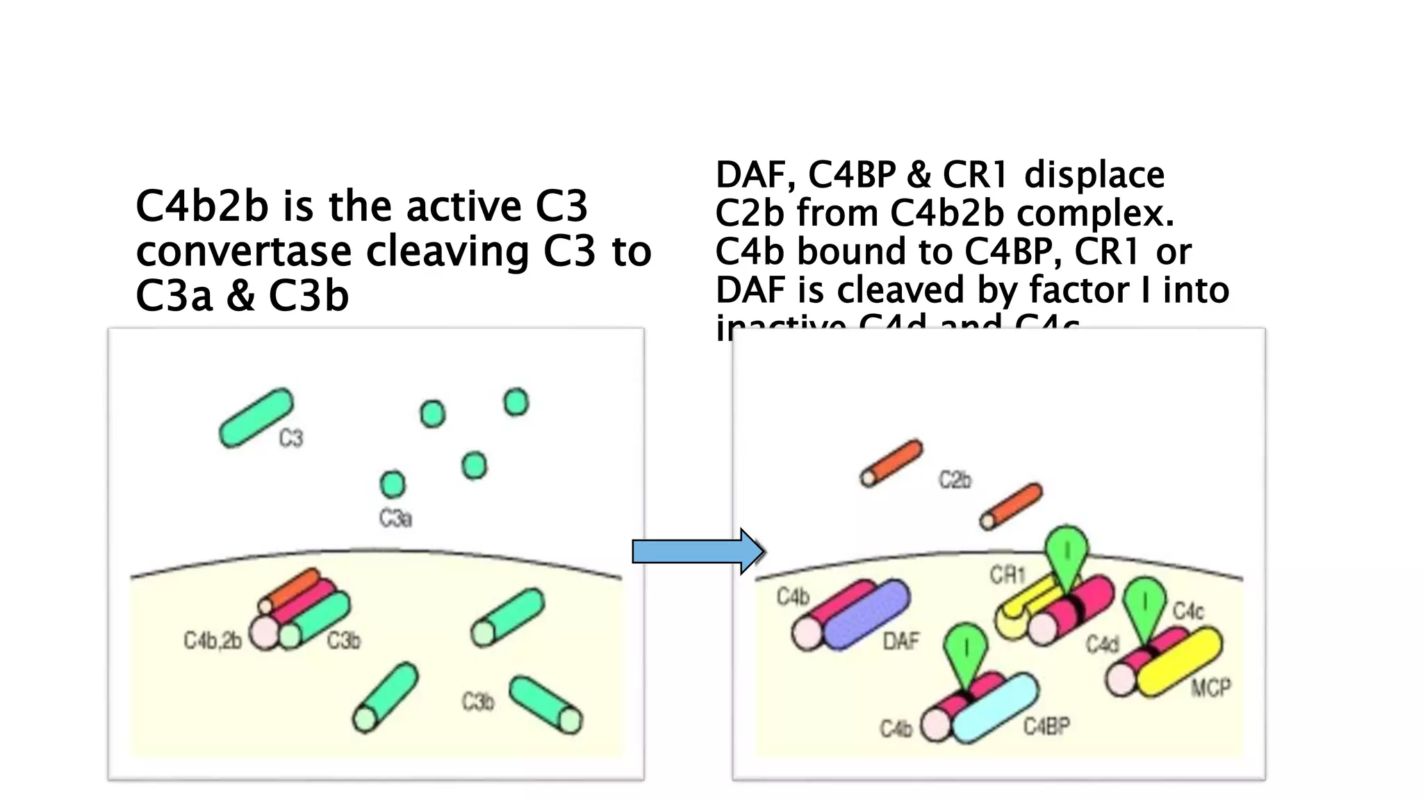
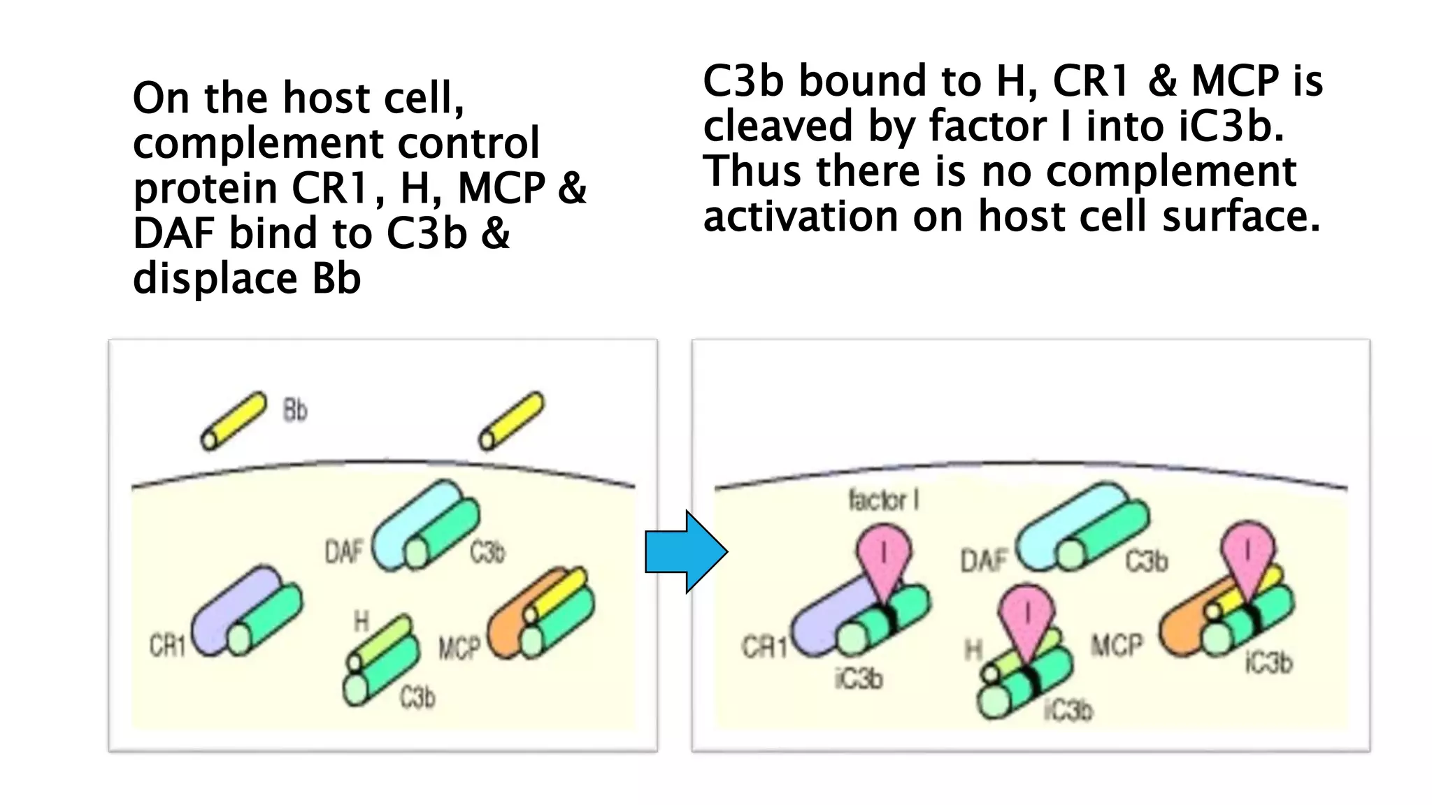
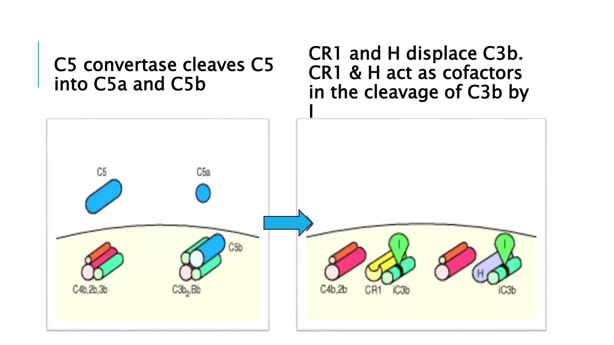
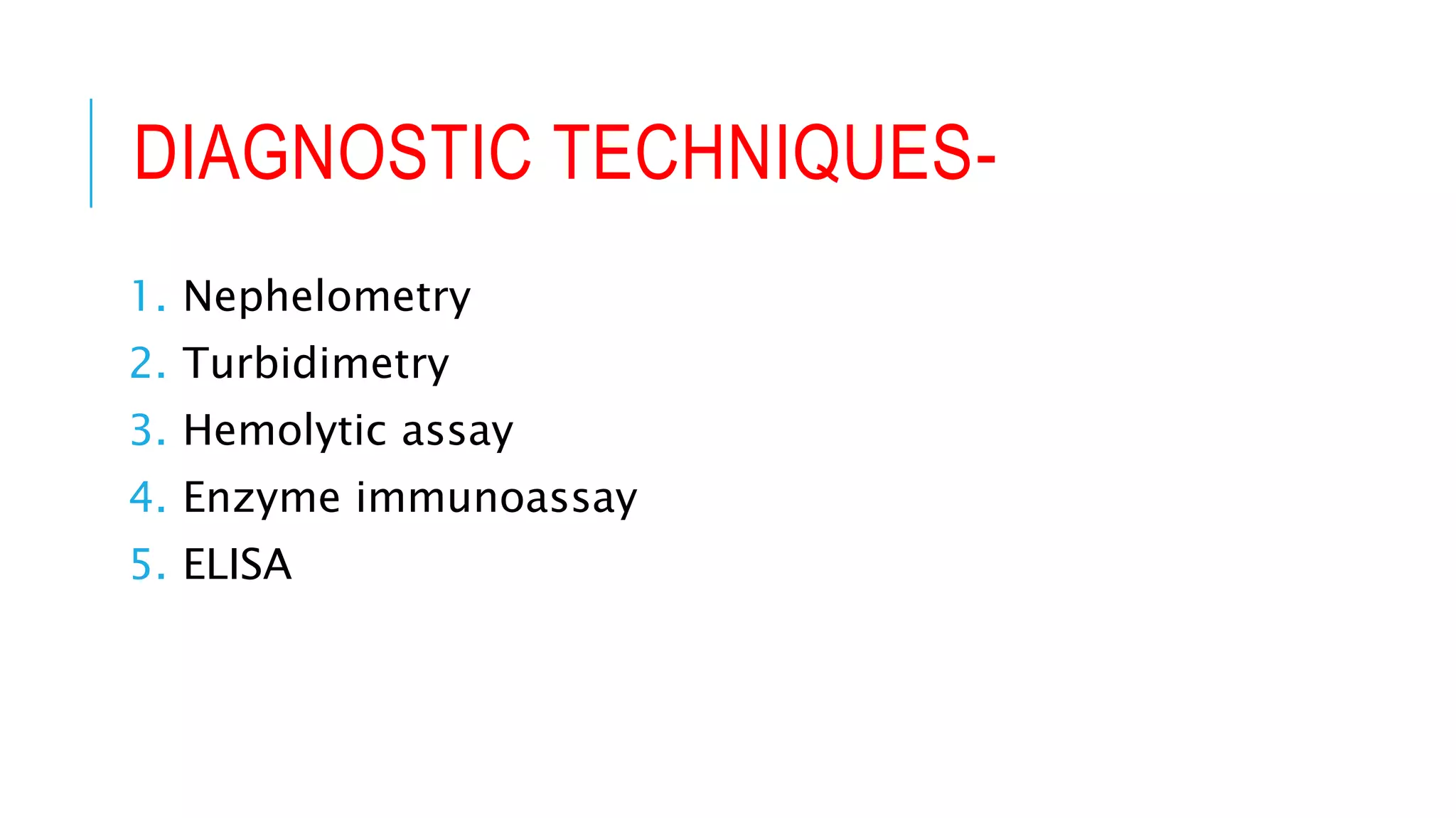
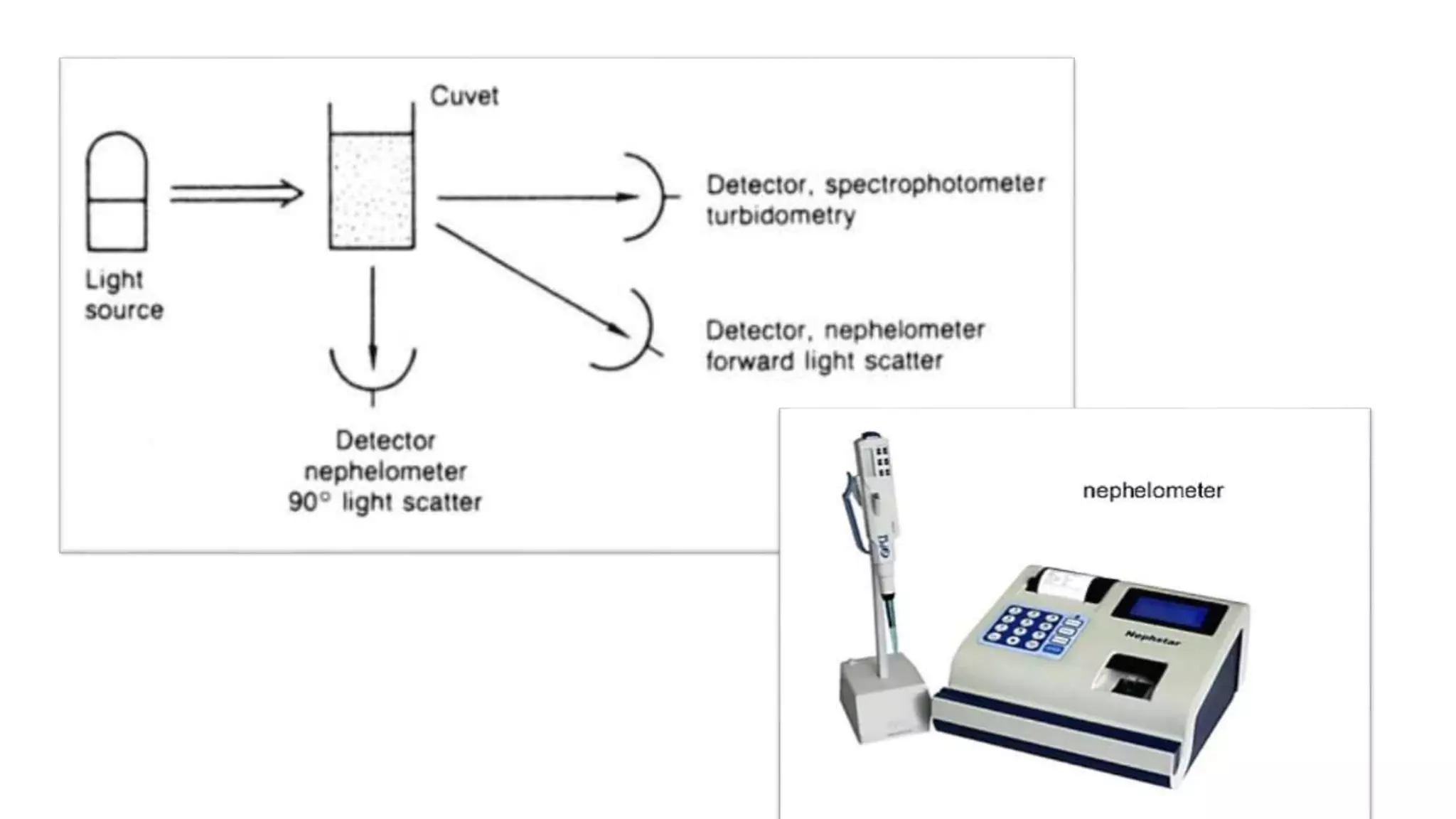
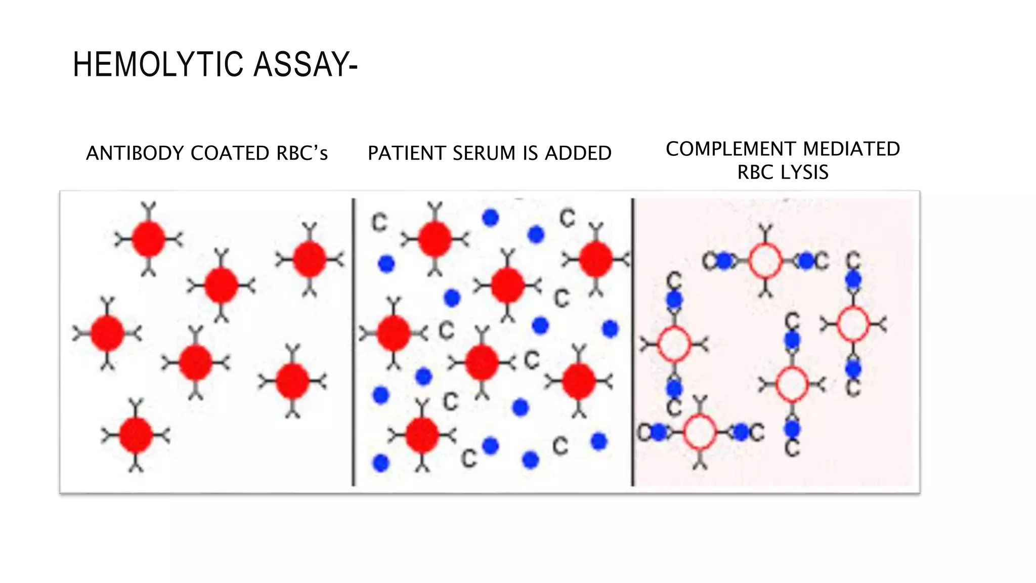
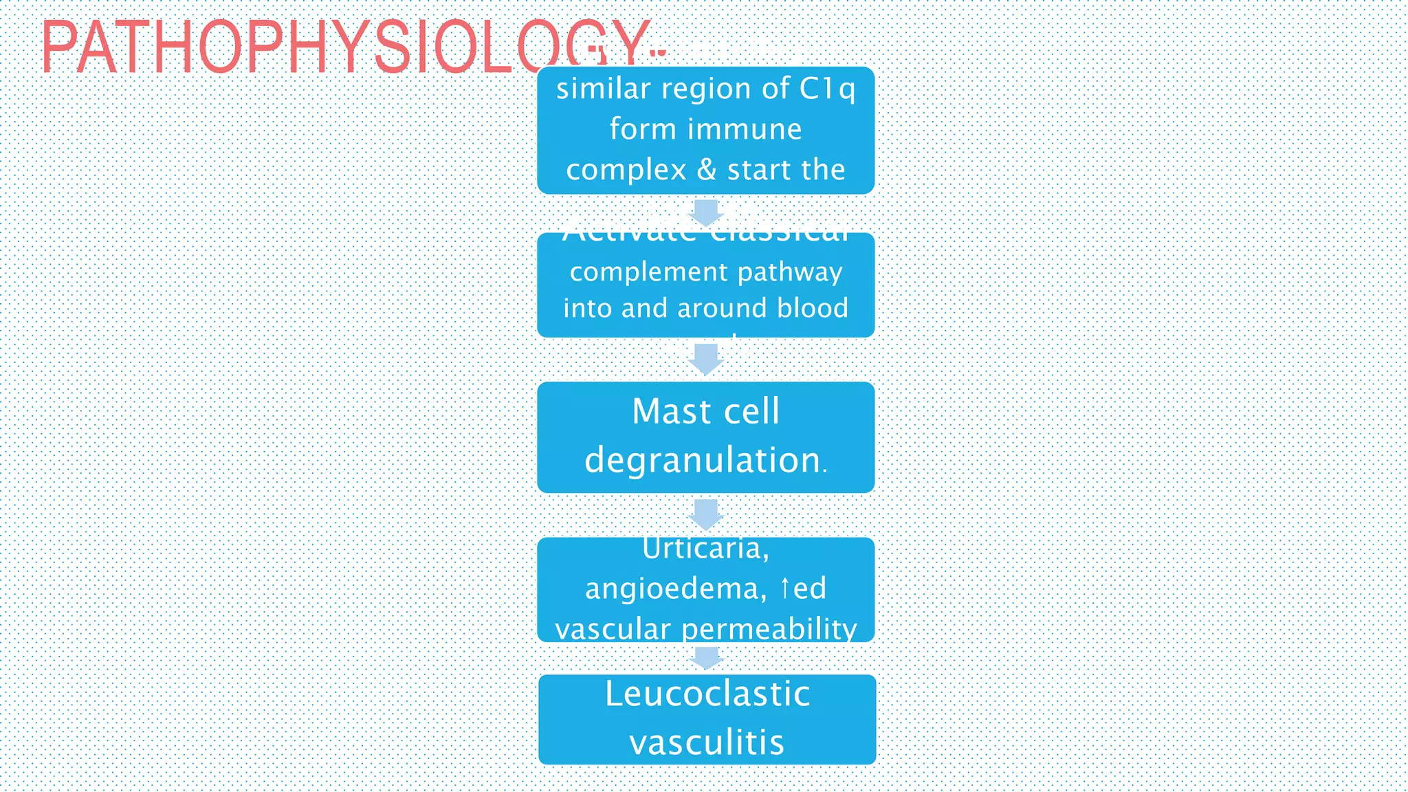
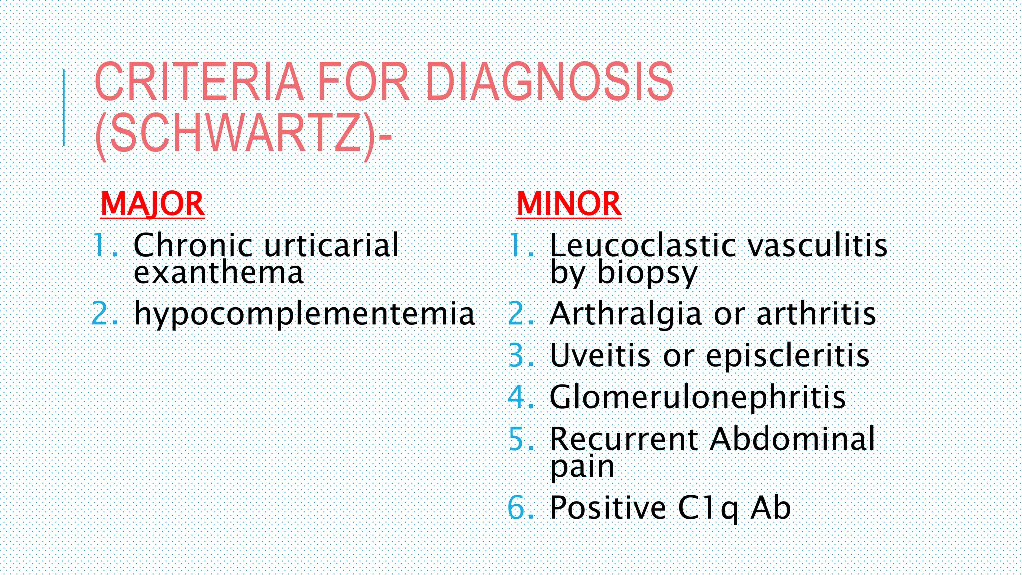
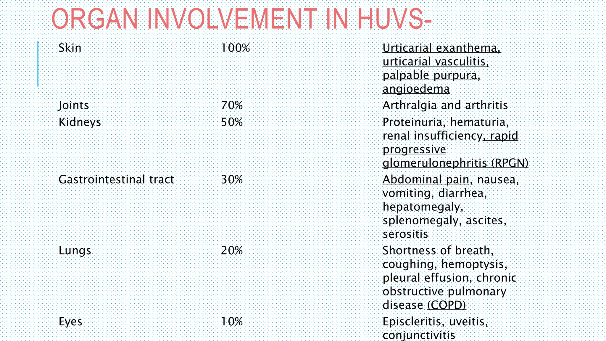
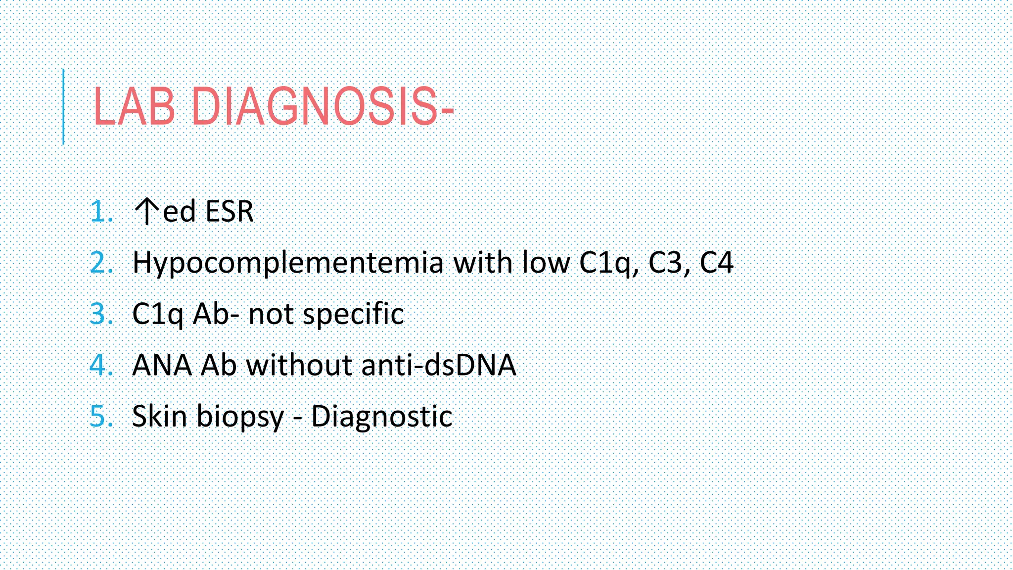
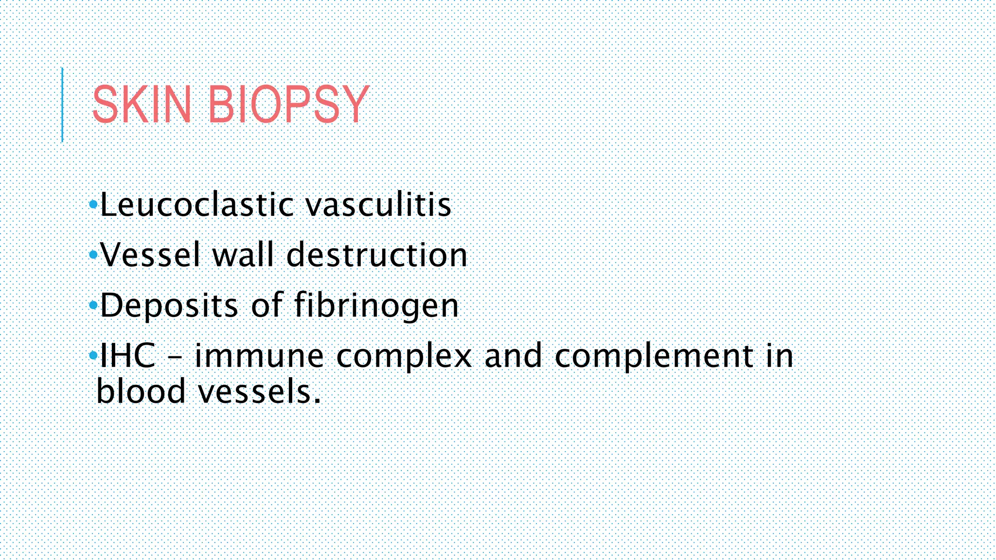
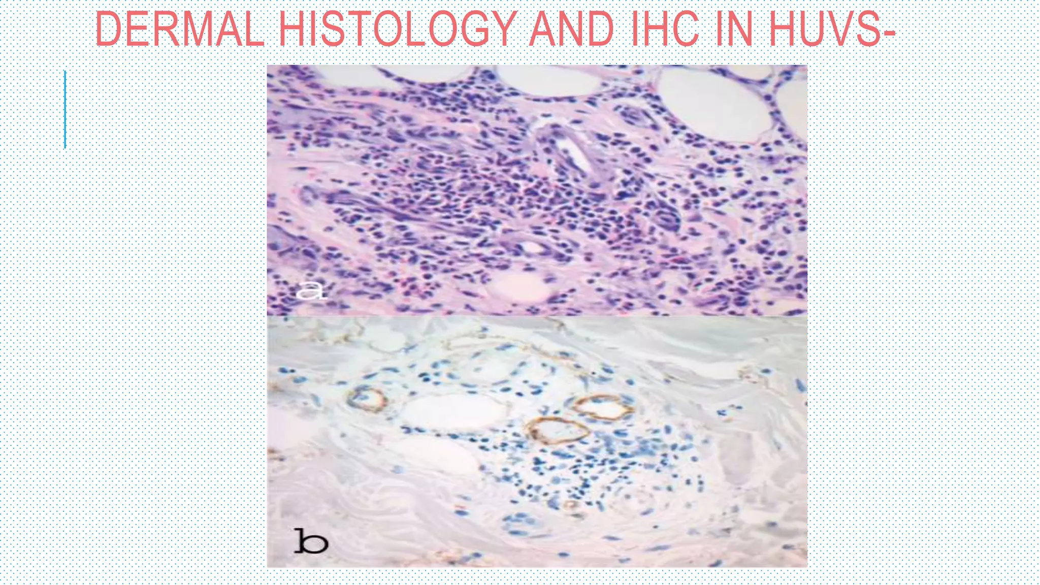
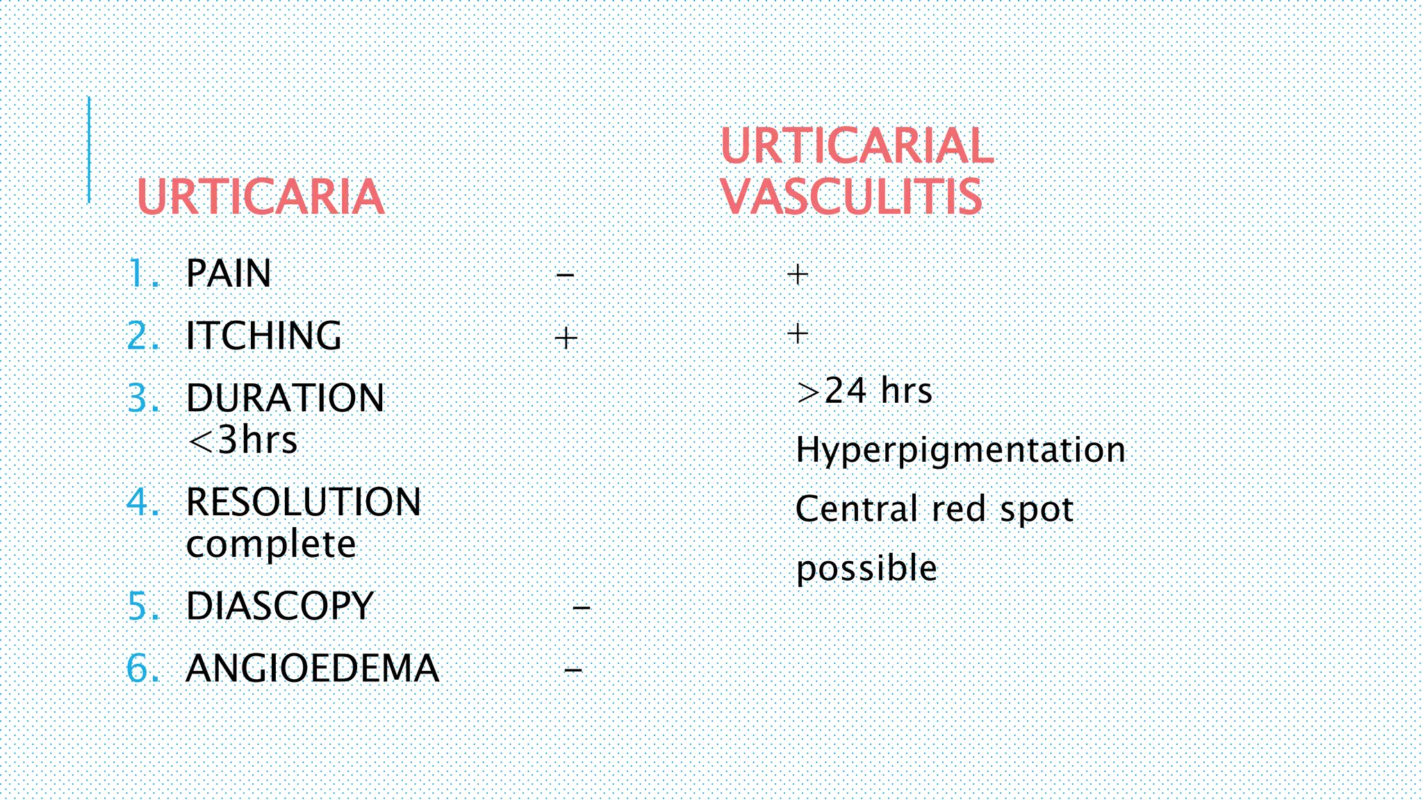
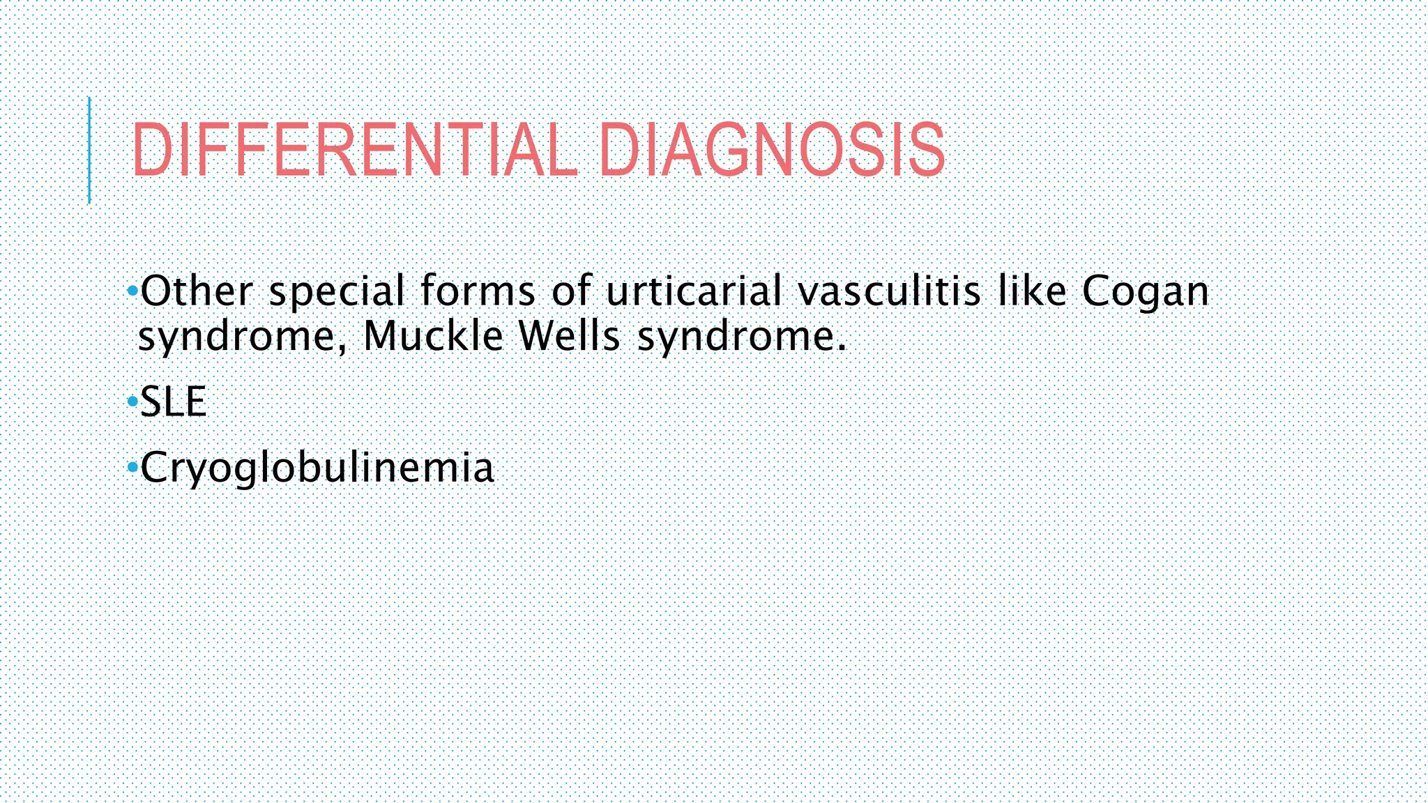
COMPLEMENT SYSTEM
&
HYPOCOMPLEMENTIC
URTICARIAL VASCULITIS
-Shivani
Singh
THE COMPLEMENT
SYSTEM
1st discovered by Jules Bordet
in 1896 as a heat labile
component of serum.
So named because of its
ability to complement the
antibacterial properties of
Ab’s in the heat labile
fraction of serum.
WHAT IS COMPLEMENT SYSTEM?
Complement is a system of ̴40 proteins in the plasma & on cell
surfaces amounting to >3g/L & constituting >15% of globular fraction of
plasma.
Arranged in a hierarchy of proteolytic cascades that starts with
identification of the pathogenic surface and leads to-
Generation of potent proinflammatory mediators
Opsonisation
Targeted lysis of pathogenic surface through assembly of MAC.
Functions in both innate and adaptive immunity.
PATHOGENIC SURFACES-
Infectious organisms
(bacteria,viruses,fungi,parasites)
Tissues damaged by physical, chemical or
neoplastic insults
Other surfaces identified as non self.
COMPLEMENT ACTIVATION
Complement proteins are present in
inactive state in plasma
Activated to become proteolytic
enzymes that degrade other
complement proteins
Forming a cascade capable of
tremendous amplification.
EARLY
REACTINGLATE
REACTING
Conversion of C₃ into C₃ₐ and C₃ by C₃ convertase is the
point at which all complement activation cascades converge.
b
Proteolysis of C₃ (most abundant component)
It can occur via 3 pathways
Classic
al
pathwa
y
Alternate
pathway
Lectin
pathway
Triggered
by fixation
of C₁ To
Ag-Ab
complex
Triggered by
microbial
surface
molecules,
complex
polysaccharide
PMBL binds to
carbohydrates of
microbes & directly
activates C₁
1. ACTIVATION
2. AMPLIFICATIO
N
3. LYSIS
CLASSICAL PATHWAY-
C1qrs complex made of
1 molecule of C1q , 2
molecules of C1r and 2
molecules of C1s.
Can bind to Fc region of
complement fixing
antibody.
C1qrs
complex
Pathogenic
surface
˄
C4
C4
b
C4a
C
2
C2
a
C2b
C3
C3
b C3a
C5
C5
b
C5
a
CLASSICAL PATHWAY
C3
ConvertaseC3/C5
Convertase
Ca²⁺,Mg²⁺
CLASSICAL PATHWAY-
Pathogenic
surface
C6
C7
C8
C9
C1qrs
C4b
C2b
C3b
C5b
C6
C7
C8
C9
C9
C9
C9 C9
C9
antibody
Membrane
Attack Complex
LECTIN PATHWAY-
Pattern Recognition Receptors (PRR)- MBL, ficolins to
conduct non self recognition.
Pathogen Associated Molecular Pattern (PAMP’s)-
Endotoxin or LPS of gram-ve bacteria
Lipoteichoic acid of gram +ve bacteria
β-glucan of fungi
MBL-
•Well characterized receptor of the collectin family.
•Synthesized in liver and secreted in plasma as a
component of acute phase response.
LECTIN BINDING PATHWAY-
Bacteria
C6
C7
C8
C9C4b
C2a
C3b
C5b
C6
C7
C8
C9
C9 C9
C9
C9
C9
MBL-MASP
MAC
ALTERNATE PATHWAY-
PATHOGENIC
SURFACE
B
C3(H₂O)
C3
Bb
Ba
Factor
D
Low Level
Spontaneou
s
Mg²⁺
ALTERNATE PATHWAY-
Pathogenic surface
C3(H₂O)
C3a
Bb
C3
C3b
Initial C3
Convertase
B
C3b
Bb
P
P
C5
C5b
C5a
ALTERNATE PATHWAY
Major C3/C5
Convertase
PATHOGENIC
SURFACE
PATHOGENIC
SURFACE
C3
b
Bb
P
C5
b
C
6 C7
C8
C9
C
6
C7
C
8 C
9
C9
C9
C9
C9C9
MAC
NOVEL PATHWAYS OF
COMPLEMENT ACTIVATION-
1. Kallikrein & Thrombin - C3 & C5 can be directly
cleaved by proteases unrelated to the complement
cascade like kallikrein and thrombin leading to
generation of anaphylotoxins (C3a & C5a).
2. C2 bypass pathway – direct cleavage of C3 by
MBL/MASP2
3. Properdin pathway - Properdin can promote de-
novo C3 convertase assembly when immobilized on
an inert surface & initiate C3 convertase formation
on microbial surface( eg Neisseria gonorrheae ).
IMPORTANCE OF C3
C
3
C3a + C3b
C3 Convertase
ANAPHYLOTOXI
N
AMPLIFICATION
OF ALTERNATE
PATHWAYC5
Convertase
OPSONISATIO
N
ROLE OF MINOR COMPONENTS-
PHYSIOLOGICAL ROLES OF
COMPLEMENT SYSTEM
REGULATION OF COMPLEMENT
SYSTEM-
TOO MUCH
ACTIVATION
TOO LITTLE
ACTIVATION
1. SERUM
PROTEINS
2. CELL SURFACE
RECEPTORS
REGULATION OF COMPLEMENT SYSTEM-
FACTOR FUNCTION DEFICIENCY
C₁ INH Blocks activation of C₁ Hereditary angioedema
DAF (Decay accelerating
factor)
Prevents formation of C3
convertase
PNH
CD59 Inhibits formation of MAC PNH
FACTOR H Spontaneous decay of C3
convertase C3bBb
HUS
FACTOR I Inhibit C3 convertase Pyogenic infections
MCP (Mebrane co factor
protein)
Inhibit C3 convertase
SCPN (serum
carboxypeptidase N)
Inactivate
anaphylotoxins.cleave C3a
& C5a
Angioedema
PROPERDIN Promotes association of
C3b with factor B
Neisseria infection
Cleaves Factor B Pyogenic infection
STAGES AT WHICH REGULATORY
PROTEINS ACT-
1. C1qrs complex
2. C3 convertase
3. C5 convertase
4. Membrane Attack Complex
C1 INH-
C1 INH
C1s
C1r
HEREDITARY
ANGIOEDEMA
C4b2b is the active C3
convertase cleaving C3 to
C3a & C3b
DAF, C4BP & CR1 displace
C2b from C4b2b complex.
C4b bound to C4BP, CR1 or
DAF is cleaved by factor I into
inactive C4d and C4c.
On the host cell,
complement control
protein CR1, H, MCP &
DAF bind to C3b &
displace Bb
C3b bound to H, CR1 & MCP is
cleaved by factor I into iC3b.
Thus there is no complement
activation on host cell surface.
C5 convertase cleaves C5
into C5a and C5b
CR1 and H displace C3b.
CR1 & H act as cofactors
in the cleavage of C3b by
I
The terminal proteins of
complement form a
membrane pore, the MAC
CD59 prevents final assembly
of MAC at the C8-C9 stage.
MIRL blocks MAC by
preventing C9 assembly.
Diseases resulting due to deficiency of complement components.
INVESTIGATIONS -
1. Quantification of individual complement
components
2. Quantification of activation products
3. Quantification of complement function
4. Quantification of autoantibodies to complement
components.
DIAGNOSTIC TECHNIQUES-
1. Nephelometry
2. Turbidimetry
3. Hemolytic assay
4. Enzyme immunoassay
5. ELISA
ANTIBODY COATED RBC’s PATIENT SERUM IS ADDED COMPLEMENT MEDIATED
RBC LYSIS
HEMOLYTIC ASSAY-
HYPOCOMPLEMENTIC URTICARIAL
VASCULITIS-
•HUVS is a severe systemic form of urticarial
vasculitis.
•Aka McDuffie syndrome.
•Female : male = 2:1
•Peak incidence 5th decade. Also seen in
children.
PATHOPHYSIOLOGY-IgG Ab to collagen
similar region of C1q
form immune
complex & start the
cascade.
Activate classical
complement pathway
into and around blood
vessels
Mast cell
degranulation.
Urticaria,
angioedema, ↑ed
vascular permeability
Leucoclastic
vasculitis
CRITERIA FOR DIAGNOSIS
(SCHWARTZ)-
MAJOR
1. Chronic urticarial
exanthema
2. hypocomplementemia
MINOR
1. Leucoclastic vasculitis
by biopsy
2. Arthralgia or arthritis
3. Uveitis or episcleritis
4. Glomerulonephritis
5. Recurrent Abdominal
pain
6. Positive C1q Ab
ORGAN INVOLVEMENT IN HUVS-
Skin 100% Urticarial exanthema,
urticarial vasculitis,
palpable purpura,
angioedema
Joints 70% Arthralgia and arthritis
Kidneys 50% Proteinuria, hematuria,
renal insufficiency, rapid
progressive
glomerulonephritis (RPGN)
Gastrointestinal tract 30% Abdominal pain, nausea,
vomiting, diarrhea,
hepatomegaly,
splenomegaly, ascites,
serositis
Lungs 20% Shortness of breath,
coughing, hemoptysis,
pleural effusion, chronic
obstructive pulmonary
disease (COPD)
Eyes 10% Episcleritis, uveitis,
conjunctivitis
LAB DIAGNOSIS-
1. ↑ed ESR
2. Hypocomplementemia with low C1q, C3, C4
3. C1q Ab- not specific
4. ANA Ab without anti-dsDNA
5. Skin biopsy - Diagnostic
SKIN BIOPSY
•Leucoclastic vasculitis
•Vessel wall destruction
•Deposits of fibrinogen
•IHC – immune complex and complement in
blood vessels.
DERMAL HISTOLOGY AND IHC IN HUVS-
URTICARIA
1. PAIN -
2. ITCHING +
3. DURATION
<3hrs
4. RESOLUTION
complete
5. DIASCOPY -
6. ANGIOEDEMA -
URTICARIAL
VASCULITIS
+
+
>24 hrs
Hyperpigmentation
Central red spot
possible
DIFFERENTIAL DIAGNOSIS
•Other special forms of urticarial vasculitis like Cogan
syndrome, Muckle Wells syndrome.
•SLE
•Cryoglobulinemia
Complement system

Complement system