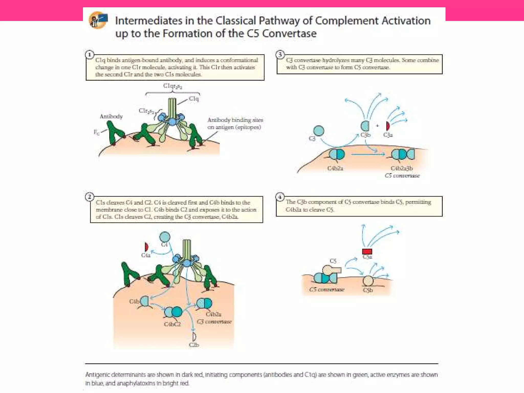
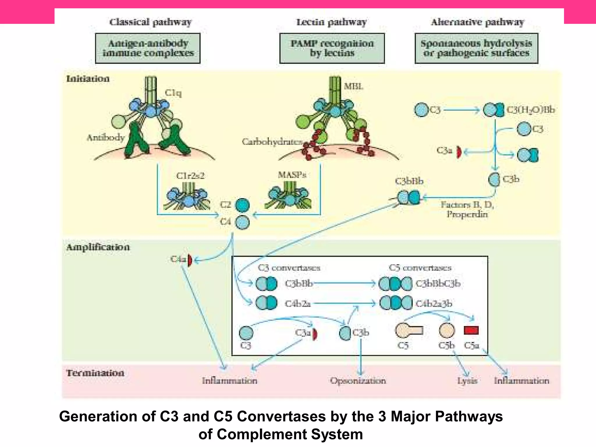
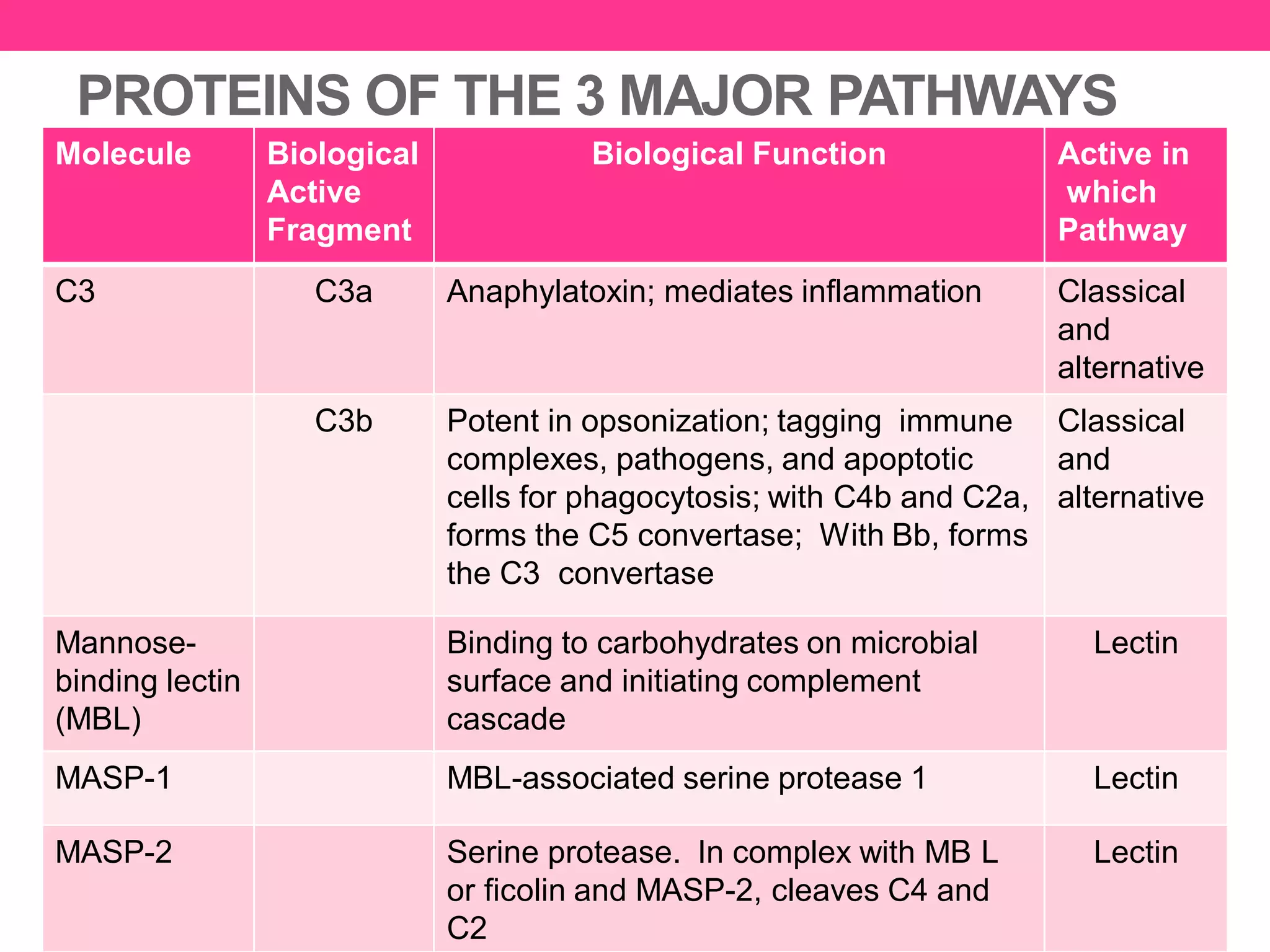
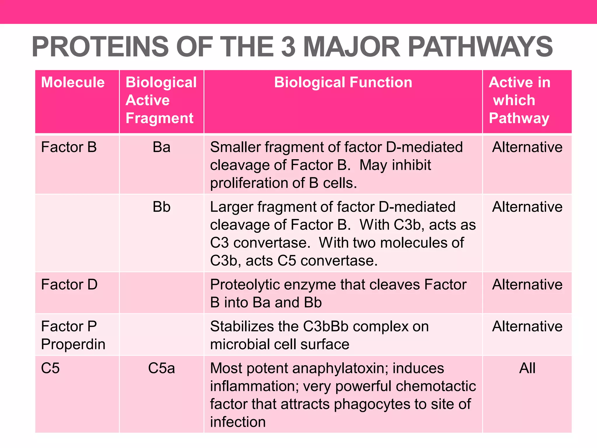
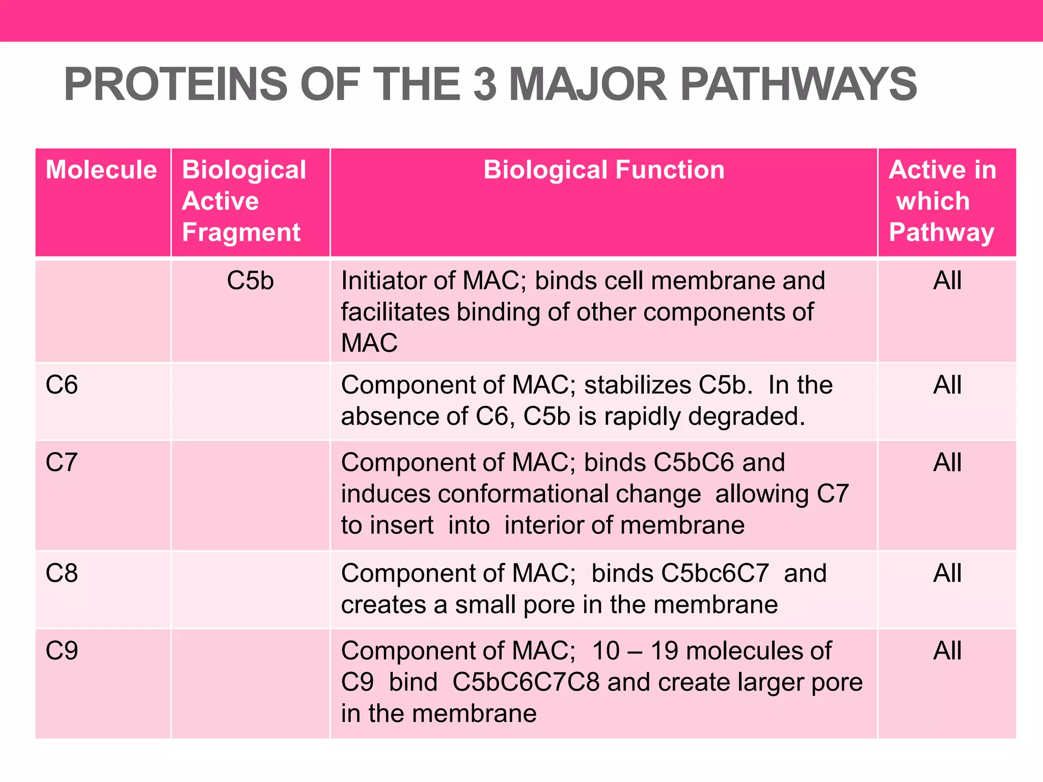
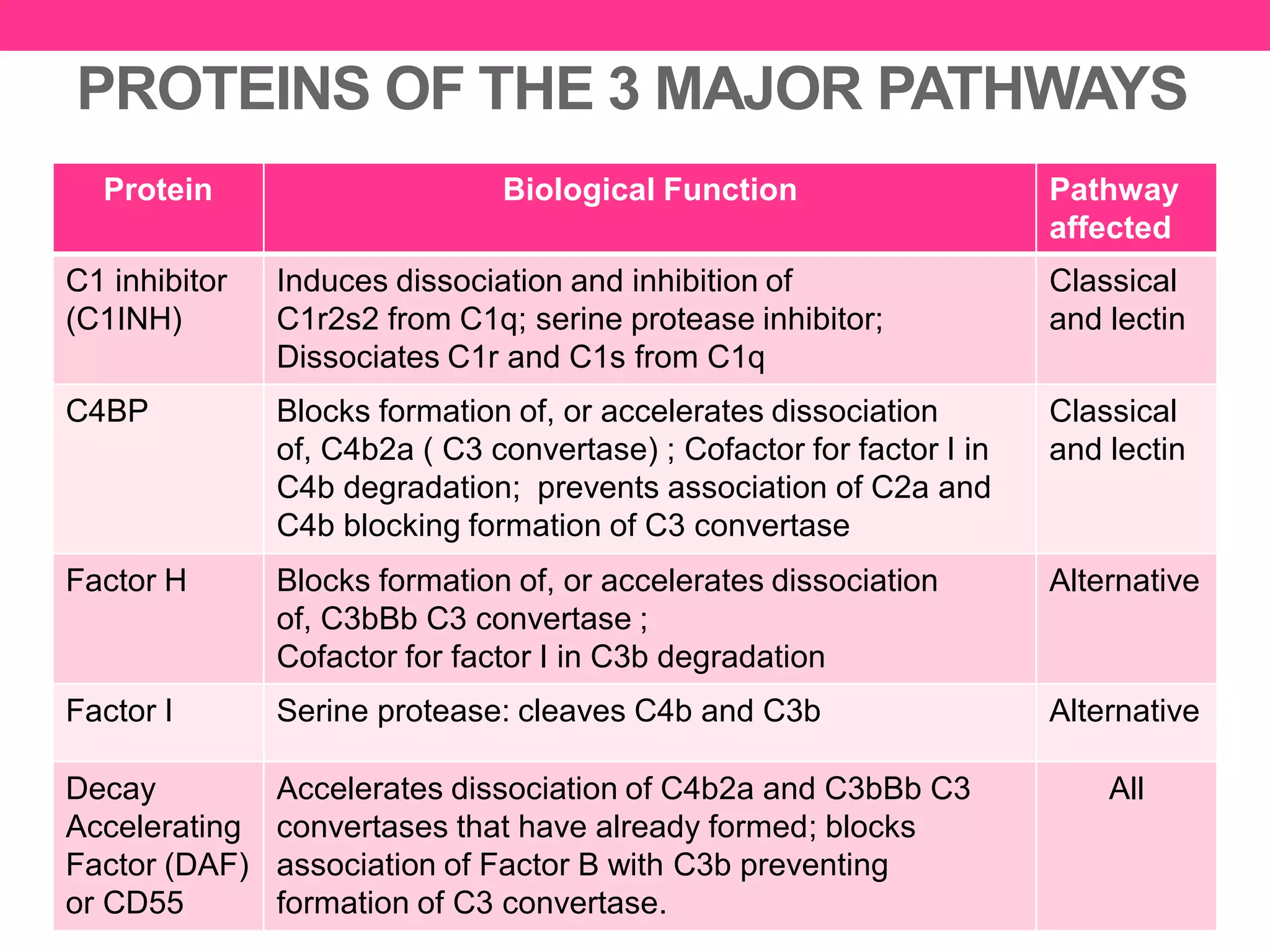
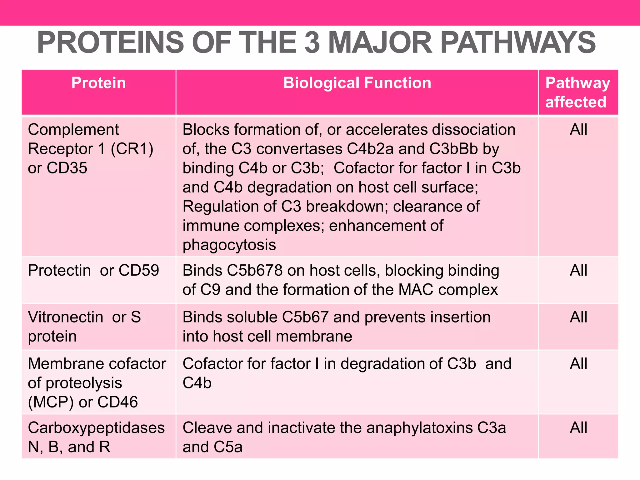
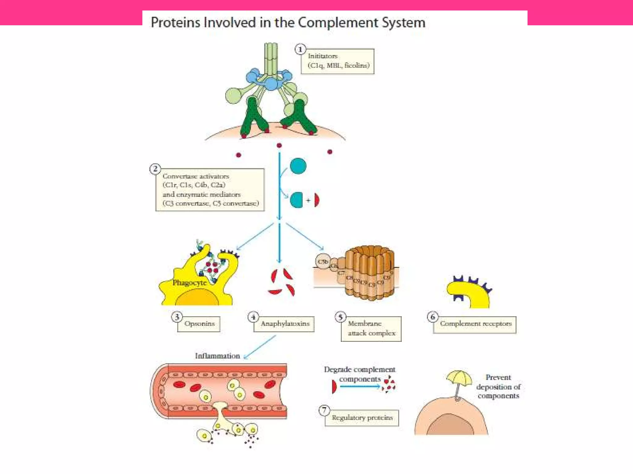
32 PROTEINS OF THE
COMPLEMENT SYSTEM
Julie S. Sy, RMT, DVM
COMPLEMENT SYSTEM
• Consists of over 30 proteins produced by the liver that
circulate in the blood serum & w/in tissues throughout the
body
• It does not change over a person’s lifetime thus it is
considered as part of the innate immune system
• However, it can be recruited into action by the adaptive
immune system.
• Together, proteins of the complement system destroy
harmful microbes by opsonisation, phagocytosis, cytolysis
& inflammation
• It also prevents excessive damage to the host’s tissues.
COMPLEMENT PROTEINS
• Exist as zymogens or proenzymes (inactive forms) in the
circulation that must first be cleaved or undergo
conformational changes prior to activation.
• Complement proteins include initiator molecules,
enzymatic mediators, membrane-binding components or
opsonins, inflammatory mediators, membrane attack
proteins, complement receptor proteins, and regulatory
components.
• Activation of one proenzyme will trigger the activation of
the next proenzyme in the pathway, and so on (cascade
process).
• More product is formed with each succeeding reaction in
the cascade, amplifying the effects
COMPLEMENT ACTIVATION
• The cascade of complement proteins that occurs during
an infection
• 3 MAJOR INITIATION PATHWAYS
i. CLASSICAL PATHWAY – activated when antibodies bind to
antigens
ii. ALTERNATIVE PATHWAY – activated by contact between certain
complement proteins with lipid carbohydrate complex found in
some microbial surfaces
iii. LECTIN PATHWAY – activated when MBL proteins bind to
carbohydrate containing mannose found on some microbial
surfaces. MBL proteins are synthesized by the liver in response to
stimulation of cytokines released by macrophages during
phagocytosis.
COMPLEMENT ACTIVATION
• Although the initiating event of each of the 3 pathways is
different, they all converge in the generation of C3
convertase.
• The classical and lectin pathways use the dimer C4b2a
for their C3 convertase.
• The alternative pathway uses C3bBb for its C3
convertase.
• The second set of convertase enzymes, C5 convertases,
are formed by the addition of a C3b component to the C3
convertases.
Membrane Attack Complex (MAC)
• Activation of the terminal components of the complement
C5b, C6, C7, C8 and C9 results in the deposition of a
membrane attack complex (MAC) onto the microbial cell
membrane.
• This complex introduces large pores in the membrane,
preventing it from maintaining osmotic integrity and
resulting in the death of the cell.
CLASSICAL PATHWAY
• Is considered as part of the adaptive immune response
since it begins with the formation of antigen-antibody
complexes.
• Complexes formed by IgM and certain subclasses of IgG
(except IgG4) only are capable of activating this pathway
• Proteins of the classical pathway are numbered in the
order in which they were discovered, which does not quite
correspond with the order in which the proteins act in the
pathway.
Steps Involved in Classical Pathway
1. Antibodies attach to antigens (for example, proteins or
large polysaccharides on the surface of a bacterium or
other cell), forming antigen-antibody complexes bind to
and activate C1.
2. Next, activated C1 activates C2 and C4 by splitting them.
C2 splits into fragments C2a and C2b, and C4 is split into
C4a and C4b.
3. C2a and C4b combine and together activate C3 by
splitting it into C3a and C3b. C3a participates in
inflammation, and C3b functions in cytolysis and
opsonisation.
4. C3b binds to the membrane in association with C4b and
C2a, and C3a is released into the microenvironment. The
resulting C4bC2aC3b is a C5 convertase.
5. C5 convertase cleaves C5, which goes on to form the
MAC.
ALTERNATIVE PATHWAY
• Activation is independent of antigen-antibody interactions
thus it is considered as part of the innate immune system
• Provides means of protection against certain pathogens
before antibody response is mounted
• Provides NON-SPECIFIC resistance against infection
without the participation of antibodies
• Hence, provides a first-line of defense against a number
of infectious agents.
• Can be activated by many Gram negative bacteria due to
presence of LPS in their cell membrane.
Steps Involved in Alternative Pathway
1. Spontaneous hydrolysis of soluble C3 to C3(H₂O)
allows binding of Factor B to C3(H₂O) and becomes
susceptible to Factor D, which cleaves Factor B into
Bb. The C3(H₂O)Bb complex acts as a fluid C3
convertase and cleaves C3 into C3a and C3b. Once
C3b is formed, Factor B will bind to it and becomes
susceptible to cleavage by Factor D. The resulting
C3bBb complex is a membrane-bound C3 convertase
that will continue to generate more C3b.
2. Addition of a second C3b molecule to the C3bBb
complex forms the C5 convertase.
3. C5 convertase cleaves C5, which goes on to form the
MAC
MANNOSE BINDING LECTIN PATHWAY
• Uses mannose-binding lectins (MBL),
proteins that recognize specific
carbohydrate components primarily
found on microbial surfaces as its
specific receptor molecules.
• MBL are capable of initiating this
pathway by binding close-knit arrays of
mannose residues that are found on
microbial surfaces such as those of
Salmonella, Listeria, Neisseria;
Cryptococcus neoformans and Candida
albicans; and even the membrane of
HIV-1 and respiratory syncytial virus.
• Like the alternative pathway, it is
considered as part of the innate immune
system.
Steps Involved in the Lectin Pathway
1. Mannose-binding lectin (MBL) molecules recognize and
bind to the carbohydrate containing mannose found in
bacterial cell walls and on some viruses.
2. Binding of MBL to the pathogen results in the association
of two serine proteases, MASP-1 and MASP-2 (MBL
associated serine proteases).
3. Formation of the MBL/MASP-1/ MASP-2 tri-molecular
complex results in the activation of the MASPs and
subsequent cleavage of C4 and C2.
4. C4b binds with C2a resulting to C4bC2a complex, a C3
convertase, which cleaves C3 into C3a and C3b.
5. C3b binds with C4bC2a resulting to C2bC2aC3b, which is
the C5 convertase. C5 convertase cleaves C5, which goes
on to form the MAC.
Generation of C3 and C5 Convertases by the 3 Major Pathways
of Complement System
PROTEINS OF THE 3 MAJOR PATHWAYS
Molecule Biological
Active
Fragment
Biological Function Active in
which
Pathway
C1 C1q Initiation of the classical pathway by
binding Ig; binding to apoptotic blebs and
initiating phagocytosis of apoptotic cells;
activates C1r
Classical
C1r Activates C1s Classical
C1s Cleaves C4 and C2 Classical
C2 C2a Prokinin; mediates inflammation;
With C1 and C4b, is a C3 convertase
Classical
and lectin
C2b Prokinin; cleaved by plasmin to yield kinin
which results in vasodilation and edema
Classical
and lectin
C4 C4a Has weak anaphylatoxin activity Classical
and lectin
C4b With C1 and C2a, is a C3 convertase;
opsonin
Classical
and lectin
PROTEINS OF THE 3 MAJOR PATHWAYS
Molecule Biological
Active
Fragment
Biological Function Active in
which
Pathway
C3 C3a Anaphylatoxin; mediates inflammation Classical
and
alternative
C3b Potent in opsonization; tagging immune
complexes, pathogens, and apoptotic
cells for phagocytosis; with C4b and C2a,
forms the C5 convertase; With Bb, forms
the C3 convertase
Classical
and
alternative
Mannose-
binding lectin
(MBL)
Binding to carbohydrates on microbial
surface and initiating complement
cascade
Lectin
MASP-1 MBL-associated serine protease 1 Lectin
MASP-2 Serine protease. In complex with MB L
or ficolin and MASP-2, cleaves C4 and
C2
Lectin
PROTEINS OF THE 3 MAJOR PATHWAYS
Molecule Biological
Active
Fragment
Biological Function Active in
which
Pathway
Factor B Ba Smaller fragment of factor D-mediated
cleavage of Factor B. May inhibit
proliferation of B cells.
Alternative
Bb Larger fragment of factor D-mediated
cleavage of Factor B. With C3b, acts as
C3 convertase. With two molecules of
C3b, acts C5 convertase.
Alternative
Factor D Proteolytic enzyme that cleaves Factor
B into Ba and Bb
Alternative
Factor P
Properdin
Stabilizes the C3bBb complex on
microbial cell surface
Alternative
C5 C5a Most potent anaphylatoxin; induces
inflammation; very powerful chemotactic
factor that attracts phagocytes to site of
infection
All
PROTEINS OF THE 3 MAJOR PATHWAYS
Molecule Biological
Active
Fragment
Biological Function Active in
which
Pathway
C5b Initiator of MAC; binds cell membrane and
facilitates binding of other components of
MAC
All
C6 Component of MAC; stabilizes C5b. In the
absence of C6, C5b is rapidly degraded.
All
C7 Component of MAC; binds C5bC6 and
induces conformational change allowing C7
to insert into interior of membrane
All
C8 Component of MAC; binds C5bc6C7 and
creates a small pore in the membrane
All
C9 Component of MAC; 10 – 19 molecules of
C9 bind C5bC6C7C8 and create larger pore
in the membrane
All
PROTEINS OF THE 3 MAJOR PATHWAYS
Protein Biological Function Pathway
affected
C1 inhibitor
(C1INH)
Induces dissociation and inhibition of
C1r2s2 from C1q; serine protease inhibitor;
Dissociates C1r and C1s from C1q
Classical
and lectin
C4BP Blocks formation of, or accelerates dissociation
of, C4b2a ( C3 convertase) ; Cofactor for factor I in
C4b degradation; prevents association of C2a and
C4b blocking formation of C3 convertase
Classical
and lectin
Factor H Blocks formation of, or accelerates dissociation
of, C3bBb C3 convertase ;
Cofactor for factor I in C3b degradation
Alternative
Factor I Serine protease: cleaves C4b and C3b Alternative
Decay
Accelerating
Factor (DAF)
or CD55
Accelerates dissociation of C4b2a and C3bBb C3
convertases that have already formed; blocks
association of Factor B with C3b preventing
formation of C3 convertase.
All
PROTEINS OF THE 3 MAJOR PATHWAYS
Protein Biological Function Pathway
affected
Complement
Receptor 1 (CR1)
or CD35
Blocks formation of, or accelerates dissociation
of, the C3 convertases C4b2a and C3bBb by
binding C4b or C3b; Cofactor for factor I in C3b
and C4b degradation on host cell surface;
Regulation of C3 breakdown; clearance of
immune complexes; enhancement of
phagocytosis
All
Protectin or CD59 Binds C5b678 on host cells, blocking binding
of C9 and the formation of the MAC complex
All
Vitronectin or S
protein
Binds soluble C5b67 and prevents insertion
into host cell membrane
All
Membrane cofactor
of proteolysis
(MCP) or CD46
Cofactor for factor I in degradation of C3b and
C4b
All
Carboxypeptidases
N, B, and R
Cleave and inactivate the anaphylatoxins C3a
and C5a
All
Proteins Involved in Complement System
1. The complement pathways are initiated by proteins that bind to
pathogens, either directly or via an antibody or other pathogen-specific
protein. After a conformational change,
2. enzymatic mediators activate other enzymes that generate the central
proteins of the complement cascade, the C3 and C5 convertases,
which cleave C3 and C5, releasing active components that mediate all
functions of complement, including
3. opsonization,
4.inflammation, and
5. the generation of the membrane attack complex (MAC). Effector
complement proteins can label an antibody-antigen complex for
phagocytosis (opsonins), initiate inflammation (anaphylatoxins), or bind
to a pathogen and nucleate the formation of the MAC. Often, these
effectors act through
6.complement receptors on phagocytic cells, granulocytes, or
erythrocytes.
7.Regulatory proteins limit the effects of complement by promoting their
degradation or preventing their binding to host cells.
Terminology
• Anaphylatoxin – binds mast cell/ basophil causing
degranulation and release of histamine and other chemicals
that increase blood vessel permeability
• Opsonins – “immune adherence” ; promote attachment of a
phagocyte to a microbe;
• promote phagocytosis by binding to complement receptors (CR1) on
phagocytic cells
• Chemotaxis – the chemical attraction of phagocytes to
microbes
• Vasodilation – blood vessels dilate and their permeability
increases following tissue damage and is responsible for the
redness and heat associated with inflammation
• Increased vascular permeability – permits defensive
substances normally retained in the blood to pass through the
walls of the blood vessels and enter the injured area.
• Permits fluid to move from the blood into the tissue spaces, is
responsible for the edema (accumulation of fluid) in inflammation
References
• Owen, Judith A., Punt, Jenny, Stranford, Sharon A.
(2013). Kuby Immunology, (7th edition). W. H. Freeman
and Company.
• Tortora, Gerard J., Funke, Berdell R., and Case, Christine
L. (2016). Microbiology An Introduction, (12th edition).
Pearson Education Limited.
• http://www.nios.ac.in/media/documents/dmlt/Microbiology/
Lesson-58.pdf

32 Proteins of the Complement System

  • 1.
    32 PROTEINS OFTHE COMPLEMENT SYSTEM Julie S. Sy, RMT, DVM
  • 2.
    COMPLEMENT SYSTEM • Consistsof over 30 proteins produced by the liver that circulate in the blood serum & w/in tissues throughout the body • It does not change over a person’s lifetime thus it is considered as part of the innate immune system • However, it can be recruited into action by the adaptive immune system. • Together, proteins of the complement system destroy harmful microbes by opsonisation, phagocytosis, cytolysis & inflammation • It also prevents excessive damage to the host’s tissues.
  • 3.
    COMPLEMENT PROTEINS • Existas zymogens or proenzymes (inactive forms) in the circulation that must first be cleaved or undergo conformational changes prior to activation. • Complement proteins include initiator molecules, enzymatic mediators, membrane-binding components or opsonins, inflammatory mediators, membrane attack proteins, complement receptor proteins, and regulatory components. • Activation of one proenzyme will trigger the activation of the next proenzyme in the pathway, and so on (cascade process). • More product is formed with each succeeding reaction in the cascade, amplifying the effects
  • 4.
    COMPLEMENT ACTIVATION • Thecascade of complement proteins that occurs during an infection • 3 MAJOR INITIATION PATHWAYS i. CLASSICAL PATHWAY – activated when antibodies bind to antigens ii. ALTERNATIVE PATHWAY – activated by contact between certain complement proteins with lipid carbohydrate complex found in some microbial surfaces iii. LECTIN PATHWAY – activated when MBL proteins bind to carbohydrate containing mannose found on some microbial surfaces. MBL proteins are synthesized by the liver in response to stimulation of cytokines released by macrophages during phagocytosis.
  • 5.
    COMPLEMENT ACTIVATION • Althoughthe initiating event of each of the 3 pathways is different, they all converge in the generation of C3 convertase. • The classical and lectin pathways use the dimer C4b2a for their C3 convertase. • The alternative pathway uses C3bBb for its C3 convertase. • The second set of convertase enzymes, C5 convertases, are formed by the addition of a C3b component to the C3 convertases.
  • 6.
    Membrane Attack Complex(MAC) • Activation of the terminal components of the complement C5b, C6, C7, C8 and C9 results in the deposition of a membrane attack complex (MAC) onto the microbial cell membrane. • This complex introduces large pores in the membrane, preventing it from maintaining osmotic integrity and resulting in the death of the cell.
  • 7.
    CLASSICAL PATHWAY • Isconsidered as part of the adaptive immune response since it begins with the formation of antigen-antibody complexes. • Complexes formed by IgM and certain subclasses of IgG (except IgG4) only are capable of activating this pathway • Proteins of the classical pathway are numbered in the order in which they were discovered, which does not quite correspond with the order in which the proteins act in the pathway.
  • 8.
    Steps Involved inClassical Pathway 1. Antibodies attach to antigens (for example, proteins or large polysaccharides on the surface of a bacterium or other cell), forming antigen-antibody complexes bind to and activate C1. 2. Next, activated C1 activates C2 and C4 by splitting them. C2 splits into fragments C2a and C2b, and C4 is split into C4a and C4b. 3. C2a and C4b combine and together activate C3 by splitting it into C3a and C3b. C3a participates in inflammation, and C3b functions in cytolysis and opsonisation. 4. C3b binds to the membrane in association with C4b and C2a, and C3a is released into the microenvironment. The resulting C4bC2aC3b is a C5 convertase. 5. C5 convertase cleaves C5, which goes on to form the MAC.
  • 10.
    ALTERNATIVE PATHWAY • Activationis independent of antigen-antibody interactions thus it is considered as part of the innate immune system • Provides means of protection against certain pathogens before antibody response is mounted • Provides NON-SPECIFIC resistance against infection without the participation of antibodies • Hence, provides a first-line of defense against a number of infectious agents. • Can be activated by many Gram negative bacteria due to presence of LPS in their cell membrane.
  • 11.
    Steps Involved inAlternative Pathway 1. Spontaneous hydrolysis of soluble C3 to C3(H₂O) allows binding of Factor B to C3(H₂O) and becomes susceptible to Factor D, which cleaves Factor B into Bb. The C3(H₂O)Bb complex acts as a fluid C3 convertase and cleaves C3 into C3a and C3b. Once C3b is formed, Factor B will bind to it and becomes susceptible to cleavage by Factor D. The resulting C3bBb complex is a membrane-bound C3 convertase that will continue to generate more C3b. 2. Addition of a second C3b molecule to the C3bBb complex forms the C5 convertase. 3. C5 convertase cleaves C5, which goes on to form the MAC
  • 12.
    MANNOSE BINDING LECTINPATHWAY • Uses mannose-binding lectins (MBL), proteins that recognize specific carbohydrate components primarily found on microbial surfaces as its specific receptor molecules. • MBL are capable of initiating this pathway by binding close-knit arrays of mannose residues that are found on microbial surfaces such as those of Salmonella, Listeria, Neisseria; Cryptococcus neoformans and Candida albicans; and even the membrane of HIV-1 and respiratory syncytial virus. • Like the alternative pathway, it is considered as part of the innate immune system.
  • 13.
    Steps Involved inthe Lectin Pathway 1. Mannose-binding lectin (MBL) molecules recognize and bind to the carbohydrate containing mannose found in bacterial cell walls and on some viruses. 2. Binding of MBL to the pathogen results in the association of two serine proteases, MASP-1 and MASP-2 (MBL associated serine proteases). 3. Formation of the MBL/MASP-1/ MASP-2 tri-molecular complex results in the activation of the MASPs and subsequent cleavage of C4 and C2. 4. C4b binds with C2a resulting to C4bC2a complex, a C3 convertase, which cleaves C3 into C3a and C3b. 5. C3b binds with C4bC2a resulting to C2bC2aC3b, which is the C5 convertase. C5 convertase cleaves C5, which goes on to form the MAC.
  • 14.
    Generation of C3and C5 Convertases by the 3 Major Pathways of Complement System
  • 15.
    PROTEINS OF THE3 MAJOR PATHWAYS Molecule Biological Active Fragment Biological Function Active in which Pathway C1 C1q Initiation of the classical pathway by binding Ig; binding to apoptotic blebs and initiating phagocytosis of apoptotic cells; activates C1r Classical C1r Activates C1s Classical C1s Cleaves C4 and C2 Classical C2 C2a Prokinin; mediates inflammation; With C1 and C4b, is a C3 convertase Classical and lectin C2b Prokinin; cleaved by plasmin to yield kinin which results in vasodilation and edema Classical and lectin C4 C4a Has weak anaphylatoxin activity Classical and lectin C4b With C1 and C2a, is a C3 convertase; opsonin Classical and lectin
  • 16.
    PROTEINS OF THE3 MAJOR PATHWAYS Molecule Biological Active Fragment Biological Function Active in which Pathway C3 C3a Anaphylatoxin; mediates inflammation Classical and alternative C3b Potent in opsonization; tagging immune complexes, pathogens, and apoptotic cells for phagocytosis; with C4b and C2a, forms the C5 convertase; With Bb, forms the C3 convertase Classical and alternative Mannose- binding lectin (MBL) Binding to carbohydrates on microbial surface and initiating complement cascade Lectin MASP-1 MBL-associated serine protease 1 Lectin MASP-2 Serine protease. In complex with MB L or ficolin and MASP-2, cleaves C4 and C2 Lectin
  • 17.
    PROTEINS OF THE3 MAJOR PATHWAYS Molecule Biological Active Fragment Biological Function Active in which Pathway Factor B Ba Smaller fragment of factor D-mediated cleavage of Factor B. May inhibit proliferation of B cells. Alternative Bb Larger fragment of factor D-mediated cleavage of Factor B. With C3b, acts as C3 convertase. With two molecules of C3b, acts C5 convertase. Alternative Factor D Proteolytic enzyme that cleaves Factor B into Ba and Bb Alternative Factor P Properdin Stabilizes the C3bBb complex on microbial cell surface Alternative C5 C5a Most potent anaphylatoxin; induces inflammation; very powerful chemotactic factor that attracts phagocytes to site of infection All
  • 18.
    PROTEINS OF THE3 MAJOR PATHWAYS Molecule Biological Active Fragment Biological Function Active in which Pathway C5b Initiator of MAC; binds cell membrane and facilitates binding of other components of MAC All C6 Component of MAC; stabilizes C5b. In the absence of C6, C5b is rapidly degraded. All C7 Component of MAC; binds C5bC6 and induces conformational change allowing C7 to insert into interior of membrane All C8 Component of MAC; binds C5bc6C7 and creates a small pore in the membrane All C9 Component of MAC; 10 – 19 molecules of C9 bind C5bC6C7C8 and create larger pore in the membrane All
  • 19.
    PROTEINS OF THE3 MAJOR PATHWAYS Protein Biological Function Pathway affected C1 inhibitor (C1INH) Induces dissociation and inhibition of C1r2s2 from C1q; serine protease inhibitor; Dissociates C1r and C1s from C1q Classical and lectin C4BP Blocks formation of, or accelerates dissociation of, C4b2a ( C3 convertase) ; Cofactor for factor I in C4b degradation; prevents association of C2a and C4b blocking formation of C3 convertase Classical and lectin Factor H Blocks formation of, or accelerates dissociation of, C3bBb C3 convertase ; Cofactor for factor I in C3b degradation Alternative Factor I Serine protease: cleaves C4b and C3b Alternative Decay Accelerating Factor (DAF) or CD55 Accelerates dissociation of C4b2a and C3bBb C3 convertases that have already formed; blocks association of Factor B with C3b preventing formation of C3 convertase. All
  • 20.
    PROTEINS OF THE3 MAJOR PATHWAYS Protein Biological Function Pathway affected Complement Receptor 1 (CR1) or CD35 Blocks formation of, or accelerates dissociation of, the C3 convertases C4b2a and C3bBb by binding C4b or C3b; Cofactor for factor I in C3b and C4b degradation on host cell surface; Regulation of C3 breakdown; clearance of immune complexes; enhancement of phagocytosis All Protectin or CD59 Binds C5b678 on host cells, blocking binding of C9 and the formation of the MAC complex All Vitronectin or S protein Binds soluble C5b67 and prevents insertion into host cell membrane All Membrane cofactor of proteolysis (MCP) or CD46 Cofactor for factor I in degradation of C3b and C4b All Carboxypeptidases N, B, and R Cleave and inactivate the anaphylatoxins C3a and C5a All
  • 22.
    Proteins Involved inComplement System 1. The complement pathways are initiated by proteins that bind to pathogens, either directly or via an antibody or other pathogen-specific protein. After a conformational change, 2. enzymatic mediators activate other enzymes that generate the central proteins of the complement cascade, the C3 and C5 convertases, which cleave C3 and C5, releasing active components that mediate all functions of complement, including 3. opsonization, 4.inflammation, and 5. the generation of the membrane attack complex (MAC). Effector complement proteins can label an antibody-antigen complex for phagocytosis (opsonins), initiate inflammation (anaphylatoxins), or bind to a pathogen and nucleate the formation of the MAC. Often, these effectors act through 6.complement receptors on phagocytic cells, granulocytes, or erythrocytes. 7.Regulatory proteins limit the effects of complement by promoting their degradation or preventing their binding to host cells.
  • 23.
    Terminology • Anaphylatoxin –binds mast cell/ basophil causing degranulation and release of histamine and other chemicals that increase blood vessel permeability • Opsonins – “immune adherence” ; promote attachment of a phagocyte to a microbe; • promote phagocytosis by binding to complement receptors (CR1) on phagocytic cells • Chemotaxis – the chemical attraction of phagocytes to microbes • Vasodilation – blood vessels dilate and their permeability increases following tissue damage and is responsible for the redness and heat associated with inflammation • Increased vascular permeability – permits defensive substances normally retained in the blood to pass through the walls of the blood vessels and enter the injured area. • Permits fluid to move from the blood into the tissue spaces, is responsible for the edema (accumulation of fluid) in inflammation
  • 24.
    References • Owen, JudithA., Punt, Jenny, Stranford, Sharon A. (2013). Kuby Immunology, (7th edition). W. H. Freeman and Company. • Tortora, Gerard J., Funke, Berdell R., and Case, Christine L. (2016). Microbiology An Introduction, (12th edition). Pearson Education Limited. • http://www.nios.ac.in/media/documents/dmlt/Microbiology/ Lesson-58.pdf